Introduction
 Free accessIntroductionFirst published February, 1989pp. 233-233

Select search scope: search across all journals or within the current journal


 Free accessIntroductionFirst published February, 1989pp. 233-233Free accessResearch articleFirst published February, 1989pp. 234-249
Endocrine glands are collections of specialized cells that synthesize, store, and release their secretions directly into the blood stream. They are sensing and signalling devices located in the extracellular fluid compartment and are capable of responding to changes in the internal and external environments to coordinate a multiplicity of activities that maintain homeostasis. Diseases of the endocrine system are encountered in many animal species and present challenging diagnostic problems. The major pathogenic mechanisms responsible for disturbances in endocrine function include: 1) primary hyperfunction of an endocrine gland; 2) secondary hyperfunction; 3) primary hypofunction of an endocrine gland; 4) secondary hypofunction; 5) endocrine hyperactivity secondary to diseases of other organs; 6) hypersecretion by nonendocrine tumors of hormone-like substances; 7) failure of fetal endocrine function; 8) endocrine dysfunction due to failure of target cell response; 9) endocrine dysfunction resulting from abnormal degradation of hormone; and 10) iatrogenic syndromes of hormone-excess. For each major category, several specific disease problems have been selected to illustrate the morphologic and functional changes that characterize the response of a particular endocrine gland to disruption of function.
Free accessResearch articleFirst published February, 1989pp. 250-255The hypophysial-portal chemotransmitter hypothesis of control of the anterior pituitary was first set forth in the 1940s on the basis of physiological studies of the effects of lesions of the hypothalamus, and of section of the pituitary stalk on pituitary function. Morphological demonstration of specific neuropeptide pathways in the hypothalamus, which project to the median eminence, and the chemical identification of releasing hormones in the hypothalamus have fully established this theory. Specific neuropeptides have been isolated which stimulate the secretion of ACTH (CRF, corticotrophin releasing hormone), TSH (TRH, thyrotropin releasing hormone), GH (GHRH, growth hormone releasing hormone), and the gonadotropins (LHRH, luteinizing hormone releasing hormone; GnRH, gonadotropin releasing hormone). Prolactin secretion is regulated by both an inhibitory hormone (dopamine), and by one or more releasing factors. A factor inhibitory to GH and TSH secretion has also been identified. All factors except for the prolactin inhibitory hormone (which is a biogenic amine) are peptides, all synthesized as part of large prohormones. These substances have all been introduced into medical and veterinary practice where they are useful for regulation of pituitary abnormalities, and study of normal physiology.
Free accessResearch articleFirst published February, 1989pp. 256-265The hypothalamus receives neuronal afferents from numerous sources including inputs from limbic structures, such as the amygdala and hippocampus, and from brainstem regions involved in the regulation of the cardiovascular system and other autonomic functions. These afferents using a vast array of neurotransmitters and neuropeptides influence the activity of the hypothalamic neurons which synthesize and secrete the hypothalamic releasing and release-inhibiting factors into the hypophyseal portal circulatory system. The afferents can modulate the activity of the hypothalamic neurons by forming synapses on the neuronal cell body, on the nerve terminals in the median eminence or both. The chemicals most frequently used as neurotransmitters are the biogenic amines, including the catecholamines (norepinephrine, dopamine and epinephrine), serotonin, acetylcholine and gamma-aminobutyric acid (GABA). The stimulatory influence of norepinephrine, serotonin, and acetylcholine on the secretion of corticotropin (ACTH) in rodents and man will be discussed, whereas GABA exerts an inhibitory effect on the secretion of ACTH in both man and rodents. These effects appear to be mediated by changes in the secretion of the corticotropin-releasing hormone (CRH) and vasopressin into the hypophyseal portal circulation. Numerous neuropeptides appear to alter the secretion of ACTH in the rat. We will discuss the stimulatory actions of neuropeptide Y (NPY), angiotensin II, and peptides of immune cell origin on the secretion of ACTH and CRH. The opioid peptides inhibit the secretion of CRH into the portal blood, however, they exert a potent stimulatory effect on prolactin secretion in the rat and man. We will discuss the receptor subtypes involved in mediating these effects and the interactions of the opioid peptides with the neurotransmitter-containing neurons which project to the hypothalamus. This discussion will focus on the factors which impinge on the releasing factor-containing neurons in the hypothalamus and how they can be studied under
Free accessResearch articleFirst published February, 1989pp. 266-293The mammalian thyroid gland is composed of 2 distinct endocrine cell populations concerned with the synthesis of 2 different classes of hormones. Follicular cells secrete the metabolically active iodothyronines whereas the C- (parafollicular) cells are concerned with the production of calcitonin, a hormone that influences blood levels of calcium and phosphorus, and bone cell metabolism. The synthesis of metabolic thyroid hormones is different than in other endocrine glands because the final assembly of hormone occurs within the follicular lumen. This extracellular synthesis of thyroid hormones is made possible by thyroglobulin, a glycoprotein synthesized by follicular cells. The secretion of thyroid hormones under the influence of pituitary thyrotrophin (TSH) from stores in the luminal colloid is initiated by elongation of microvilli and formation of pseudopods. FD&C Red No. 3 is a tetraiodinated derivative of fluorescein which in lifetime studies increases the incidence of thyroid follicular cell adenomas in male Sprague-Dawley rats. The striking changes in circulating levels of thyroid hormones and morphologic evidence of follicular cell stimulation are the result of alterations in the peripheral metabolism of thyroxine. An inhibition by FD&C Red No. 3 of 5′-deiodinase in the liver and kidney would explain the lower serum triiodothyronine (T3) levels. The pituitary, sensing the lowered circulating levels of T3, increased the secretion of thyroid stimulating hormone which resulted in the morphologic evidence of follicular cell stimulation in the long-term studies. Other xenobiotics increase the incidence of thyroid tumors in rodents by a direct effect on the thyroid gland to disrupt 1 of 3 or more possible steps in the biosynthesis of thyroid hormones. Physiologic perturbations alone, such as iodine deficiency or partial thyroidectomy, can disrupt thyroid hormone economy in rodents and, if sustained, increase the development of thyroid tumors. The wide variety of drugs, chemicals, and physiologic perturbations which increase thyroid tumor development appear to act through a secondary (indirect) mechanism to promote tumor development by causing a long-standing hypersecretion of thyroid stimulating hormone. Nodular and/or diffuse hyperplasia of C-cells occurs with advancing age in many strains of laboratory rats and in response to long-term hypercalcemia in certain animal species and human beings. Focal or diffuse hyperplasia of ten precedes the development of C-cell neoplasms. Radiation and the feeding of diets high in vitamin D resulting in hypercalcemia have been reported to increase the incidence of C-cell tumors in rats.
Free accessResearch articleFirst published February, 1989pp. 294-306Hepatic microsomal enzymes play an important role in thyroid hormone homeostasis. Glucuronidation of thyroxine is the rate limiting step in the biliary excretion of thyroxine in the rat; the monodeiodinases are important in the conversion of T4 to T3 and reverse T3 and subsequent deiodinations. Phenobarbital is known to affect thyroid function in rats due to an alteration of hormone disposition. We have further characterized these effects and have demonstrated that phenobarbital increases the biliary excretion of thyroxine glucuronide primarily as a result of an induction of hepatic thyroxine glucuronyltransferase. Studies on the mode of action for phenobarbital promotion of thyroid follicular neoplasia were conducted using an initiation-promotion model established by Hiasa et al (35). In this model, we demonstrated that supplemental administration of thyroxine blocked the promoting effect of phenobarbital and furthermore, using various dosages of thyroxine, we observed that the tumor promoting effect of phenobarbital was directly proportional to the level of plasma TSH. The results of these studies support the hypothesis that the the tumor promoting effect of phenobarbital in the thyroid gland is mediated via increased secretion of pituitary TSH as a compensatory response to the known effects of phenobarbital on peripheral thyroid hormone disposition. Since a number of microsomal enzyme inducing agents have increased the incidence of thyroid follicular neoplasia in rat carcinogenicity studies, thyroid function should be assessed and a secondary mechanism of hormone imbalance should be considered in the interpretation of the significance of these findings in rodents.
Free accessResearch articleFirst published February, 1989pp. 307-316Cells which express morphological and functional markers originally described in neurons and neural crest-derived endocrine cells are now known to originate from both ectodermal and endodermal progenitors. These cells are organized to secrete peptides, amines, and other regulatory products in response to neurogenic or chemical stimulation. Individual products may function in endocrine, paracrine, neurotransmitter, or neuromodulatory roles. Multiple products are of ten produced by individual cells and stored in the same secretory granules. The hormonal profiles of particular types of neuroendocrine cells can, to varying degrees, be changed by environmental signals during development or in adult life. These changes are caused both by transcriptional and post-transcriptional mechanisms. Hormonal profiles are also altered during the development and progression of neoplasia. Signals which stimulate hormone secretion produce a number of ancillary effects, including activation and induction of enzymes which replenish hormone stores, activation of cellular oncogenes, and stimulation of cell proliferation. The effects of environmental signals on neuroendocrine cells are mediated by intracellular transduction pathways which involve cyclic AMP, phosphatidylinositol, calcium, and receptor protein kinase activity. These effects can be potentiated, inhibited, or qualitatively altered by exogenous agents.
Free accessResearch articleFirst published February, 1989pp. 317-329The adrenal cortex is the target of a surprisingly large number of exogenous chemicals. Until recently, the toxic action of these chemicals was discovered serendipitously. Following our observations that acrylonitrile, cysteamine or pyrazole induces hemorrhagic adrenocortical necrosis in the rat, we recently recognized a structure-activity correlation which predicts the adrenocorticolytic property of alkyl chemicals, i.e., 2–3 carbons with double or triple bonds and with nucleophilic terminal radicals (e.g., -CN, -SH, -NH2). On the basis of our results obtained with electron microscopic, histochemical and biochemical studies as well as those of others, we propose the following sequence of events in the pathogenesis of chemically induced adrenocortical necrosis: 1) Depletion of glutathione and increased dopamine concentration in the adrenals; 2) Endothelial damage and rupture of capillary walls in the adrenal cortex due to either direct attack by the chemicals (metabolites) and/or released monoamines; 3) Retrograde embolization of medullary tissue fragments into the cortical capillaries; 4) Enhanced destruction of cortical vascular walls with subsequent platelet aggregation, fibrin deposition which is of ten associated with a systemic drop in platelet counts, and changes in blood coagulation; 5) Escape of plasma and cellular elements of blood into extravascular spaces and damage of adrenocortical parenchymal cells; and 6) Hemorrhage and necrosis in the adrenal cortex. This pathogenetic sequence was investigated in detail with acrylonitrile, and studied in various aspects with thioguanine, cysteamine and pyrazole.
Free accessResearch articleFirst published February, 1989pp. 330-332Adult adrenal medullary cells, in many strains of rats, develop diffuse and nodular hyperplasia and neoplasia under a variety of conditions. Both endogenous and exogenous factors affect the development of these proliferative changes. The former include the animals's train, age, and sex. The latter include drugs and other environmental agents, diet, and perhaps stress. Adrenal medullary neoplasms which arise under diverse circumstances of ten closely resemble each other both morphologically and functionally, and exhibit characteristics of immature chromaffin cells. Recent data indicate that normal, mature-appearing epinephrine-and norepinephrine-type chromaffin cells are able to divide, and suggest that signals which regulate chromaffin cell function also regulate cell proliferation. Prolongation of these signals or superimposed abnormalities might initiate pathological proliferative states. It remains to be determined whether the mechanisms which promote or prevent cell proliferation in the adult adrenal are related to those involved in normal development.
Free accessResearch articleFirst published February, 1989pp. 333-345Parathyroid glands in animals consist of a single basic type of secretory cell concerned with the elaboration of one hormone. Parathyroid hormone is the principal hormone involved in the minute-to-minute fine regulation of blood calcium in mammals. A larger biosynthetic precursor of parathyroid hormone is first synthesized on ribosomes of the endoplasmic reticulum in chief cells. Pre-proparathyroid hormone is rapidly converted to proparathyroid hormone in the Golgi apparatus. Active parathyroid hormone is packaged into membrane-limited secretory granules that are stored in the cytoplasm until secretion is stimulated. Parathyroid cells in most animals store relatively small amounts of preformed hormone but are capable of responding to minor fluctuations in calcium ion concentration by rapidly altering the rate of hormonal synthesis and secretion. Recently synthesized and processed active parathyroid hormone may be released directly in response to increased demand and bypass the storage pool of mature secretory granules. Relatively few chemicals or experimental manipulations significantly increase the incidence of parathyroid tumors. Irradiation increases the development of parathyroid adenomas in rats and the incidence is modified by feeding diets with variable amounts of vitamin D. Parathyroid adenomas have been encountered infrequently following the administration of a variety of chemicals in 2-year bioassay studies in Fischer rats; however, the incidence increases dramatically when comparing 2-year studies to lifetime data.
Free accessResearch articleFirst published February, 1989pp. 346-356Neoplasms of the parathyroid glands are uncommon in all species of laboratory and domestic animals, but occur in low incidence in rats, Syrian hamsters, and dogs and rarely in mice. Proliferative lesions of the parathyroid gland include hyperplasia (diffuse and focal), adenomas, and carcinomas. The tumors may be functional or nonfunctional. Trophic atrophy of remaining parathyroid tissue is present around functional tumors. Humoral hypercalcemia of malignancy (HHM) is a syndrome that occurs in human and animal patients with certain malignant neoplasms and is characterized by hypercalcemia, hypophosphatemia, and increased osteoclastic bone resorption. The syndrome is thought to be due to the release of parathyroid hormone (PTH)-like factors by the tumor cells which bind to PTH receptors in bone and kidney and result in the clinical manifestations of HHM. Parathyroid hormone-related protein (PTHrP) is a newly purified and sequenced protein which originated from human tumors associated with HHM. PTHrP has been shown to stimulate
Free accessResearch articleFirst published February, 1989pp. 357-363Diabetes mellitus is a collection of diseases that culminate in defects in carbohydrate metabolism and result in inappropriate hyperglycemia. Most individuals with this disease can be categorized into 1 of 2 groups, depending upon their insulin requirements. Individuals who require insulin therapy to maintain life have been designated as having insulin-dependent diabetes mellitus (IDDM), while individuals who can live without insulin treatment have been classified as having noninsulin-dependent diabetes mellitus (NIDDM). The single most important pathological finding in IDDM is a substantial reduction in the number of insulin-secreting pancreatic beta cells. Compelling experimental and epidemiological evidence indicates that, at least in some forms of IDDM, environmental factors, such as chemical toxins, play an important role in the etiology of this disease. Chemical toxins can precipitate IDDM through a variety of mechanisms. They can poison beta cells directly and cause the destruction of a critical mass of beta cells; alternatively, they can trigger autoimmune processes directed against beta cells; or, finally, they can augment the diabetogenic properties of another agent, such as a virus, to hasten the onset of clinical manifestations. In NIDDM, impaired beta cell function appears to be the initial event observed at the onset of this syndrome. Functional defects similar to those seen in NIDDM can be produced in laboratory animals using a specific beta cell toxin. These animals develop permanent glucose intolerance and insulin resistance as a consequence of beta cell alteration. A variety of other chemicals also have been found to produce glucose intolerance; however, this condition is transient and is resolved when the chemical is removed. This review will focus on a discussion of chemicals that are known to cause IDDM, NIDDM or transient glucose intolerance and their putative mechanisms of action. Additionally, new methods for identifying and evaluating potential toxins will be considered.
Free accessResearch articleFirst published February, 1989pp. 364-376Successful human reproduction is a complex process which requires normal function of 2 individuals. Reproductive toxicants can impair reproduction by acting in the male, female or both. Reproductive toxicants can produce their adverse effects by several direct and indirect mechanisms. The mechanisms by which reproductive toxicants impair reproduction are reviewed.
Free accessResearch articleFirst published February, 1989pp. 377-384Since their introduction in the early 1960s, the oral contraceptive (OCs) steroids have been subjected to preclinical and clinical investigations unprecedented in medical history. As a result of such extensive studies, it is now possible to make a comprehensive review of preclinical and clinical data on oral contraceptives. The OCs were introduced at a time when the Food and Drug Administration (FDA) was undergoing drastic changes as a result of the thalidomide tragedy, the introduction of the Kefauver-Harris Amendment, and, the desire for greater control over the pharmaceutical industry. The initial requirements for the safety evaluation of OCs were identical to those of other drugs. There were no explicit requirements for OCs although it was generally felt that the requirements should be more stringent because the OCs were being used in otherwise healthy women for long periods of time and with minimal medical supervision. In the 1960s when it became apparent from ongoing studies that there was an increased incidence of mammary tumors in dogs treated with some progestins, the FDA made the decision to terminate clinical studies and established the requirement for 7- and 10-yr studies in dogs and monkeys, respectively. The primary purpose of this paper is to present an historical perspective of the evolution of the preclinical requirements for the evaluation of the safety of OCs prior to their use in the various phases (I, II, III) of clinical trials. Some proposed changes in the requirements are discussed. This information will form the basis for other presentations dealing with the safety assessment of OCs in rats, dogs, and monkeys.
Free accessResearch articleFirst published February, 1989pp. 385-388Current regulatory guidelines for testing contraceptive drugs in long-term rodent studies have established dosages based on multiples of the proposed human usage level. These multiples in rodents are 1–2, 10, and 50. The estrogen/progestogen ratio for most human contraceptive drugs ranges from 1/5 to 1/80. One of the biological endpoints in arriving at the human estrogen/progestogen ratio is the development of an endometrial decidualization response. The ratio necessary to achieve a similar uterine response in the rat is 1:10,000 to 1:20,000. Thus, dosages in the rodent, when based only on a multiple of the proposed human usage level, result in a highly estrogenic combination with estrogen being completely dominant. Continuously elevated estrogen in the rat is toxic to dopaminergic neurons in the hypothalamus which secrete prolactin inhibiting factor (PIF). Hyperplasia of pituitary lactotrophs occurs from both the direct stimulatory effect of estrogen and the uninhibited secretory activity of lactotrophs related to depressed PIF secretions. Prolactinomas result. Increased levels of prolactin lead to mammary gland stimulation and tumor development. Dosage levels for future rodent studies of contraceptive drugs should be based on pharmacokinetics, endocrine profiles, and biological endpoints rather than on multiples of the human usage level.
Free accessResearch articleFirst published February, 1989pp. 389-395The effects of oral contraceptives have been studied in the beagle bitch for periods up to 7 yr. High doses of these potent estrogen: progestogen (E:P) combinations have been shown to promote tumors in the mammary glands, smooth muscle of the tubular genitalia, and occasionally in the transitional epithelium of the neck/trigone area of the urinary bladder. The contraceptive formulations used in humans are balanced with an E:P ratio of about 1:5 to 1:80 to produce a desired decidual response in the uterus. The corresponding ratio for producing the decidual reaction in the dog is 1:1,000 to 1:3,000 with the result that the dog is grossly overdosed with estrogens when given the human formulation at the usual multiples of up to 25 times the human dose. Smooth muscle tumors of the tubular reproductive tract are common sequelae to estrogen overstimulation in the dog and are known to occur in other species, including the human. The dog also has major differences in hormonal control and sensitivity when compared to humans. Progestogens stimulate synthesis and release of growth hormone (GH) in dogs which in turn is the major stimulant (with progestogens) of mammary growth and tumors. Evidence is accumulating which indicates that most if not all progestogens can produce mammary tumors in the dog if given by the correct route and at high enough dosage. In contrast, GH in humans is not increased nor does it have any significant mammotrophic role. Mammary tumors in dogs related to oral contraceptives are now widely considered to be irrelevant as a model or predictor for human tumors. Transitional cell tumors in the urinary bladder seem to be a species specific phenomenon seen on occasion in the dog, but not in the rat, monkey, or human. The usual location in the neck/trigone area may be related to the embryologic origin of this portion of the bladder, which derives from tissues more closely related to the genital organs than does the rest of the bladder.
Free accessResearch articleFirst published February, 1989pp. 396-400Combination oral contraceptives have been available since 1960. They contain both an estrogen and a progestogen and have been studied extensively in both lower animals and humans and have been the subject of special regulatory requirements for toxicological and clinical studies. The initial oral contraceptives, by today's standards, contained very high levels of both hormones. There has been a continuous decrease in the dose of both the estrogen and the progestogen during the past quarter century, with continued maintenance of high degree of effectiveness. This decrease of dosage has been stimulated by findings from prospective clinical trials and retrospective case control trials. As additional information has been gained with oral contraceptives, new benefits beyond their effectiveness as contraceptives have been realized. Today's oral contraceptives provide a high degree of effectiveness, low incidence of nuisance side effects, and low incidence of major adverse effects.
Free accessResearch articleFirst published February, 1989pp. 401-410The use of the non-human primate in long-term studies of contraceptive steroids has been questioned because of time, expense and apparent lack of results predictive for humans. Controversies have arisen primarily over the occurrence of mammary nodules in studies of different contraceptive steroids and the occurrence of uterine tumors in 2 high-dose group monkeys in the Depo-Provera study. The long-term studies have been criticized because of the experimental design and the small number of monkeys per dose group. Individual studies by themselves did not reveal lesions other than those expected from an exaggerated pharmacologic response of target tissues; however, a pattern may emerge from reviewing and combining results of different studies that indicate the results of these studies are in agreement with the clinical findings in man. Effects of contraceptive steroids on the mammary gland and genital organs will be discussed. Data from 17 contraceptive steroid studies involving 264 untreated control and 733 treated non-human primates were available.
Free accessResearch articleFirst published February, 1989pp. 411-445This presentation reviews the male reproductive system, concentrating on newer advances in our knowledge of its physiology, biochemistry, and regulation, and introduces the topic of male reproductive toxicology. GnRH is the hypothalamic peptide responsible for the stimulation of LH and FSH release from the pituitary. It is synthesized as a pro-hormone, processed in the hypothalamus and released into the portal system in a pulsatile fashion. The timing of these pulses is critical to the release of LH and FSH into the general circulation. While LH and FSH are the main trophic hormones for the testis, we now realize the importance of not only endocrine control, but also of paracrine and autocrine regulation. Specifically, the local control of Leydig cells, Sertoli cells, and germ cells appears to be modulated by numerous growth factors and local regulators arising from within the testis. This point is emphasized both during a discussion of the interaction of the various cell types in the testis and during a discussion of spermatogenesis, where techniques which show stage-specific secretions are highlighted. Newest advances in the mechanism of action of steroidal and peptide hormones are also emphasized with special reference to the possible interaction between toxicants and endocrine control of the reproductive system. This update of the reproductive system “sets the stage” for an in-depth examination of the site and mechanism of action of reproductive toxicants.
Free accessResearch articleFirst published February, 1989pp. 446-451This review will expand on the themes presented by Heindel and Treinen (1988). As a prelude to describing where selected compounds act on the endocrine regulation of the testis and the theories about their mechanisms, we will briefly review some of the central pathways that underlie this control. After reviewing some studies that define the site of action of lead on the reproductive system, we will discuss the “signature” lesion caused by androgen deficiency, and then move on to an evaluation of the effects of an antiandrogen (flutamide) on the male reproductive system. Finally, some consideration will be given to alterations in hepatic function which modify circulating levels of androgens.
Free accessResearch articleFirst published February, 1989pp. 452-456Toxicants which affect the male reproductive system can act indirectly via alteration of the complex hormonal control of the testes, or directly by virtue of their chemical reactivity. Both categories of agents may require metabolic biotransformation to attain activity, and the site of activation may play a significant role in the modulation of testicular toxicity. The modes of action of 2 well known direct-acting male reproductive toxicants are discussed here. The use of a combination of
Free accessEditorialFirst published February, 1989pp. 457-457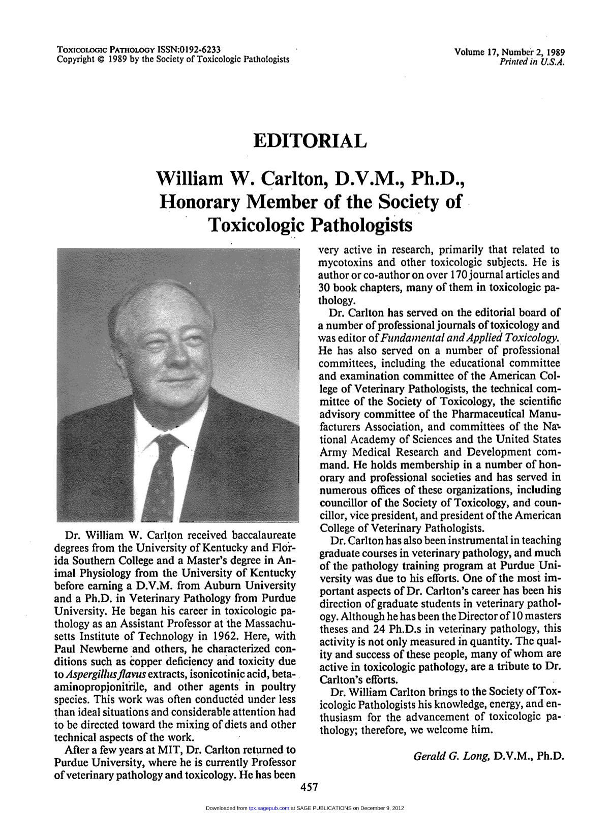 Free accessResearch articleFirst published February, 1989pp. 458-459
 Free accessOtherFirst published February, 1989pp. 460-460
 Free accessOtherFirst published February, 1989pp. 463-464