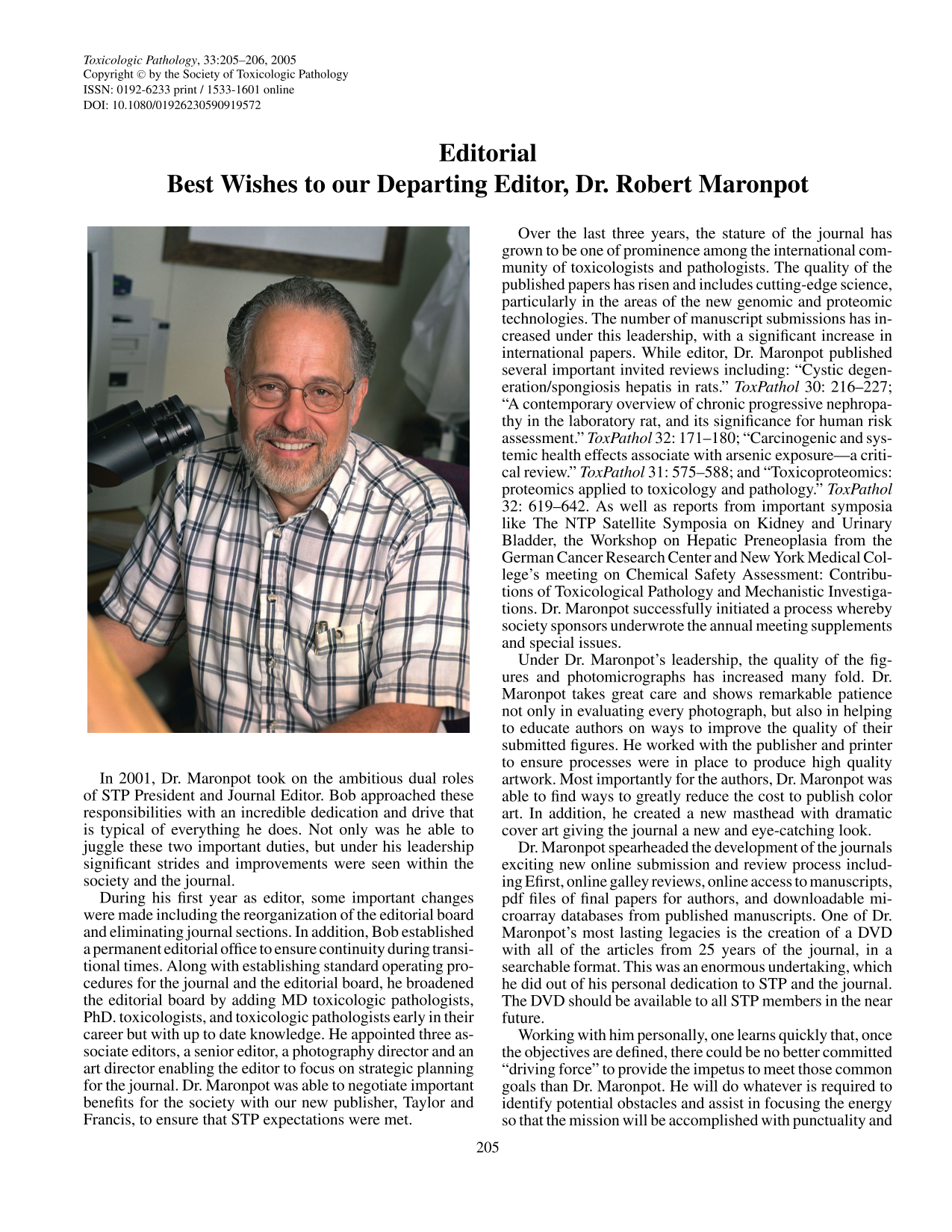
Editorial
 Free accessEditorialFirst published February, 2005pp. 205-206
Best Wishes to our Departing Editor,Dr. Robert Maronpot
View All


Select search scope: search across all journals or within the current journal

 Free accessEditorialFirst published February, 2005pp. 205-206Free accessResearch articleFirst published February, 2005pp. 207-217
Cisplatin (CDDP)-induced renal lesions in rats prove a useful model for analysis of the pathogenesis of post-tubular injury-renal interstitial fibrosis. This study investigated the histopathological changes in 10-day-old neonatal rats induced by a single injection of CDDP (4.5 mg/kg). Compared with age-matched controls, on postinjection (PI) days 1 to 6, the number of apoptotic cells, demonstrable with TUNEL method, was significantly increased in CDDP-treated neonates, and there was no marked epithelial necrosis nor fibrotic lesions. Fibrotic lesions began to be developed solitarily around some nephrons with dilated ducts in the corticomedullary junction on PI day 10 and the lesions became more prominent until PI day 20. The α-SMA-positive myofibroblastic cells were seen exclusively in the fibrotic lesions. Additionally, the numbers of macrophages reacting with ED1 (specific for exudate macrophages), ED2 (for resident macrophages), and OX6 (recognizing MHC class II antigens expressed in antigen-presenting macrophages/dendritic cells) were significantly increased around the affected renal tubules. A greater immunoreaction for TGF-β1 was seen mostly in the renal epithelial cells of CDDP-treated neonates. These findings indicated that macrophage populations and myofibrolastic cells as well as TGF-β1 may be responsible for the production of neonatal renal interstitial fibrosis. Compared with CDDP-injected adult rats that develop extensive interstitial fibrosis (Yamate et al.,
Free accessResearch articleFirst published February, 2005pp. 218-224Repeated administration of theophylline, a phosphodiesterase inhibitor, induces the enlargement of the salivary glands in rats. Time-course changes after a single administration of theophylline were examined in the salivary glands, including phosphodiesterase enzyme activity, and the expression of aquaporin 5 (AQP5), a water channel. We also examined the contribution of β-adrenergic receptors to theophylline-induced salivary changes. Male F344 rats were given 50 mg/kg of theophylline intraperitoneally either alone or concurrently with a 10 mg/kg subcutaneous injection of propranolol. After treatment with theophylline alone, the weight and histology of the submaxillary and parotid glands were examined. Phosphodiesterase activity and AQP5 were detected by enzyme- and immuno-histochemistry, respectively. At 4 hours, 8 hours, or both, organ weights were decreased with depletion of secretory vesicles in the acinar cells. In the submaxillary glands, reduced activity of phosphodiesterase and increased expression of AQP5 in the intercalated ducts were observed at 4 hours. When co-administered, propranolol partially abolished theophylline-induced glandular reduction. These results suggest that the theophylline-induced transient reduction in size of the salivary glands is attributable not only to phosphodiesterase inhibition but also to β-adrenergic receptor activation and that the intercalated ducts in submaxillary glands play a role in the production of saliva.
Free accessResearch articleFirst published February, 2005pp. 225-229Accidental inhalation of selenium (Se) derivatives, such as dimethyl selenide (DMSe), has been associated with damage of respiratory tissues. However, systemic effects of inhaled Se have not been thoroughly established. We have investigated whether mouse kidney and liver show cellular pathology as a result of a single intratracheal instillation of two different doses of DMSe (0.05 and 0.1 mg Se/kg BW). The animals were sacrificed 1, 7, 14, and 28 days after either 1 of the 2 DMSe treatments; samples were studied by light microscopy. Instillation of the low DMSe dose resulted in acute and transient tubular disease of the kidney expressed by swelling and vacuolation of epithelial cells of proximal tubules; in some mice, tubular necrosis was observed. After 14 days of the DMSe treatment, these lesions were ameliorated and, by day 28, the kidney tubular epithelium depicted a normal morphology. The same low dose of DMSe caused sustained damage to centrilobular hepatocytes characterized by swollen and vacuolized liver cells. After the instillation of the high DMSe dose, the mice presented sustained liver and kidney focal necrosis. Our data suggest that inhalation of DMSe results in: (i) acute tubular injury of the kidney and damage to centrilobular liver cells and (ii) this systemic pathology induced by DMSe is a dose-dependent phenomenon.
Free accessResearch articleFirst published February, 2005pp. 230-238Parathyroid hormone related peptide (PTHrP) was discovered as a causative factor of humoral hypercalcemia of malignancy (HHM). In the present study using HHM model rats, the time course of odontoblastic response to PTHrP and its relation to incisal fracture were elicited. Nude rats were implanted with PTHrP-expressing tumor (LC-6) cells, mandibular incisors were collected at several time points. Microscopically 3 distinctive types of odontoblastic/dentin lesions were observed. Hypercalcfied dentin, which was reported as hypercalcemia-induced lesion in previous reports, observed in all areas of the dentin from week 5–10 samplings. Dentin niche, observed solely in week-10 sampling point, exhibited a nature identical to that of reparative odontoblast reported in the literatures of various cytotoxic agents. Since cytotoxicites were neither observed prior to the lesions nor reported as a role of PTHrP, the reparative response may have derived from highly sustained levels of PTHrP. Loss of columnar odontoblasts height was initially observed at week-5 time point in the middle section of the incisor. This primary loss of cell height prior to incisor fracture was considered to be the earliest response to the increased PTHrP levels of this model.
Free accessResearch articleFirst published February, 2005pp. 239-245The aim of this study was to use immunohistochemistry with morphometry to investigate COX-1 and COX-2 expression in the normal rat gastrointestinal (GI) tract and examine if sites of ulceration previously observed with long-term COX-2 inhibitor administration in mice correlate with differential COX-1/COX-2 expression. COX-2 positive cells were observed predominantly in the apical lamina propria of intestinal villi with fewer cells in the mucosal epithelium. The highest level of COX-2 expression was observed at the ileocaecal junction (ICJ). COX-2 expression was also present in parasympathetic ganglia of the submucosa and muscularis. In the stomach, the highest grade of COX-2 expression was observed in the apical lamina propria of the fundus adjacent to the junctional ridge. In contrast, COX-1 positive cells within the lamina propria were evenly distributed along the GI tract but were present in higher numbers than COX-2 positive cells. The mean level of COX-1 expression at the ICJ was not significantly different from the ileum and caecum. Evidence that the highest level of COX-2 expression in normal rats is located on the ileal side of the ICJ provides the first mechanism to explain spontaneous ulceration and perforation of the distal ileum in COX-2−/− animals.
Free accessResearch articleFirst published February, 2005pp. 246-257Simvastatin and cerivastatin have been used to investigate the development of statin-induced muscle necrosis in the rat. This was similar for both statins and was treatment-duration dependent, only occurring after 10 days had elapsed even if the dose was increased, and still occurring after this time when dosing was terminated earlier as a result of morbidity. It was then widespread and affected all areas of the muscular system. However, even when myotoxicity was severe, particular individual muscles and some types of fibres within affected muscles were spared consistently. Fibre typing of spared muscles and of acutely necrotic fibres within affected muscles indicated a differential fibre sensitivity to statin-induced muscle necrosis. The fibres showed a necrotic response to statin administration that matched their oxidative/glycolytic metabolic nature: Least sensitive →
Free accessResearch articleFirst published February, 2005pp. 258-266Despite widespread exposure to military jet fuels, there remains a knowledge gap concerning the actual toxic entities responsible for irritation observed after topical fuel exposure. The present studies with individual hydrocarbon (HC) constituents of JP-8 jet fuel shed light on this issue. To mimic occupational scenarios, JP-8, 8 aliphatic HC (nonane, decane, undecane, dodecane, tridecane, tetradecane, pentadecane, hexadecane) and 6 aromatic HC (ethyl benzene,
Free accessResearch articleFirst published February, 2005pp. 267-271In the present study, susceptibility of CB6F1 mice carrying the human prototype c-Ha-
Free accessResearch articleFirst published February, 2005pp. 272-282We recently proposed a chemically induced rat model for human hemolytic disorders associated with thrombosis. The objective of the present investigation was to apply a noninvasive, high-magnification X-ray analysis, the Faxitron radiography system, to characterize the protracted bone damage associated with this 2-butoxyethanol model and to validate it by histopathology. Groups of female Fischer 344 rats were given 0, 250, or 300 mg of 2-butoxyethanol/kg body weight daily for 4 consecutive days. Groups were then sacrificed 2 hours or 26 days after the final treatment. The treated animals displayed a darkened purple-red discoloration on the distal tail. Histopathological evaluation, including phosphotungstic acid-hematoxylin staining of animals sacrificed 2 hours after the final treatment, revealed disseminated thrombosis and infarction in multiple organs, including bones. The Faxitron MX-20 specimen radiography system was used to image selected bones of rats sacrificed 26 days posttreatment. Premature thinning of the growth plate occurred in the calcaneus, lumbar and coccygeal vertebrae, femur, and ilium of the treated animals. Areas of decreased radiographic densities were seen in the diaphysis of the femur of all treated animals. The bones were then examined histologically and showed a range of changes, including loss or damage to growth plates and necrosis of cortical bone. No thrombi were seen in the animals sacrificed at 30 days, but bone and growth plate changes consistent with prior ischemia were noted. The Faxitron proved to be an excellent noninvasive tool that can be used in future studies with this animal model to examine treatment modalities for the chronic effects of human thrombotic disorders.
Free accessResearch articleFirst published February, 2005pp. 283-291Following recent clarification in Europe that a single control group is now acceptable for rodent carcinogenicity studies, the use of dual controls may be reduced or disappear. To date, virtually nothing has been published on whether this latter situation has improved the identification of tumorigenic risk potential in these studies. In this paper, the results of 13 rat carcinogenicity studies, performed between 1991 and 2002, with 2 control groups, are presented. Although no major differences in tumor incidences between these dual control groups were found, some interstudy variation occurred. In cases where a notable difference was seen, the use of 2 control groups, as well as robust, contemporary background data, allowed an easier interpretation of findings in drug-treated groups. Thus, the continued use of dual control groups has a vital role in the assessment of tumoriogenic risk. The paper also presents an update on survival, on the range and extent of background spontaneous neoplasms, and comments on genetic drift in this commonly used rat strain.
Free accessResearch articleFirst published February, 2005pp. 292-299Treatment with 8-methoxypsoralen (8-MOP) and ultraviolet radiation (primarily UVA), called PUVA therapy, has been used to treat different chronic skin diseases but led to a significant increased risk for skin cancer. The National Toxicology Program (NTP) performed a study in mice treated with PUVA that showed a significant increase in squamous cell carcinomas of the skin. In the present study, we evaluated the protein expression of
Free accessResearch articleFirst published February, 2005pp. 300-304Histopathological examination of the testes of exposed fetuses and neonates is important in assessing the developmental effects of environmental toxins, including sex hormone modulators. Modified Davidson’s fluid (mDF) has been suggested as a superior substitute for Bouin’s fluid for fixation of adult animal testes. We compared the morphology of fetal rat testes stained with hematoxylin and eosin (H&E) or immunochemically after fixation in 10% neutral buffered formalin (NBF), Bouin’s fluid, or mDF. Fixation in mDF resulted in more sharply defined nuclear detail and better preservation of cellular cytoplasm on H&E-stained sections of rat testes on gestation day 19. Use of Bouin’s fluid did not allow satisfactory detection of apoptotic cells by fluorescent terminal deoxynucleotide transferase-mediated deoxy-UTP nick labeling. Staining with the immunoperoxidase system and the conventional chromogen diaminobenzidine tetrahydrochloride to visualize 5-bromo-2-deoxyuridine-positive cells demonstrated that the number of positive nuclei and intensity of staining were similar with all 3 fixatives. Immunostaining for cytoskeletal protein vimentin was more intense and provided better details of the Sertoli cell cytoplasm with formalin fixation than with mDF. Our study demonstrates that fixation in mDF provided better morphologic detail in the fetal rat testis compared with 10% NBF and Bouin’s fluid and illustrates the importance of establishing the correct fixation conditions for each immunostaining protocol.