Editorial
 Free accessEditorialFirst published September, 2009pp. 789-789

Select search scope: search across all journals or within the current journal


 Free accessEditorialFirst published September, 2009pp. 789-789Free accessEditorialFirst published September, 2009pp. 790-791
 Free accessResearch articleFirst published September, 2009pp. 792-799
Pathogenic species of
Free accessResearch articleFirst published September, 2009pp. 800-809Class II major histocompatibility complex (MHCII) is required for the presentation of antigens to CD4 helper T cells. During nephritis, not only primary antigen presenting cells such as histiocytes and lymphocytes, but also cytokine-stimulated tubular epithelial cells express MHCII. Leptospirosis in fattening pigs is characterized by several degrees of nephritis, from absence of lesions to severe multifocal tubulo-interstitial inflammation. Renal tissue from 20 8-month-old pigs with spontaneous nephritis and 6 control pigs without renal lesions were investigated for leptospirosis by indirect immunohistochemistry (IHC) and polymerase chain reaction (PCR). IHC for MHCII also was performed on renal samples. Serum samples were tested for different serovars of
Free accessResearch articleFirst published September, 2009pp. 810-816Transmissible spongiform encephalopathies (TSEs) are a group of diseases that result in progressive and invariably fatal neurologic disease in both animals and humans. TSEs are characterized by the accumulation of an abnormal protease-resistant form of the prion protein in the central nervous system. Transmission of infectious TSEs is believed to occur via ingestion of prion protein–contaminated material. This material is also involved in the transmission of bovine spongiform encephalopathy (“mad cow disease”) to humans, which resulted in the variant form of Creutzfeldt-Jakob disease. Abnormal prion protein has been reported in the retina of TSE-affected cattle, but despite these observations, the specific effect of abnormal prion protein on retinal morphology and function has not been assessed. The objective of this study was to identify and characterize potential functional and morphologic abnormalities in the retinas of cattle infected with a bovine-adapted isolate of transmissible mink encephalopathy. We used electroretinography and immunohistochemistry to examine retinas from 10 noninoculated and 5 transmissible mink encephalopathy–inoculated adult Holstein steers. Here we show altered retinal function, as evidenced by prolonged implicit time of the electroretinogram b-wave, in transmissible mink encephalopathy–infected cattle before the onset of clinical illness. We also demonstrate disruption of rod bipolar cell synaptic terminals, indicated by decreased immunoreactivity for the alpha isoform of protein kinase C and vesicular glutamate transporter 1, and activation of Müller glia, as evidenced by increased glial fibrillary acidic protein and glutamine synthetase expression, in the retinas of these cattle at the time of euthanasia due to clinical deterioration. This is the first study to identify both functional and morphologic alterations in the retinas of TSE-infected cattle. Our results support future efforts to focus on the retina for the development of new strategies for the diagnosis of TSEs.
Free accessResearch articleFirst published September, 2009pp. 819-835The use of murine models to investigate human diseases has been an invaluable tool. In the areas of inflammation and oncogenesis, such models have provided unique insights into pathogenesis and mechanisms to evaluate potential therapy. As such, one facet of these disease processes links inflammation and cancer. Inflammation is associated with at least 15% of the world's malignancies. One example of this relationship is documented in the association between colitis and colorectal cancer. To date, the precise molecular events linking inflammation and cancer remain unclear. A new paradigm that may bridge these processes includes the cancer stem cell hypothesis. In this review, murine models of colitis, colon cancer, and colitis-associated cancer are discussed in reference to the potential of this paradigm to clarify the relationship of these devastating diseases.
Free accessResearch articleFirst published September, 2009pp. 836-845Fifty-one meningiomas obtained from 28 dogs and 23 cats were selected for this study to investigate the immunohistochemical expression of matrix metalloproteinase (MMP)-2 and MMP-9 and to compare it to the reverse transcriptase subunit of human-telomerase, progesterone receptor expression, and the proliferative index of the tumors, expressed by Ki67 and proliferating cellular nuclear antigen. Paraffin-embedded tumor tissue was obtained from biopsy samples (28 cases) and at necropsy (23 cases). The most common histotype was malignant in dogs (12/28) and transitional in cats (12/23). Slides immunolabelled for MMPs showed a diffuse cytoplasmic pattern. Twenty-one cases (19 dogs and 2 cats) did not express MMP-2, while only 2 cases were completely negative for MMP-9. The highest values of MMP-2 and MMP-9 were observed in a psammomatous and meningothelial tumor, respectively. On statistical analysis, MMP-2 expression did not show a significant correlation with MMP-9. Moreover, both MMP expressions failed to show significant variance among histologic patterns of the tumor and correlation with the proliferative index. MMP immunolabeling showed an inconstant correlation with progesterone receptor expression. No significant correlation was found between MMP and reverse transcriptase subunit of human-telomerase expression. In feline meningiomas, the MMP-2 value was significantly higher than in canine tumors and the MMP-9 value tended to be low for meningiomas with a follow-up duration from the 23rd month to the 44th month. In cats, the longer the time from surgery, the lower the proliferative index seemed to be. In dogs, we failed to find a correlation between MMP expression and the follow-up duration.
Free accessResearch articleFirst published September, 2009pp. 846-859Previous studies showed that intercellular communication by gap junctions has a role in bone formation. The main connexin involved in the development, differentiation, and regulation of bone tissue is connexin (Cx) 43. In addition, Cx46 is also expressed, mostly localized within the trans-Golgi region. Alterations in the expression pattern and aberrant location of these connexins are associated with oncogenesis, demonstrating a deficient gap junctional intercellular communication (GJIC) capacity in neoplastic tissues. In this study, we evaluated normal and neoplastic bone tissues regarding the expression of Cx43 and Cx46 by immunofluorescence, gene expression of these connexins by real-time PCR, and their correlation with cell proliferation index and deposition of collagen. Fourteen neoplastic bone lesions, including 13 osteosarcomas and 1 multilobular tumor of bone, were studied. The mRNA levels of Cx43 were similar between normal and neoplastic bone tissue. In normal bone tissue, the Cx43 protein was found mainly in the intercellular membranes. However, in all bone tumors studied here, the Cx43 was present in both cell membranes and also aberrantly in the cytoplasm. Regarding only tumor samples, we determined a possible inverse correlation between Cx43 expression and cellular proliferation, although a positive correlation between Cx43 expression and collagen deposition was also noted. In contrast, Cx46 had lower levels of expression in neoplastic bone tissues when compared with normal bone and was found retained in the perinuclear region. Even though there are differences between these two connexins regarding expression in neoplastic versus normal tissues, we concluded that there are differences regarding the subcellular location of these connexins in normal and neoplastic dog bone tissues and suggest a possible correlation between these findings and some aspects of cellular proliferation and possibly differentiation.
Free accessResearch articleFirst published September, 2009pp. 860-868Phosphatase and tensin homolog (PTEN) belongs to the group of gatekeeper tumor suppressor genes and is involved in multiple mechanisms leading to cellular defense against neoplastic transformation and progression. Twenty-four dogs and 17 cats were submitted to a 2-year follow-up study, and clinicopathologic features were recorded and compared with immunohistochemical PTEN staining. PTEN-negative status occurred in 33% of canine and 76% of feline mammary carcinomas. In canine mammary carcinomas, there was a significant (
Free accessResearch articleFirst published September, 2009pp. 869-877Hepatocyte growth factor (HGF) and the proto-oncogenic receptor c-Met are implicated in growth, invasion, and metastasis in human cancer. Little information is available on the expression and role of both gene products in canine osteosarcoma. We hypothesized that the expression of c-Met is associated with malignant histologic characteristics, a short survival time, and a reduced disease-free interval in canine osteosarcoma. Quantitative real-time polymerase chain reaction was used to analyze the messenger RNA (mRNA) expression of both HGF and c-Met in 59 canine osteosarcoma samples. The relationship between HGF and c-Met expression, patient outcome, and histologic characteristics of the tumor were studied. Western blot analysis was performed to investigate the presence of active HGF protein. The expression pattern of c-Met in 16 slides of canine osteosarcoma was identified by immunohistochemistry. Coexpression of HGF and c-Met mRNA in all canine osteosarcoma samples suggested autocrine or paracrine receptor activation. A significant, moderately positive correlation was found between c-Met and HGF mRNA expression. c-Met mRNA expression was not associated with survival time or disease-free interval. Expression of c-Met was significantly associated with metastasis via the lymphogenic route. Immunolabeling with c-Met revealed a cytoplasmic staining pattern in all osteosarcoma cell types. In this study, c-Met mRNA expression in canine osteosarcoma was found to be of no influence on survival time and disease-free interval. Further studies are necessary to confirm the involvement of the c-Met pathway in the lymphogenic route of metastasis.
Free accessResearch articleFirst published September, 2009pp. 878-883Twenty-seven feline cutaneous mast cell tumors (MCTs) were selected for this retrospective study. Samples were routinely processed and stained with hematoxylin and eosin (HE) and toluidine blue, and tumors were classified as well-differentiated (19/27), atypical or poorly granulate (7/27), and pleomorphic (1/27). Immunohistochemistry to detect KIT protein was performed on all samples. The immunoreactivity was recorded by distribution within the tumor, cellular location, and intensity. Well-differentiated MCTs were predominantly characterized by diffuse cytoplasmic (8/19) and membranous stain (7/19); a diffuse distribution of KIT positive cells was displayed in most of these tumors as well (15/19). Atypical MCTs showed diffuse distribution of labeled cells (4/7), and diffuse cytoplasm immunostaining was seen most (5/7). The pleomorphic MCT showed diffuse cytoplasmic KIT stain, with moderate labeling intensity, typically displaying focal distribution in deeper areas of the neoplasm. According to the results, there was no correlation between the type of MCTs and KIT expression, although the use of feline KIT immunohistochemistry could be useful to assess the mast cell origin.
Free accessResearch articleFirst published September, 2009pp. 884-892Meningiomas are common primary brain tumors in dogs; however, little is known about the molecular genetic mechanisms involved in their tumorigenesis. Several tumor suppressor genes have been implicated in meningioma pathogenesis in humans, including the neurofibromatosis 2 (
Free accessResearch articleFirst published September, 2009pp. 893-896GATA-4 is a transcription factor expressed in Sertoli cells and less commonly in Leydig (interstitial) cells but not germ cells in adult human beings, cattle, pigs, and mice. We examined GATA-4 in 76 formalin-fixed, paraffin-embedded canine testicular tumors, including 21 Sertoli cell tumors (SCT), 28 Leydig (interstitial) cell tumors (LCT), 24 seminomas (GCT), and 3 mixed germ cell sex cord-stromal tumors (MGSCT). Our hypothesis was that immunohistochemistry for GATA-4 could discriminate between germ cell and sex cord-stromal tumors of the canine testis. SCTs (21/21) had strong diffuse nuclear and weak and inconsistent cytoplasmic staining. LCTs (27/28) also had strong diffuse nuclear staining and much weaker and granular cytoplasmic staining. GCTs were negative for this marker. Sex cord-stromal cells of MGSCT were also positive. These results indicate that GATA-4 is mainly expressed in sex cord-stromal tumors and not in germ cell tumors of the canine testis.
Free accessResearch articleFirst published September, 2009pp. 897-903The adenomatoid tumor is an uncommon benign lesion, thus far described only in humans. Adenomatoid tumors typically arise in the genital tract, exceptionally in the heart, and usually represent an incidental finding. Microscopically, they are constituted by epithelioid cells that form tubular structures and anastomosing channels within a fibrous stroma. Mesothelial origin of these lesions is suggested by their immunohistochemical characteristics. In cattle, previously reported myocardial epithelial inclusions are morphologically similar in that the cells are immunoreactive for both cytokeratins and vimentin, and bear surface microvilli. Myocardial lesions found incidentally at slaughter in 8 cattle histologically resembled the so-called bovine myocardial epithelial inclusions and had morphologic and immunohistochemical features consistent with human adenomatoid tumor. All lesions were in the left ventricular myocardium, adjacent to the epicardium, and composed of epithelioid cells that formed cords and tubules, and were immunoreactive for pan-cytokeratins, cytokeratin 5/6, vimentin, calretinin, Wilms' tumor 1 suppressor gene, and CD30 antigen. By electron microscopy, numerous long slender microvilli were associated with desmosomes and tonofibrils. The immunohistochemical and ultrastructural features were considered consistent with mesothelial origin. These lesions, corresponding to the previously described myocardial epithelial inclusions in cattle, might be considered embryologic rests and could represent the bovine counterpart of the human adenomatoid tumor.
Free accessResearch articleFirst published September, 2009pp. 904-915In humans, neurofibroma and schwannoma are distinct entities within the group of benign peripheral nerve sheath tumors. In the veterinary literature, these tumors are often classified together simply as benign peripheral nerve sheath tumors, and diagnostic criteria for their subclassification are not well established. We describe peripheral nerve sheath tumors with microscopic, immunohistologic, and ultrastructural features similar to those in subtypes of human neurofibroma in 12 dogs, 2 horses, and 1 chicken. Dogs were of different breeds and were aged 2 months to 15 years. The canine tumors were located in the skin, peripheral nerve, tongue, and large intestine. The 2 horses were 11 and 12 years old. The equine tumors were located in the subcutis of the neck and axilla. The chicken was a mature white Leghorn chicken with an ocular neoplasm. Neurofibromas of this study had localized, plexiform, diffuse and combined plexiform and diffuse growth patterns, and microscopic features similar to those in classic, collagenous, cellular, myxoid, and pigmented neurofibromas of humans. One diffuse neurofibroma contained areas of schwannian differentiation (hybrid neurofibroma-schwannoma). Two plexiform neurofibromas occurred together with diffuse ganglioneuromatosis in the large intestine of young dogs, as has also been reported in humans. This investigation shows the existence of identical subtypes of neurofibroma in animals and humans and identifies similarities in tumor location and patient age between animals and humans. This report will allow a more discriminating classification of benign peripheral nerve sheath tumors and probably has a bearing on epidemiology, pathogenesis and prognosis.
Free accessResearch articleFirst published September, 2009pp. 916-927Submissions to the University of Tennessee pathology service from June 1999 to June 2008 were searched for feline cases of tumors involving the eyelids or nictitans. Forty-three tumors were identified. The average age at diagnosis was 10.4 years. Significantly more males than females had eyelid tumors. There were 12 squamous cell carcinomas (SCCs), 11 mast cell tumors (MCTs), 6 hemangiosarcomas (HSAs), 4 adenocarcinomas (ACAs), 3 peripheral nerve sheath tumors (PNSTs), 3 lymphomas, 3 apocrine hidrocystomas (AHCs), and 2 hemangiomas. Cats with MCTs were significantly younger than cats with all other tumor types combined. In contrast, cats with SCCs were significantly older than cats with other tumor types. The HSAs and SCCs were significantly more likely than other tumors to occur in nonpigmented areas. The MCTs, HSAs, AHCs, and hemangiomas did not recur after surgical excision. In contrast, the lymphomas, ACAs, SCCs, and PNSTs frequently recurred and/or resulted in death or euthanasia of the cat. The SCCs were significantly more likely to recur than the MCTs. The average survival time for cats with SCCs was 7.4 months. Although eyelid MCTs have been reported in cats, the prevalence in this study is much higher than previously described.
Free accessResearch articleFirst published September, 2009pp. 928-933Local recurrence of marginally excised subcutaneous soft tissue sarcomas is variable and difficult to predict. This study aimed to identify predictors of local recurrence after excisional biopsy. Medical records of 236 dogs from which tumors had been received between 2004 and 2007 were analyzed. Medium- to large-breed dogs, median age 10 years, were most commonly affected. A total of 139 tumors were graded histologically: 71 were grade 1 (51%); 59, grade 2 (42%); and 9, grade 3 (7%). Of these, 34 tumors (25%) were completely excised, and 104 (75%) were marginally excised. None of 30 completely excised tumors with follow-up information recurred. Three of 41 grade 1 tumors (7%), 14 of 41 grade 2 tumors (34%), and 3 out of 4 grade 3 tumors recurred after marginal excision. Kaplan-Meier survival curves were generated to evaluate survival and the tumor-free interval. The log-rank test and log-rank test for trend were used for comparisons. Tumor recurrence-free intervals for dogs with grade 1 and 2 tumors and for those with grade 1 and 3 tumors differed significantly (
Free accessResearch articleFirst published September, 2009pp. 934-939C-KIT is the cellular homolog of the feline sarcoma viral oncogene v-KIT, which encodes the tyrosine kinase receptor protein KIT. Mutations and varied expression of this gene have been demonstrated within multiple neoplasms in people and domestic animals. The purpose of this study was to determine if KIT protein is expressed in feline soft tissue fibrosarcomas (ST FSA) using immunohistochemistry (IHC). The computer database at the Oklahoma Animal Disease Diagnostic Laboratory was searched from January 1, 2006, to December 31, 2007, for any domestic cat with an ST FSA. Routinely stained slides from 46 feline ST FSAs were reviewed and graded based on the scale outlined by Kuntz et al. Immunohistochemistry for KIT protein was performed on one representative section from each cat. There were a total of 12/46 (26%) cats that were immunoreactive for KIT. Immunoreactivity was detected in greater than 80% of the neoplastic cells in 4/46 (9%) cats. Immunoreactivity was detected in less than 10% of the neoplastic cells in 8/46 (17%) cats. Immunoreactivity was characterized by evenly distributed cytoplasmic stippling within the neoplastic spindle-shaped cells and/or multinucleated giant cells. Based on these results, KIT immunoreactivity can be detected within feline ST FSAs using IHC. The results of this study also indicate that KIT immunoreactivity in feline ST FSA does not correlate with the histologic grade (
Free accessBrief reportFirst published September, 2009pp. 940-944The genus
Free accessCase reportFirst published September, 2009pp. 945-948Ectopic infection with
Free accessResearch articleFirst published September, 2009pp. 949-951A primary intracerebral plasmacytoma was identified in a 7-year-old spayed female Boston Terrier. Grossly, a well-demarcated, 2 cm in diameter, roughly spherical tumor was in the rostral aspect of the left cerebral hemisphere. Histologically, the neoplasm was composed of sheets of round cells with distinct plasmacytoid features and marked anisocytosis and anisokaryosis. Cells were positive for vimentin, CD18, CD79a, and lambda light-chain, and negative for kappa light chain, cytokeratin, lysozyme, glial fibrillary acidic protein, and S100 protein. Clonally rearranged B-cell antigen receptor genes were detected by PARR (polymerase chain reaction for antigen receptor rearrangements), confirming clonal proliferation of B lymphocytes. Although primary solitary intracerebral plasmacytoma is rare in dogs and other species, it should be included in the differential diagnosis for central nervous system round-cell neoplasms. Clonality testing can be utilized to support the histological diagnosis of this neoplasm type.
Free accessResearch articleFirst published September, 2009pp. 952-959Lymphomas were reported to be induced in rats in bioassays of aspartame, methyl-
Free accessBrief reportFirst published September, 2009pp. 960-965
Free accessResearch articleFirst published September, 2009pp. 966-970Susceptibility of Canada geese (
Free accessResearch articleFirst published September, 2009pp. 971-976The largest recorded outbreak of highly pathogenic avian influenza virus of the subtype H7N7 occurred in the Netherlands in 2003. We describe the immunohistochemical and histopathologic findings of 3 chickens naturally infected during this outbreak. Influenza virus antigen occurred in endothelial cells and mononuclear cells of all tissues examined and occurred in parenchymal cells of heart, lung, kidney, pancreas, and trachea, often associated with multifocal inflammation and necrosis. These findings are consistent with the acute stage of highly pathogenic avian influenza from other subtypes. In the severely edematous wattle skin, most endothelial cells contained virus antigen, while in all other tissues virus antigen was only detected in a few endothelial cells. Virus histochemistry showed that this H7N7 virus attached to more endothelial cells in wattle skin than in other vascular beds. This might explain, at least partly, the tropism of the virus and the associated severity of lesions in this tissue.
Free accessResearch articleFirst published September, 2009pp. 977-984Marked renal vascular changes, suggestive of hypertension, were present in adult western gray kangaroos (
Free accessBrief reportFirst published September, 2009pp. 985-991Nine juvenile ferrets (
Free accessResearch articleFirst published September, 2009pp. 992-999Rat respiratory virus (RRV) is the working name for a novel respiratory pathogen of laboratory rats in North America, Europe, and Asia. Although the agent has not been definitively identified, evidence supports a viral etiology. Because no serologic or molecular assays for RRV are available, diagnosis depends on histopathologic evaluation of the lung. We introduced 104 Wistar Han rats, free of known pathogens and of RRV-associated lesions, into a rat production colony positive for RRV-type lesions, but free of other histologic, serologic, or microbiologic evidence of infectious disease. Lungs of 8 of the naïve rats were examined grossly and microscopically each week, weeks 0–13. Irregular gray-white lesions suggestive of interstitial pneumonia were grossly evident from weeks 6 through 13. Primary histopathologic evaluation of all lungs by one pathologist found multifocal, lymphohistiocytic interstitial pneumonia or prominent perivascular lymphoid cuffing from weeks 5 through 13. Based on results of the initial evaluation, diagnostic criteria for RRV infection (i.e., changes seen only after exposure to the RRV-positive colony) were tentatively selected and used by 2 other pathologists to classify each lung as RRV positive, RRV equivocal, or RRV negative. The secondary evaluation found 95% concordance in RRV diagnosis between pathologists, and correlated well with the initial evaluation, thus confirming the consistency of the criteria. These data show that RRV-naïve rats introduced into an RRV-endemic colony develop equivocal microscopic lesions of RRV by 5 weeks of exposure, and positive diagnostic lesions by 7 weeks. Interstitial pneumonia becomes grossly evident after 6 weeks of exposure.
Free accessCase reportFirst published September, 2009pp. 1000-1002A juvenile dwarf rabbit (
Free accessResearch articleFirst published September, 2009pp. 1003-1014Infection with human T-cell leukemia virus type 1 (HTLV-1) leads sometimes to the development of adult T-cell lymphoma/leukemia (ATL), which is invariably fatal and often associated with humoral hypercalcemia of malignancy. The transformation of infected CD4 T cells and the pathogenesis of leukemia have been studied with great limitation in tissue culture and patients. To better understand the pathogenesis and perform preclinical drug studies, animal models of ATL are urgently needed. In mice, inoculation of HTLV-1 cell lines mostly leads to development of localized lymphomas. To develop an ATL animal model with leukemic spread of ATL cells, mouse strains with different well-defined immune deficiencies were inoculated intraperitoneally with different HTLV-1infected cell lines (ACH.2, C8166, MT-2, MET-1). Inoculation of MET-1 cells into NOD/SCID mice provided the best model system for slowly developing T-cell leukemia with multiple organ involvement. In leukemic mice, an increase in serum calcium levels correlated with expression of receptor activator of nuclear factor kappa-light-chain-enhancer of activated B cells ligand on leukemic cells and secretion of parathyroid hormone-related protein and interleukin-6. In contrast to the other cell lines that did not spread systemically, MET-1 expressed both the adhesion molecules CDlla (LFA-lot) and CD49d (VLA-4ot) and produced or induced expression of matrix metalloproteinases 1, 2, 3, and 9, thus underlining the importance of these molecules in the spread of adult T-cell leukemia cells. The MET-1/NOD/SCID model will be useful for developing interventions against invasion and spread of leukemic cells and subsequent humoral hypercalcemia of malignancy.
Free accessResearch articleFirst published September, 2009pp. 1015-1017A backgrounding operation for calves in Wyoming identified a disease syndrome presenting as lethargy, fever, and death between November and January each year. An unfixed heart was submitted for examination, along with samples of lung. There was focal red discoloration in papillary muscle of the left ventricular myocardium. Histologically, the lesion corresponded to acute necrotizing myocarditis with myriad intravascular and intralesional Gram-negative coccobacilli.
Free accessBook reviewFirst published September, 2009pp. 1018-1018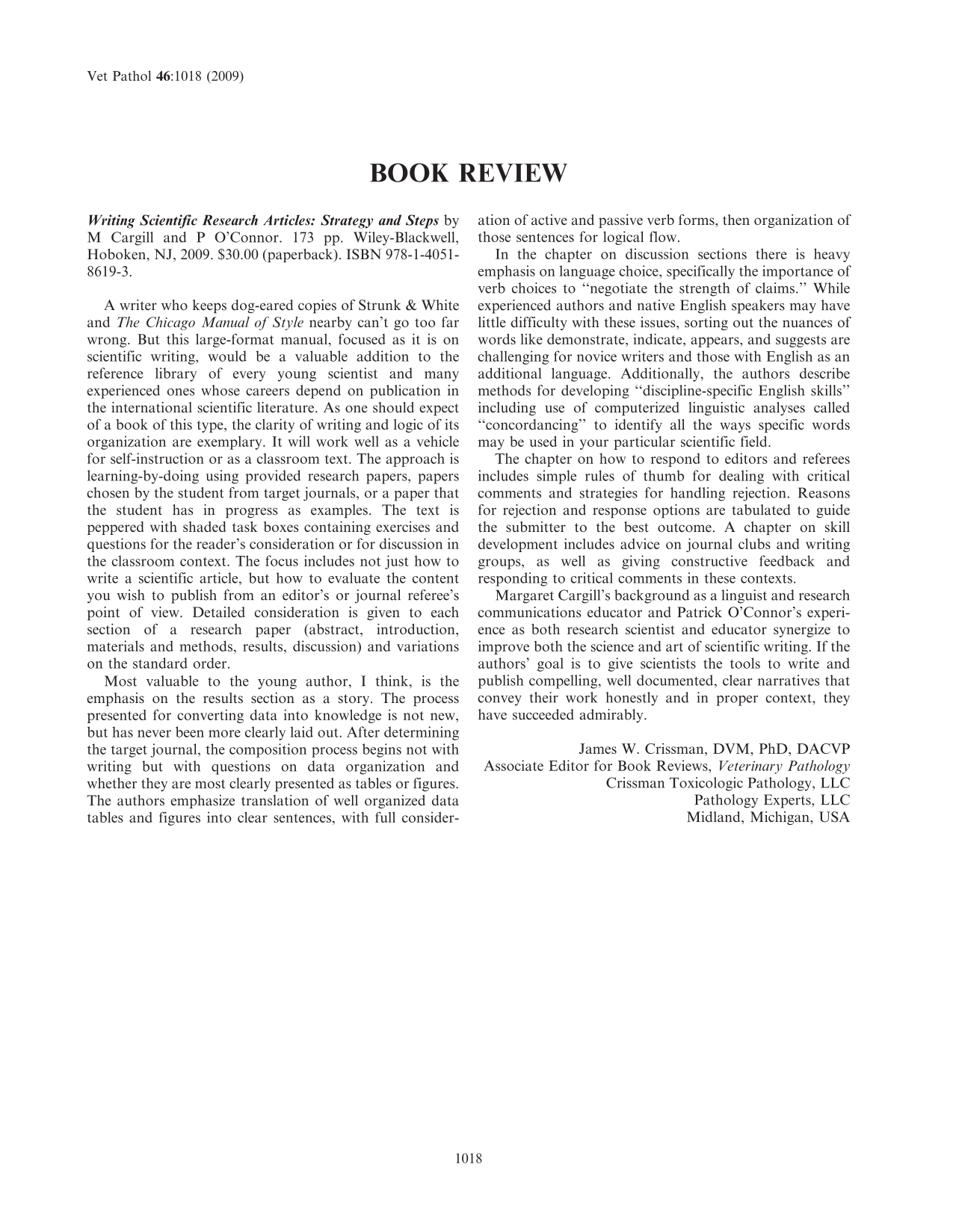 Free accessOtherFirst published September, 2009pp. 1019-1019
 Free accessAbstractFirst published September, 2009pp. 1020-1083
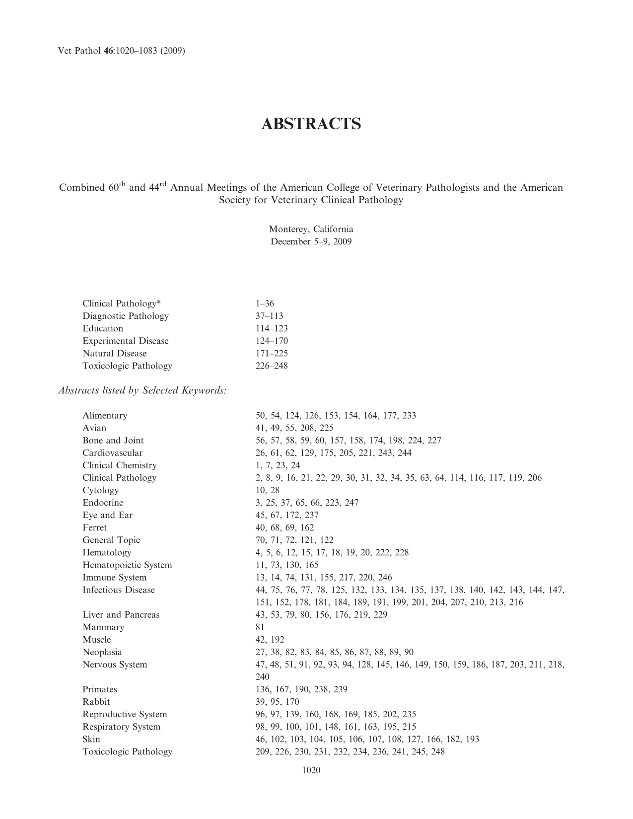 Free accessOtherFirst published September, 2009pp. 1084-1089