
Abstract
Bioink, a key element of three-dimensional (3D) bioprinting, is frequently engineered to achieve improved printing performance. Viscoelasticity related to rheological properties is correlative of the printability of bioink for extrusion bioprinting, which affects the complexity of printing 3D structures. This article shows the use of hydroxyethyl cellulose (HEC) as a rheological additive for engineering bioink to improve the printability without reducing the biocompatibility. Different concentrations of HEC were added to four types of bioink, namely, reagent-crosslinked, temperature-dependent phase change, ultraviolet-polymerized, and composite hydrogel bioinks, to investigate the effect on the viscoelasticity properties, print fidelity, and other printed scaffold properties. The results indicate that HEC is able to increase the rheological properties by 100 times to stabilize complex structures and improve the printing fidelity to narrow the gap between the design value and theoretical value, even converting nonviscous ink into directly printable ink, as well as tune the swelling ratio for better molecular permeability. The degradation of bioink can also be tuned by the addition of HEC. Moreover, this bioink is biocompatible for cell lines and primary cells. HEC is expected to be widely used in 3D extrusion-based bioprinting.
Introduction
Three-dimensional (3
The bioink for extrusion bioprinting is a non-Newtonian fluid; viscosity affects the deposition of extruded fibers, and viscoelasticity affects shape fidelity.13,14 Thus, the way to improve printability is to modify the rheological properties. One method is to control the crosslinking degree of hydrogels during printing, such as by changing the printing temperature to adjust the rheological properties of temperature-sensitive hydrogels,15–17 exposing them to ultraviolet (UV) light before or during extrusion to partially crosslink the photopolymerization hydrogels,18–21 and adding a certain concentration of ionic solutions to improve the viscoelasticity of ionic crosslinking hydrogels.22,23 However, this method is a “crosslinking-then-printing” approach, and changing the printing conditions is only suitable for specific bioprinters and bioinks. In addition, the partial crosslinking of hydrogels may result in nozzle clogging.
Another method to improve printability is adding self-supporting rheological additives, which is a “printing-then-crosslinking” approach, with high universality for manifold bioinks. Currently, the most common self-supporting rheological additive used for bioprinting is nanoclay. 24 Laponite can not only improve the printability of biomaterial inks to construct complex 3D structures with high shape fidelity25–29 but can also be applied in four-dimensional printing.30,31 Unfortunately, laponite is a dispersant that is insoluble in gel aqueous solution. The presence of nanoparticles can fill the pores of scaffolds, resulting in a reduction in the scaffold swelling 29 and the permeability of the active factors 32 ; these issues may affect the activity of the cells encapsulated in the bioinks and the effect of drugs, which are not amenable to printing bioinks for embedding cells. Therefore, it is necessary to develop water-soluble rheological additives to improve the printability of various types of bioinks.
In this work, we introduced hydroxyethyl cellulose (HEC) as a rheological additive to improve the rheological properties of bioinks. HEC is a nonionic ether derivative of natural cellulose that can be regenerated sustainably; it is widely used in cosmetics, cleaning solutions, and other household products as a gelling and thickening agent. It has good rheological properties, such as shear-thinning behavior and thixotropy,33–35 meeting the requirements of extrusion bioprinting. HEC has good water solubility and is soluble in both cold and hot water. In addition, this compound will not ionize in aqueous solution, has good salt tolerance, and is compatible with most water-soluble polymers.36–38 Moreover, HEC is biocompatible 39 and has been used in drug delivery40–42 and tissue engineering.43,44
In this article, HEC was added into calcium ion-crosslinked sodium alginate (SA), enzyme-crosslinked FB, temperature-dependent phase change gelatin (G), UV-polymerized PEGDA, and composite SA/gelatin to improve the extrusion printability. We systematically investigated the effect of different concentrations of HEC on bioink in terms of viscoelasticity, printing temperature, printing fidelity, swelling, permeability, degradation, and biocompatibility.
Materials and Methods
Material preparation
SA/HEC ink and G/HEC ink were prepared by adding HEC (434965; Aldrich) into 1.5% (w/v) SA (A0682; Sigma) and 6.5% (w/v) G (G1890; Sigma), respectively, with continuous stirring at 70°C for 2 h, resulting in SA/HEC ink and G/HEC ink with four concentrations of HEC: 0%, 1.5%, 3%, and 6%. SG/HEC ink was prepared by adding HEC into SG (1.5% SA and 6.5% gelatin), resulting in SA/HEC ink with different concentrations of HEC, namely, 0%, 1.5%, 3%, and 6%, as well as different mass ratios of HEC/SG: 0, 1:20, 1:10, 1:4, and 1:2. FB/HEC ink and PEGDA/HEC ink were prepared by adding HEC into 4% (w/v) FB (F8630; Sigma) and 10% (w/v) PEGDA (455008; Sigma), respectively, resulting in FB/HEC and PEGDA/HEC ink with three concentrations of HEC: 0%, 3%, and 6%.
Rheological characterization
Rheological properties of SA/HEC, G/HEC, and SG/HEC were measured by a rheometer (AR2000ex; TA Instruments) equipped with a Peltier plate with a diameter of 40 mm and a truncation gap distance of 100 μm. To measure the storage modulus (G′) and loss modulus (G′′), frequency sweep tests were performed by varying the angle frequency from 0.1 to 100 rad·s−1 at a fixed strain of 1%. To measure the viscosity, these inks were loaded with steady rate sweeps within a shear rate range of 0.1–100 s−1. Temperature sweep tests of SG/HEC were conducted at a rate of 1°C·min−1 from 40°C to 5°C, at a fixed strain and frequency of 1% and 1 Hz. All dynamic experiments were performed at 25°C.
Fabrication of scaffolds
The extrusion system was a homemade bioprinter, and the driving force for fluid distribution was mechanical force generated by a piston. The bioprinter had an electric-driven 3D moving system with a speed of 2–50 mm·s−1 and a cartridge for disposable sterile syringes. The predesigned STL file was imported into the Repetier host software to convert the STL 3D model into G code with 100% filling. The biomaterial inks were loaded into the cartridge with a 25-gauge (250 μm) print head. Then, 3D structures were fabricated according to the design. Finally, for SG/HEC printing, the printed structures were immersed in 1.5% CaCl2 solution for 10 min for crosslinking.
Scaffold property characterization
Swelling test
To measure the swelling rate of SG/HEC, crosslinked cylindrical samples with a diameter of 3 cm and a height of 5 mm were completely lyophilized, and the dry weight of each sample was recorded (Wd). Then, these samples were soaked in 3 mL phosphate-buffered saline (PBS) at 37°C. After 48 h, the swelling reached equilibrium, and the swollen weight of each sample was recorded (Ws). The equilibrium swelling ratio was calculated as Ws/Wd.
Permeability test
To measure the permeability of SG/HEC, crosslinked cylindrical samples with a diameter of 1 cm and a height of 3 mm were soaked in fluorescein sodium PBS solution, FITC-inulin PBS solution, and FITC-70 kDa dextran solution for 48 h. Then, the samples were removed and immersed in 1.5 mL PBS. At 10, 20, 30, and 60 min, the PBS solution was completely replaced, the volume was recorded as V, and the fluorescence intensity was measured. The molecular concentration of each PBS sample was obtained according to the fluorescence intensity standard curve and recorded as C. The molecular permeation content was calculated as C × V.
Degradation test
To measure the degradation ratio of SG/HEC, crosslinked cylindrical samples with a diameter of 1 cm and height of 4 mm were fabricated, and the weight of each sample was recorded as Wo. The static degradation of samples was evaluated by soaking in PBS and incubating on a 37°C shaker without shaking. On days 1, 2, 3, 4, and 5, the weights of samples were recorded as Wsd. The static degradation ratio was calculated as (Wo − Wsd)/Wo × 100.
In addition, the dynamic degradation of samples was evaluated by being soaked in PBS and incubated on a 37°C shaker at 120 rpm·min−1 for 24 h. After being removed from PBS, the weights of the samples were recorded as Wdd. The dynamic degradation ratio was calculated as (Wo − Wdd)/Wo × 100.
Gel formation test
SA/HEC inks were loaded into a 2 mL syringe, followed by extrusion into a 1.5% CaCl2 solution. The formation of fibrous gel was immediately observed. FB/HEC inks were gently mixed with 20 U·mL−1 thrombin (T4648; Sigma) in PBS at a volume ratio of 1:1, followed by pouring into PDMS round molds, and then, the gel formation was observed after culturing at 37°C for 30 min. HEC was dissolved in 10% (w/v) PEGDA containing 1.0% (w/v) Irgacure 2959 (H823463; Macklin) and then poured into PDMS round molds, followed by exposure to UV light for 10 min. Finally, the PDMS molds were removed to observe gel formation.
Biocompatibility test
Cell culture
Human umbilical vein endothelial cells (HUVECs) and human aortic smooth muscle cells (HAVSMCs) were donated by Dalian Medical University and maintained in Dulbecco's modified Eagle's medium (DMEM; Gibco) with 10% fetal bovine serum (FBS; Gibco) and 1% penicillin/streptomycin (Gibco). Cells were cultured at 37°C and 5% CO2. The medium was replaced every 2 days.
Primary hepatocytes were isolated from male Sprague-Dawley rats weighing 100–120 g according to the Seglen two-step perfusion protocol. We obtained hepatocytes with 85–95% viability. The primary hepatocytes were cultured in William's medium E supplemented with CM4000 (Gibco), 5% FBS, and 1% penicillin/streptomycin. Primary neuronal cells were harvested from newborn Sprague-Dawley rats within 24 h of birth. After the brain was exposed, the cortical tissue was collected and digested with collagenase for 10 min at 37°C. Then, FBS was added. The tissue was mechanically dissociated, filtered through a 100 μm cell strainer, and centrifuged at 800 rpm·min−1. Cells were resuspended in Neurobasal media with 2% B27 neural supplement and 1% penicillin/streptomycin.
Biocompatibility evaluation
To measure cell direct contact biocompatibility of SG/HEC, crosslinked cylindrical samples with a diameter of 1 cm and a height of 3 mm were fabricated. HUVECs and HAVSMCs were separately seeded on SG/HEC and cultured in DMEM. Cell viability was tested with live–dead assay. Then, 5 μM calcein AM solution and 1 μM propidium iodide (PI) were added to the medium and incubated at 37°C for 15 min. The samples were washed with PBS and imaged with a fluorescence microscope. Living and dead cells were counted using ImageJ software.
To evaluate cell indirect biocompatibility of SG/HEC, crosslinked cylindrical samples with a diameter of 1 cm and a height of 3 mm were fabricated and soaked in 2 mL culture media for 24 h according to the ISO 10993-5-biological evaluation of medical devices. Then, the extract liquid was collected. HUVECs, HAVSMCs, primary hepatocytes, and primary neuronal cells were plated on 96-well plates at 5000 cells well−1. Cultures were incubated with 100% extract liquid. The relative cell viability was calculated by taking the absorbance of cells cultured in the extract of HEC/SG with a ratio of 0 as 100%.
Statistical analysis
All data are presented as the mean ± standard deviation of independent replicates (n = 4). Statistical analysis was performed by analysis of variance using IBM SPSS Statistics, and p-values of <0.05 were considered statistically significant (*[0.01 < p < 0.05], **[0.001 < p < 0.01], ***[p < 0.001]).
Results
HEC to alter the printability of the bioink
HEC to alter the viscoelasticity of the bioink
The viscoelasticity of the bioinks was assessed using rheology at 25°C. This study investigated the effects of different concentrations of HEC on the rheological properties of ion-crosslinked SA bioink, temperature-dependent phase transition G bioink, and composite SG bioink. As shown in Figure 1A, the HEC composite inks at different concentrations of HEC exhibited different flow behaviors, and the viscosities of the HEC composite inks were higher than those of inks without HEC. In addition, the viscosities of SA, G, and SG at 25°C were all lower than 10 Pa·s, while the presence of HEC increased the viscosities to 100 Pa·s. As shown in Figure 1Aa, when the HEC concentration was 0%, the viscosity of S/HEC hardly changed with the shear rate, indicating that SA was a nonviscous ink. When HEC concentration was increased to 6%, it demonstrated obvious shear-thinning behavior. As shown in the viscosity-shear rate curves in Figure 1Ab and Ac, G/HEC and SG/HEC both exhibited shear-thinning behavior with increasing shear rates at different concentrations of HEC, which was true even if the HEC concentration was 0, indicating that G and SG were viscous inks.

Effect of HEC concentration on shear thinning properties and viscoelasticity modulus.
The viscoelastic properties of HEC composite inks are shown in Figure 1B and C. With increasing HEC concentration, the storage modulus (G′) and loss modulus (G′′) of S/HEC, G/HEC, and SG/HEC were increased, that is, the viscosity and elasticity of the bioink were increased. The viscoelastic modulus of different inks was ∼100 or <100 Pa when the HEC concentration was lower than 6%. By comparing the values of G′ (Fig. 1Ba) and G′′ (Fig. 1Bb) of S/HEC, it was found that G′ < G′′ when the HEC concentration was 0% and 1.5%, indicating that S/HEC exhibited a fluid-like property. When the HEC concentration was 3%, G′ and G′′ of S/HEC were equivalent only at high angular frequency. When the HEC concentration in S/HEC inks increased to 6%, G′ was similar to G′′ at all angular frequencies, indicating that S/HEC exhibited semisolid-like properties. For G/HEC (Fig. 1Bb, Cb) and SG/HEC (Fig. 1Bc, Cc) inks, G′ was higher than G′′ in the presence of HEC, indicating that the inks exhibited solid-like properties after adding HEC.
HEC to alter the printing temperature of the bioink
When SG was mixed with the cell suspension, the bubbles were removed at 37°C, and then, it was loaded in the ink cartridge at printing temperature. Changing the temperature can affect the gel properties. We measured the sol/gel transition temperature of SG/HEC by illustrating the modulus/temperature sweeping curves in which the intersection point (circle mark in Fig. 2A) indicates the gelling temperature. The sol/gel transition temperature of SG/HEC increased with increasing HEC concentration. When the concentration of HEC was 0% and 1.5%, the sol/gel transition temperature was lower than 20°C. When the concentration of HEC was 3% and 6%, the sol/gel transition temperature exceeded 20°C. We then printed grid scaffolds with SG/HEC (3%) and SG at 25°C. As shown in Figure 2A, printing SG/HEC (3%) was successful, but otherwise, printing SG failed.
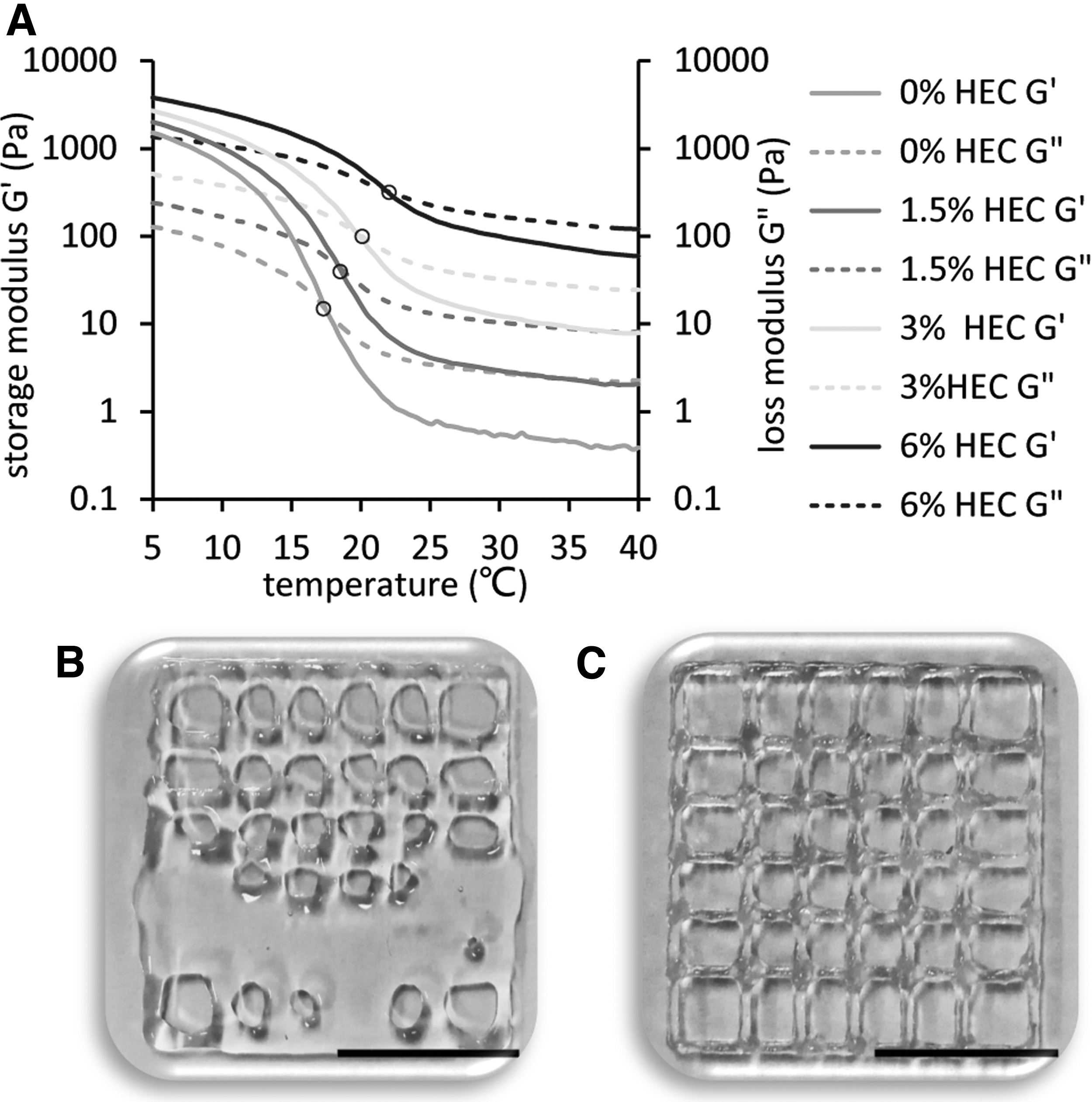
Effect of HEC on printing temperature of SG.
HEC to alter the printing fidelity of the bioink
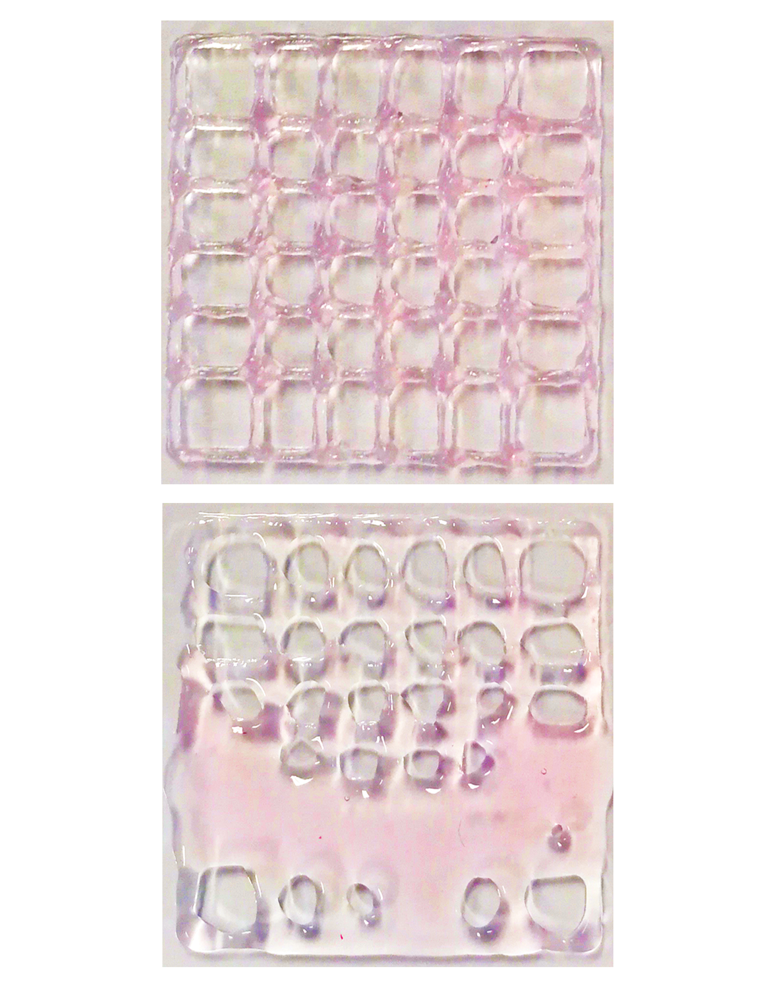
During the printing process, gravity and diffusion effects affected the height of the tube (Fig. 3A) and the pore area of the lattice (Fig. 3B). The ratios of actual height to theoretical height of SG with different concentrations of HEC are shown in Figure 3E; the higher the HEC concentration, the closer the ratio was to 1. The ratios of actual area to theoretical area of SG with different concentrations of HEC are shown in Figure 3F; the higher the HEC concentration, the higher the ratio. An increase in the HEC concentration reduced the structural deformation and improved the printing fidelity of SG.
Effect of HEC on the printing fidelity of bioink.
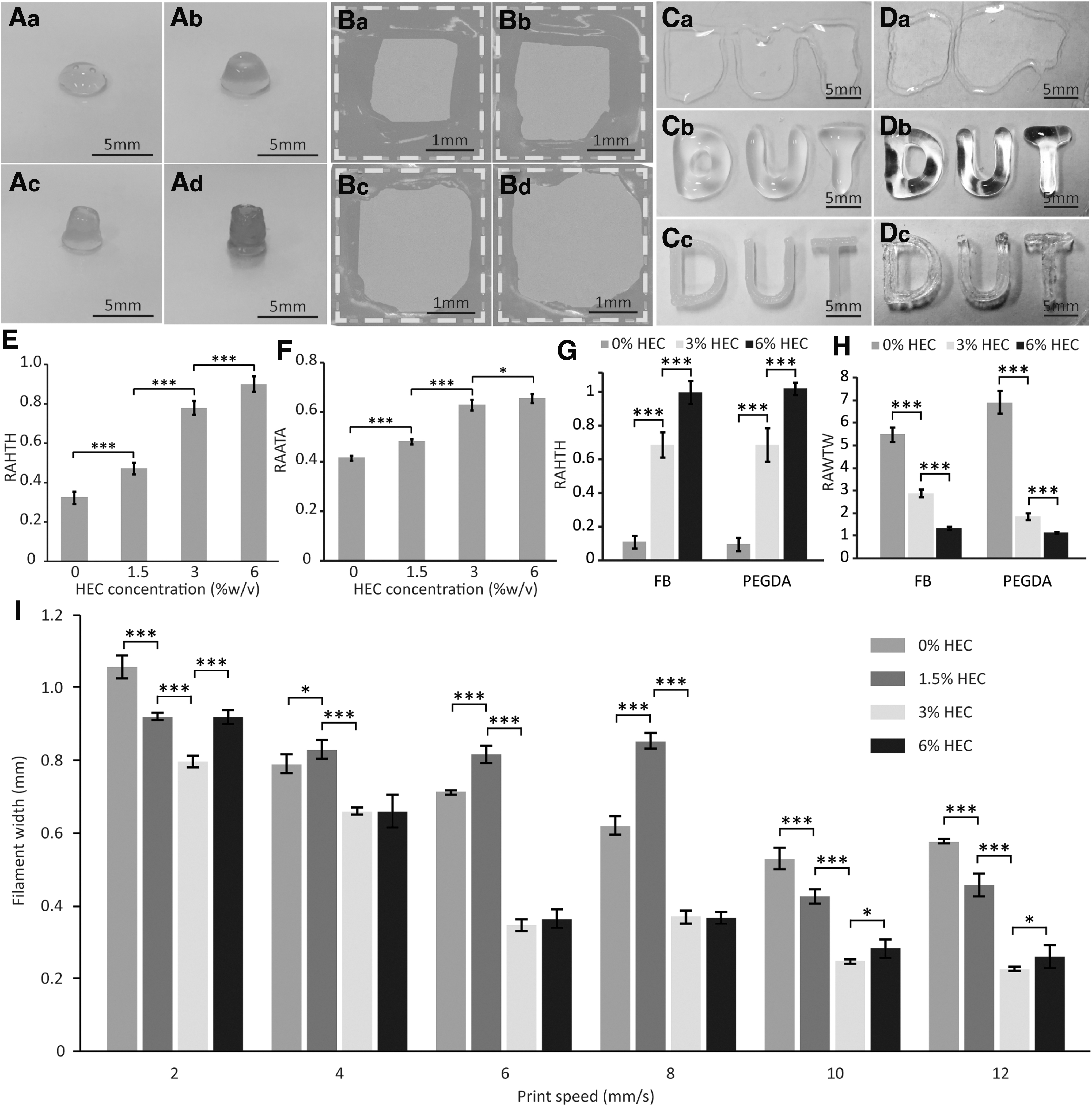
The UV-crosslinked PEGDA bioink and enzyme-crosslinked FB bioink both exhibited fluid-like behavior, resulting in unsuccessful direct printing, as shown in Figure 3Ca and Da. With the addition of 3% HEC (Fig. 3Cb, Db) and 6% HEC (Fig. 3Cc, Dc), the 3D structure was formed and printed successfully. With increasing HEC concentration, the ratio of actual width to theoretical width (Fig. 3G) and the ratio of actual height to theoretical height (Fig. 3H) were closer to 1 or 100%. This was because the addition of HEC increased the storage modulus of FB and PEGDA to hold the 3D structure and reduce the flow of the bioink.
During the printing process, the moving speed of the 3D platform affects the stacking of bioink, resulting in filament widths changing with printing speed. The effect of different concentrations of HEC and printing speed on the filament width is shown in Figure 3I. With increasing printing speed, the filament was gradually stretched, resulting in thinning of the filament width. At the same printing speed, the filament width of SG with 3% HEC added was thinner than that of SG with 0% HEC and 1.5% HEC added. This was because the bioink with a higher HEC concentration needed to bear stronger extrusion pressure, and the resulting filament width would be changed greatly. However, the filament width of SG with 6% HEC added was significantly larger than that of SG with 3% HEC added, which may be due to its high viscosity, which made the printing lag effect stronger than the strain effect. Therefore, we chose the SG/HEC at HEC concentrations lower than 6% to study the effect of HEC on scaffold properties.
Bioprinted 3D structures
Figure 4 displays 3D structures printed with SG/HEC (3%). The printed Khufu Pyramid structure included a sharp vertex and inclined plane, which maintained high printing fidelity. We also fabricated a cylindrical array structure, which retained the matrix distance of the design structure. The high thin-walled structure with a height thickness ratio of more than 25 was successfully printed, which provided the possibility for the direct printing of large perfusable tissue.
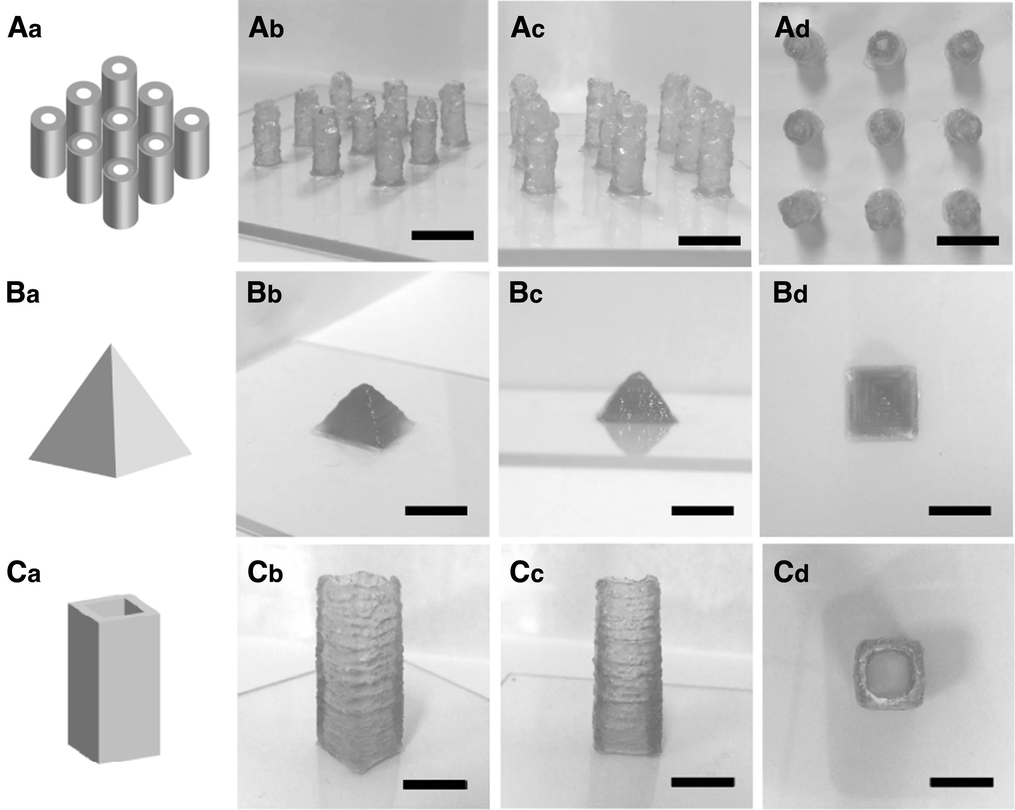
Photograph of printed 3D structures.
HEC to alter the swelling and permeability of scaffolds
Figure 5A shows the equilibrium swelling ratio of the SG/HEC ink at different HEC concentrations for 48 h. With increasing HEC concentration, the equilibrium swelling ratio of SG/HEC also increased, which was possibly because the HEC enlarged the pores of the SG scaffold.

Effect of HEC on the SG swelling rate, permeability, and degradation rate.
Figure 5B shows variation of permeability in SG/HEC scaffolds at different concentrations of HEC. The presence of HEC increased the permeability of molecules at different molecular weights in the SG scaffolds. However, there was no significant difference in the permeability of fluorescein sodium (Fig. 5Ba) at different concentrations with 1.5% and 3% HEC. The permeabilities of FITC-inulin (Fig. 5Bb) and FITC-70 kDa dextran (Fig. 5Bc) of SG/HEC scaffolds with 3% HEC were higher than those with 1.5% HEC. The penetration rates of fluorescein sodium in SG scaffolds slowed after 20 min, while those of FITC-inulin and FITC-70 kDa dextran in SG scaffolds slowed after 30 min, indicating that the SG scaffolds were selective to molecular weight, and the penetration rate of large molecules was slower. It can also be observed that the presence of HEC did not affect the change in penetration rates over time.
Alteration of scaffold degradation by HEC
Figure 5C shows the degradation of SG/HEC ink at different concentrations. There was no significant difference in the degradation rate at different concentrations of SG/HEC under static conditions (Fig. 5Ca), and the degradation rate of SG/HEC decreased with increasing HEC concentrations under dynamic conditions (Fig. 5Cb). Figure 5Cc presents the cylinder scaffold shape after dynamic degradation. The 3D structure-based SG quickly collapsed, while the presence of HEC reduced the collapse rate of the 3D structure.
Effect of HEC on gel formation of nonviscous liquids
We also explored the influence of the presence of HEC on the final gel formation of nonviscous liquids. After SA/HEC ink (Fig. 5Da) was extruded into the calcium chloride solution, it could immediately form a fibrous gel, regardless of whether the concentration of HEC was 0%, 3%, or 6%. There was no significant difference in the filament width (Fig. 5Ea) at different concentrations of SA/HEC. PEGDA/HEC inks (Fig. 5Db) at different concentrations were all crosslinked by photopolymerization to form gels in the presence of a photoinitiator and UV irradiation, and the diameters of cylinder gel formed (Fig. 5Eb) were all 10 mm, regardless of the concentration of HEC. Figure 5Dc shows the gel formation of FB/HEC inks at different concentrations by thrombin crosslinking, and the increasing HEC concentration did not affect fibrin gel formation and there was no significant difference in the diameters of cylinder gel formed (Fig. 5Ec) at different concentrations of FB/HEC.
Effect of HEC on scaffold biocompatibility
Cell lines (HUVECs and HAVSMCs) and primary cells (primary hepatocytes and primary neuronal cells) were used to evaluate the SG/HEC biocompatibility of different concentrations of HEC. The mass ratios of HEC/SG corresponding to HEC concentrations of 0% (w/v), 0.4% (w/v), 0.8% (w/v), 2% (w/v), and 4% (w/v) were 0, 1:20, 1:10, 1:4, and 1:2, respectively. To evaluate the direct biocompatibility of SG/HEC, HUVECs and HAVSMCs were seeded on scaffolds, and the viability was detected by the live–dead assay. HUVECs (Fig. 6Aa, Ba) and HAVSMCs (Fig. 6Ab, Bb) seeded on SG/HEC with different concentrations of HEC maintained up to 95% cell viability during a 7-day culture period. We also evaluated the indirect biocompatibility of SG/HEC. The effects of the medium extract of SG/HEC with different concentrations of HEC on cell growth were tested, and the results are shown in Figure 6C. The growth of cell lines (HUVECs [Fig. 6Ca] and HAVSMCs [Fig. 6Cb]) and primary cells (primary hepatocytes [Fig. 6Cc] and primary neuronal cells [Fig. 6Cd]) was not affected by medium extract of SG/HEC with increasing HEC concentrations. The results showed that HEC hardly affected the biocompatibility of the scaffolds.

Effect of HEC on scaffold biocompatibility.
Discussion
There are many factors that affect the printability of bioinks, such as the environmental temperature, the material properties, and the 3D structure design. Reagent-crosslinked, temperature-dependent phase change, and UV-polymerized hydrogels are the commonly used bioinks in extrusion printing6,8,9; these materials include alginate, FB, Matrigel, gelatin, Pluronic, gelatin methacryloyl (GelMA), PEGDA, and so on. To investigate the universality of HEC in the extrusion bioprinting application of bioinks, we added HEC at different concentrations to calcium ion-crosslinked SA, enzyme-crosslinked FB, temperature-dependent phase change G, UV-polymerized PEGDA, and composite SG, and then evaluated the effects of HEC on the printability, fidelity, permeability, degradation rate, and biocompatibility of these bioinks.
The nonviscous inks, such as the polymerized PEGDA precursor, uncrosslinked FB, and low-concentration SA, are not self-supporting and are unprintable without a secondary intervention. Figure 1A shows that 1.5% (w/v) SA was a nonviscous ink that did not show shear-thinning behavior. Correspondingly, the viscoelasticity analysis also showed that the ink was liquid, indicating that SA cannot be printed successfully, which was shown by the printed 3D structure-based nonviscous inks (PEGDA and FB) (Fig. 3Ca, Da). However, the presence of HEC can improve the viscosity and flow behavior. The printing of nonviscous inks succeeded after the addition of 3% HEC, but the shape fidelity was not good enough. The addition of 6% HEC made the nonviscous ink exhibit obvious shear-thinning behavior, and the viscoelasticity indicated semisolid-like properties, facilitating the shape fidelity of the printed scaffolds, which was confirmed by the printing of PEGDA/HEC (Fig. 3Cc) and FB/HEC (Fig. 3Dc). It is worth mentioning that the presence of HEC did not affect the crosslinking mechanism of nonviscous inks, and gels (Fig. 5D) could still be formed via ions, enzymes, or UV irradiation. Thus, HEC has the potential as a rheological additive to make nonviscous inks self-supporting.
Figure 1A shows that the SG and G inks were viscous inks exhibiting shear-thinning behavior, and the effects of HEC on the rheological properties of G and SG were similar. Therefore, the next focus was on the impact of HEC on the printability of SG. Figure 2B shows that grid scaffold printing based on SG at 25°C failed because the shear viscosity was lower than 10 Pa·s, making it impossible to form a stable structure. 45 The shear viscosity of SG can be increased 100 times by adding HEC. When the HEC concentration was 3%, the shear viscosity of SG/HEC could be as high as 100 Pa·s, which can hold shapes of 3D structures, as shown in Figure 4. The presence of HEC made the SG/HEC inks exhibit solid-like properties, facilitating the excellent shape fidelity of the printed scaffolds. 29 With increasing HEC concentration, the viscoelastic modulus of SG increased, resulting in improved shape fidelity (Fig. 3A, B). HEC can be added to viscous inks to improve printability and shape fidelity.
SG is the composite ink of SA and G, combining the temperature sensitivity of G and the calcium ion crosslinking property of SA. 22 The viscosity and viscoelasticity of G and SG will change as the temperature changes, and so, the printing temperature affects the printability of the extrusion and ultimately affects the printing fidelity.46,47 The addition of HEC improved the sol/gel transition temperature of SG. With increasing HEC concentration, the sol/gel transition temperature of SG gradually increased from low temperature to room temperature, which improved the printing temperature, reduced the requirements of environmental temperature and printer equipment parameters, preserved cell viability and phenotype during the printing process, 48 and widened the application range of low-temperature-dependent gels such as SG as bioinks.
In 3D culture, the diameter of the microspheres will affect the oxygen concentration, and the viability and functional activity of cells in the large-diameter microspheres will decrease.49–51 Therefore, the printed filament width also needs to be maintained within a relatively narrow range to maintain the activity and function of the long-term culture of the central cells. The addition of 3% HEC reduced the requirement of 3D printer printing speed for fine print width, improving the stability of the printing process during printing, and may affect printing fidelity.
In summary, HEC, as a rheological additive, can not only improve the printability of bioinks and the shape fidelity of 3D constructs, but can also make bioinks self-supporting. To compare the efficiency of printability improvement of HEC and existing rheological additives mentioned in other reports, such as laponite and methylcellulose, SA was chosen as the basic ink. The concentration of HEC added to increase the shear viscosity of SA by 100 times was 6% (w/v), while that of laponite was 2% (w/v) 26 and that of methylcellulose was 9% (w/v), 52 which indicated that the efficiency of HEC on printability was higher than that of methylcellulose and lower than that of laponite. On the contrary, the addition of laponite reduced the swelling rate, 29 but the addition of HEC increased the swelling rate, ensuring the water environment of the scaffold and porosity. The addition of HEC increased the permeability of fluorescein sodium, inulin, and 70 kDa glucan, indicating that the presence of HEC did not reduce the mass transfer efficiency of compounds such as small molecules, protein growth factors, and biological macromolecules. Thus, the addition of HEC can ensure the mass transfer efficiency while improving printability.
Conclusion
In conclusion, this article shows the great potential of HEC in the field of 3D extrusion bioprinting. HEC is able to adjust the printability properties of the bioinks for the construction of complex 3D structures, even converting nonviscous inks into directly printable inks. The addition of HEC can improve scaffold swelling to increase the hydrophilicity for better cell adhesion, molecular permeability, and mass transport. It can also tune scaffold degradation. Moreover, this compound is biocompatible with different kinds of cells. HEC is expected to be widely used for engineering bioinks in 3D extrusion bioprinting.
Footnotes
Author Disclosure Statement
The authors declare no conflict of interest.
Funding Information
This work was supported by the National Natural Science Foundation of China (NNSFC; No. 21675017), the State Key Laboratory for Managing Biotic and Chemical Threats to the Quality and Safety of Agro-products (No. KF20190108), and the National Key Research and Development Program of China (No. 2017YFC1702001).