
Abstract
The demand for biomimetic and biocompatible scaffolds in equivalence of structure and material composition for the regeneration of bone tissue is relevantly high. This article is investigating a novel three-dimensional (3D) printed porous structure called bone bricks with a gradient pore size mimicking the structure of the bone tissue. Poly-ɛ-caprolactone (PCL) combined with ceramics such as hydroxyapatite (HA), β-tricalcium phosphate (TCP), and bioglass 45S5 were successfully mixed using a melt blending method and fabricated with the use of screw-assisted extrusion-based additive manufacturing system. Bone bricks containing the same material concentration (20 wt%) were biologically characterized through proliferation and differentiation tests. Scanning electron microscopy (SEM) was used to investigate the morphology of cells on the surface of bone bricks, whereas energy dispersive X-ray (EDX) spectroscopy was used to investigate the element composition on the surface of the bone bricks. Confocal imaging was used to investigate the number of differentiated cells on the surface of bone bricks. Proliferation results showed that bone bricks containing PCL/HA content are presenting higher proliferation properties, whereas differentiation results showed that bone bricks containing PCL/Bioglass 45S5 are presenting higher differentiation properties. Confocal imaging results showed that bone bricks containing PCL/Bioglass 45S5 are presenting a higher number of differentiated cells on their surface compared with the other material contents.
Introduction
As the human body has capacity to repair and regenerate, but tissue regeneration is extremely limited, tissue engineering emerged in the 1990s aiming to address the organ shortage problem through the development of substitutes to restore, repair, or enhance tissue/organ functions.1–3 Relevant techniques include cell therapy and the use of biodegradable three-dimensional (3D) porous structures, called scaffolds, designed to support cell attachment, differentiation, and proliferation, allowing the formation of a new tissue.4–8 Recently, 3D bioprinting strategies based on the use of bioinks (hydrogel-based inks containing cells) have been also investigated, but this strategy is limited to soft tissues such as skin, cartilage, and cardiac tissues.9–12 Cell therapy, based on the use of autologous, allogenic, or heterologous cells, is a simple technique but presents some limitations in terms of guaranteeing that cells remain in the desired location for a clinically relevant period of times.13–15
For bone tissue engineering, the scaffold-based approach is the most relevant strategy. It is based on the use of biocompatible and biodegradable materials (e.g., polymers, ceramics, or polymer/ceramic composites) to produce 3D porous structures that support cell attachments, differentiation, and proliferation, providing a proper biomechanical environment for the formation of a new tissue.16–20 Initially, techniques such as electrospinning, particulate leaching, solvent casting and gas foaming, and freeze-drying were widely used.21–25 However, these techniques, despite being simple and relatively low cost, require significant human intervention, most of them require the use of toxic organic solvents, and do not allow to control pore size, pore shape, pore distribution, and pore interconnectivity.26–30
Additive manufacturing significantly contributed to further develop this tissue engineering scaffold-based approach by providing good topological control over the 3D printed scaffolds allowing for high reproducibility and repeatability, and to control material composition.31–33 For tissue engineering applications polymeric scaffolds (e.g., poly-ɛ-caprolactone [PCL], PLA) reinforced with bioactive ceramic materials (e.g., hydroxyapatite [HA], β-tricalcium phosphate [TCP], and bioglass) are commonly used to guarantee good mineralization and tissue integration.34–38
However, there are no conclusive in vitro and in vivo studies comparing the biological characteristics of scaffolds containing different types of bioactive ceramic materials.39–43 Most studies focused also on rectangular or cylindrical scaffolds with uniform pore shapes and sizes and despite the positive biological results these scaffolds did not present proper mechanical properties compromising their use for load-bearing applications.44–47 Moreover, several studies used chemical methods and organic solvents to prepare the polymer-ceramic blends, but as observed, these methods usually led to a decrease in the mechanical properties of the scaffolds due to poor interfacial adhesion with the reinforcement particles acting as defects in the polymer matrix.48,49
We previously demonstrated that a simple melt blending approach not only allows to have a good control over the incorporation of reinforcement particles on the polymeric matrix but also the fabrication of scaffolds with improved mechanical properties.50–52 Moreover, by designing scaffolds based on the anatomical characteristics (shape and pore size gradient) of bone, we reported for the first time the use of additive manufacturing to create geometrically graded scaffolds (scaffolds presenting a gradient of pore sizes).52–54 As observed, this approach allowed to obtain PCL/HA, PCL/TCP, and PCL/bioglass scaffolds with mechanical properties suitable for trabecular and cortical bone applications.52–54
This article considers again the concept of geometrically graded composite scaffolds (PCL/HA, PCL/TCP, and PCL/bioglass), mimicking the architecture of bone, investigating in detail the in vitro biological performance of these scaffolds. The effect of the pore size gradient on both cell proliferation and differentiation is investigated showing that it is possible to control/tailor the biological response through a proper control over the 3D topological characteristics of a scaffold. The most suitable polymer-based composite material is also investigated allowing to identify the most suitable anatomically designed scaffolds for the final in vivo evaluation.
Materials and Methods
Materials
PCL (CAPA 6500, molecular weight = 50,000 Da, melting point = 60°C, glass transition temperature = −60°C) was provided by Perstorp Caprolactones (Cheshire, United Kingdom) in the form of pellets. HA (molecular weight = 502.31 g/mol, melting point = 1100°C) in a nanopowder form (<20 nm particle size) and TCP (molecular weight = 310.18 g/mol, melting point = 1391°C) in powder form (ranging between 20 and 30 μm) were provided by Sigma-Aldrich (St. Louis, MO). Bioglass 45S5, with a composition of 45 wt% SiO2, 24.5 wt% CaO, 24.5 wt% Na2O, and 6 wt% P2O5, was supplied by CeraDynamics Ltd. James Kent Group (Stoke, United Kingdom) in a powder form (<10 μm particles size).
MesenPRO RSTM basal media, 2% (v/v) growth supplement, 1% (v/v) glutamine, and 1% (v/v) penicillin/streptomycin were provided by STEMPRO, Invitrogen. Dulbecco's Modified Eagle Medium (DMEM), 3-[4,5-dimethylthiazol-2-yl]-2,5-diphenyl tetrazolium bromide assay (MTT), osteocalcin (OCN) assay kit, bicinchoninic acid assay kit (BCA protein assay kit), Alexa Fluor 488-conjugated phalloidin, and 4′′,6-diamidino-2-phenylindole (DAPI) were provided by Thermo Fisher Scientific. The SensoLyte para-nitrophenyl phosphate (pNPP) Alkaline Phosphatase (ALP) assay kit was supplied by AnaSpec (Fremont, CA) and Dulbecco's phosphate buffered saline (PBS) was supplied by Merck Life Science.
Scaffolds fabrication
Anatomically designed scaffolds, with different material compositions (PCL scaffolds, PCL scaffolds containing 20 wt% of HA, PCL scaffolds containing 20 wt% of TCP, and PCL scaffolds containing 20 wt% of bioglass), were fabricated using the screw-assisted extrusion-based additive manufacturing 3D Discovery system (RegenHU), and a continuous path algorithm based on 38 zigzag double filaments and 14 spiral filaments. Scaffolds were designed to have an overall porosity of 52%. Blends were prepared using a melt blending process following a procedure previously described by our group, 55 and the scaffolds were printed using the following processing parameters: 90°C of melting temperature, 20 mm/s of deposition velocity, and 12 rpm of screw rotational velocity. The filaments were extruded using a 0.33 mm diameter needle.
Surface chemical composition
The surface chemical composition of the printed filaments before and after cell seeding was analyzed using energy dispersive X-ray (EDX) spectroscopy (scanning electron microscopy [SEM] FEG FEI Quanta 200; FEI Company), to determine the concentration of calcium (Ca), carbon (C), oxygen (O), and phosphorous (P). Scaffolds were gold coated prior imaging. The obtained SEM images were analyzed using the Oxford AZtec software (Oxford Instruments).
In vitro biological studies
In vitro studies were conducted to investigate
cell seeding efficiency and proliferation (MTT assay) and
osteogenic differentiation (SensoLyte pNPP ALP and OCN assay).
Tests were conducted using human adipose-derived stem cells (hADSCs; STEMPRO; Invitrogen; passage 6) and samples with the following dimensions: 11.25 × 4.11 × 5 mm (length × width × height). Before cell seeding scaffolds (n = 4) were sterilized, with 20/80 wt% of deionized water/ethanol, for 4 h inside 50 mL tubes and washed twice with PBS to remove any dead microorganisms and dust particles left on the surface of the scaffolds. The hADSCs were cultured in T75 sterilized flasks using MesenPRO RSTM basal media, which was replaced with fresh media until the number of cells have a confluency of ∼90% to cover all scaffolds. A suspension of ∼50,000 cells was used for all tests.
MTT studies
MTT assay was used to assess cell attachment and proliferation at days 3, 7, and 14 after cell seeding. This is a calorimetric assay used to determine the metabolic activity of cells by measuring the absorbance of formazan that is reduced from MTT by the cells mitochondria.56–58 In brief, DMEM media of 200 μL were placed in each well containing the scaffolds. On day 3, scaffolds were transferred to new well plates and 150 μL of MTT reagent and 150 μL of DMEM were added into each well. Afterward, the plates were placed in an incubator at 37°C for 3 h, and then the culture media was replaced with 450 μL of MTT reagent. After shaking in the dark at 37°C for 15 min, the absorbance was measured at 590 nm using the microplate reader Infinite 200 Tecan (Mannedorf, Switzerland).
Osteogenic differentiation studies
Osteogenic differentiation, at days 3, 7, and 14 after cell seeding, was investigated using the ALP assay according to the manufacturer's instructions. In brief, 1 × ALP dilution assay buffer was used to wash twice the bone scaffolds (n = 4), which were lysed with the use of 0.2% (v/v) Triton X-100 solution in 1 × assay buffer. Scaffolds were vortexed for 1 min, sonicated for 3 min, and freeze–thawed twice at a temperature of −80°C. Then, scaffolds were centrifugated at 2500 g for 15 min at a temperature of 4°C. Afterward, the supernatant that was mixed with pNPP substrate was extracted and the samples were incubated for 1 h at room temperature. A microplate reader was used to measure the absorbance at 450 nm. The ALP concentration was determined based on a standard curve and normalized to the total protein concentration using the BCA.
OCN, produced by osteoblasts, was considered to investigate the bone formation process. Tests were conducted following the manufacturer's instructions and results determined at days 3, 7, and 14 after cell seeding. Scaffolds (n = 4) were lysed, cell lysis solution centrifuged, the supernatant was added followed by the addition of an antibody cocktail (50 μL), and incubated for 1 h at room temperature. Then, scaffolds were washed three times and mixed with TMB substrate solution (100 μL) and shaken for 10 min in the dark. Finally, 100 μL of stop solution was added into the liquid and read at 450 nm with the use of microplate reader. OCN concentration was evaluated with a standard curve and normalizing to the total protein concentration.
Cell morphology
An SEM machine, FEI ESEM Quanta 200 (FEI Company), was used to investigate the morphological characteristics of attached cells. Scaffolds were coated (platinum coating) with the use of EMITECH K550X sputter coater (Quorum Technologies) before imaging.
Confocal laser scanning microscopy was used to study cell spreading and cell shape on the scaffolds surface. Alexa Fluor 488-conjugated phalloidin and DAPI were used for staining, the actin cytoskeleton, and cellular nuclei of differentiated cells, respectively. Leica TCS SP5 (Leica, Milton Keynes, United Kingdom) confocal microscope was used for imaging.
Data analysis
Statistical analysis was conducted with the use of one-way analysis of variance with Tukey's post hoc test in GraphPad Prism software supplied by GraphPad Software, Inc. (San Diego, CA). Statistically significant differences were considered at *p < 0.05, **p < 0.01, ***p < 0.001, and ****p < 0.0001.
Results
Figure 1A shows the 3D printing system used to create the anatomically designed scaffolds that present a pore size gradient (Fig. 1B). Three different regions across the scaffold corresponding to different pore sizes were considered in this research (Fig. 1C).

Chemical composition
The element composition at the surface of the printed filaments before and after 14 of cell differentiation is presented in Table 1 and Figures 2 and 3. Results show that the increase of calcium, oxygen, and phosphorous elements and the reduction of carbon elements from day 0 to 14 can explain the rapid proliferation and differentiation of cells on scaffolds surface. Studies have shown that during differentiation studies an apatite layer, mostly contained calcium and phosphorus elements, is created on the filaments surface of all different material contents, similar to that of human bone, which occurs due to the degradation of the scaffolds.59–64
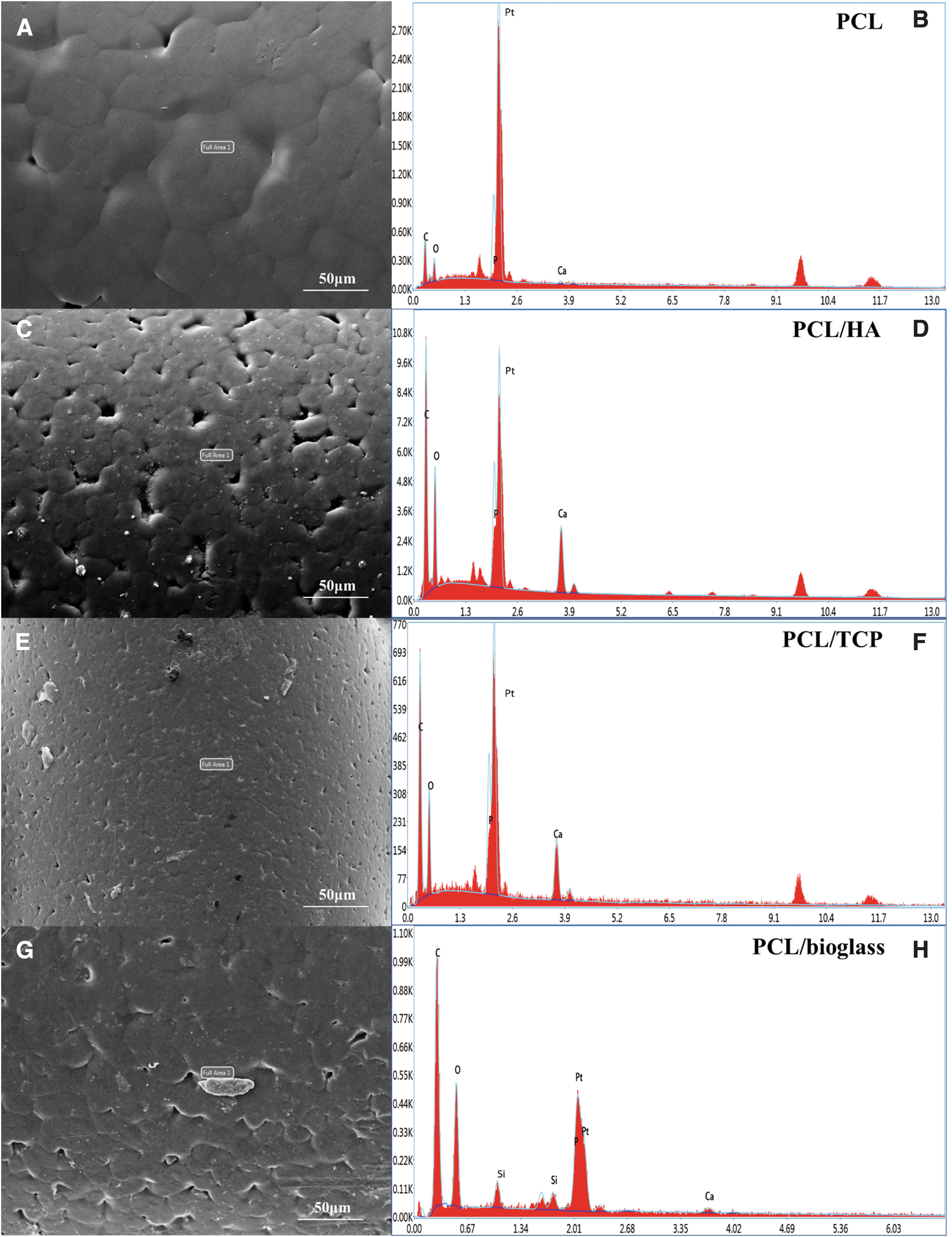
EDX spectra and SEM images of scaffolds at day 0.
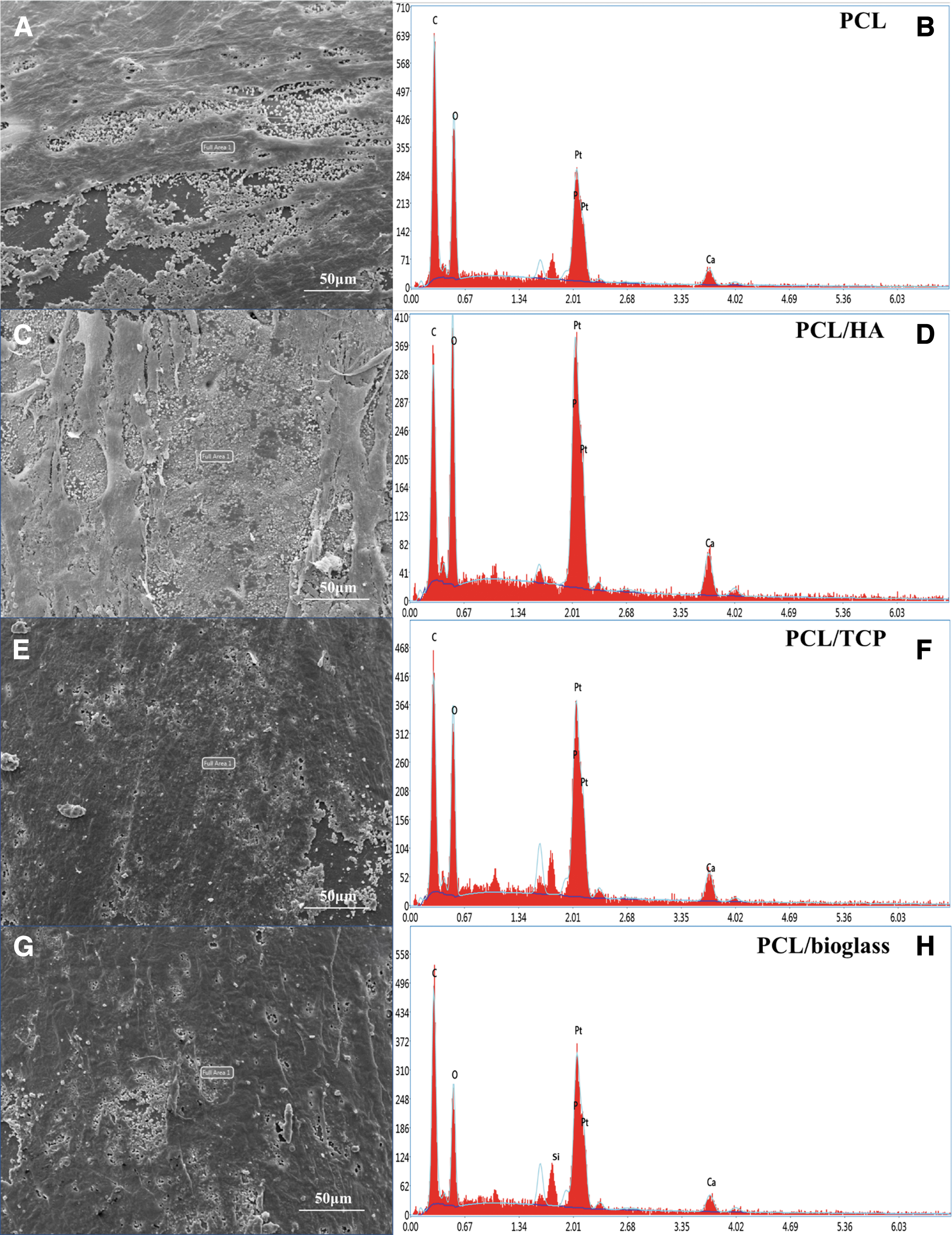
EDX spectra and SEM images of scaffolds after 14 days of cell seeding.
Element Composition Before and After Cell Seeding on Scaffolds Containing Different Material Content
HA, hydroxyapatite; PCL, poly-ɛ-caprolactone; TCP, β-tricalcium phosphate.
However, on the surface of PCL/bioglass scaffolds, except from the apatite layer, presence of silicon elements can be observed before and after cell seeding.65–67 The presence of silicon on PCL/bioglass scaffolds is enhancing cell growth proliferation and differentiation, which can explain the high ALP and OCN expression of PCL/bioglass scaffolds compared with PCL, PCL/HA, and PCL/TCP scaffolds.65–67
Cell proliferation
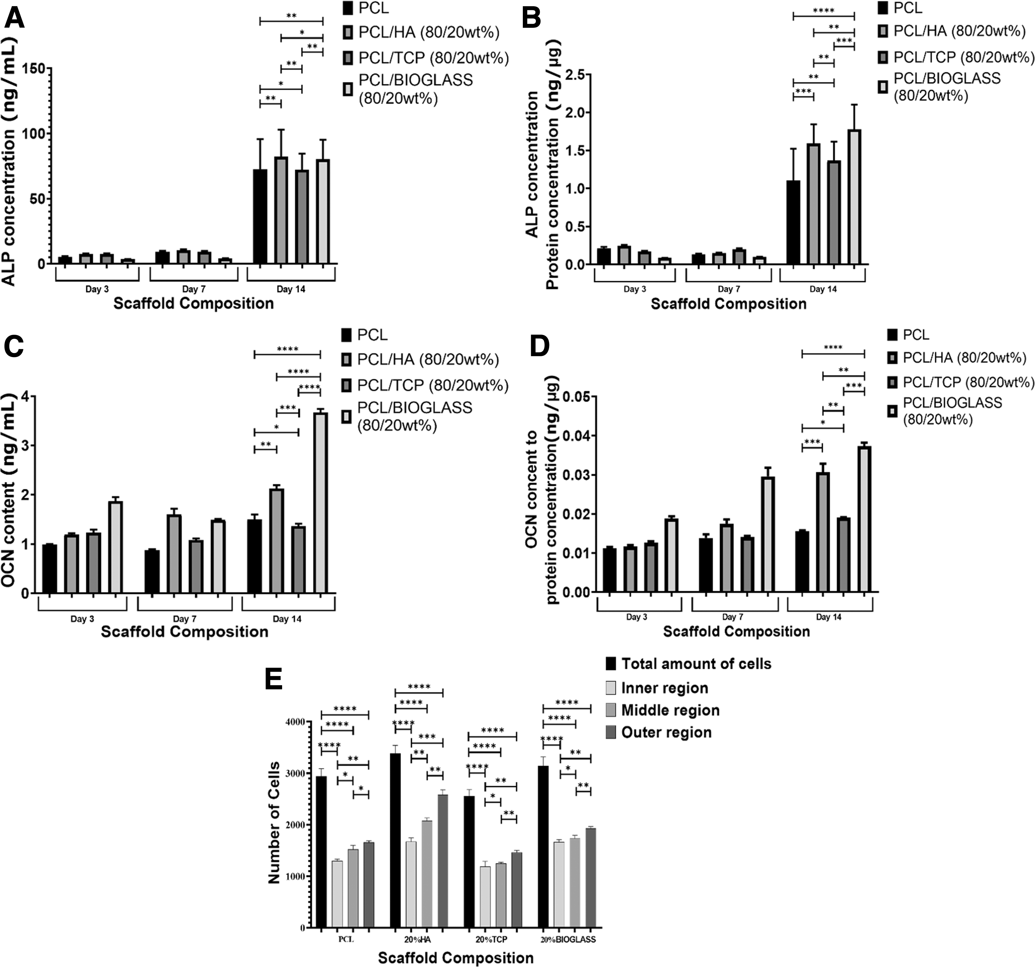
Overall cell proliferation results on the printed scaffolds are presented in Figure 4 and Table 2. At day 14 after cell seeding, results show that scaffolds containing nano HA particles exhibit high overall cell attachment and proliferation (243,413.1 ± 22,483.7), in comparison with the PCL/TCP (210,253.3 ± 8875.6), PCL/bioglass (240,890.9 ± 15,085.5), and PCL (186,578.9 ± 14,221.4) scaffolds, with PCL scaffolds showing the lowest values. However, no statistically significant differences were observed between PCL/HA and PCL/bioglass scaffolds. Cell proliferation slightly increases between days 3 and 7 and significantly increases between days 7 and 14.

Cell proliferation results (MTT) on the 3D printed scaffolds at different time points. Statistically significant differences were considered at *p < 0.05, **p < 0.01, ***p < 0.001, and ****p < 0.0001. The * is small difference and while more * are added the differences between the results are higher. The **** refers to Tukey's post hoc test. MTT, 3-[4,5-dimethylthiazol-2-yl]-2,5-diphenyl tetrazolium bromide assay.
p-Values of Proliferation Test (MTT) for Scaffolds Containing Different Material Content
MTT, 3-[4,5-dimethylthiazol-2-yl]-2,5-diphenyl tetrazolium bromide assay.
Between days 7 and14, cell proliferation kinetics is particularly high in the case of bioglass scaffolds suggesting a longer-term beneficial effect associated to the addition of bioglass particles. Contrary to other studies that reported that the bioactive effect of bioglass was hindered when loaded at low concentrations to PCL scaffolds,68,69 this study shows that the considered material preparation method and fabrication strategy allows to produce bioactive PCL/bioglass scaffolds. Overall, the results also show that all considered scaffolds can support cell attachment and proliferation and do not induce any cytotoxic effect.
Cell differentiation
Figure 5A, B and Table 3 present the ALP activity results on scaffolds containing different material compositions at days 3, 7, and 14 after cell seeding. ALP is an important marker of osteoblast activity playing an important role in starting the extracellular matrix (ECM) mineralization and calcification due to the accumulation of calcium ions and phosphates.70–72 As expected, the addition of bioactive reinforcements increase as a positive effect on the ALP expression, with PCL scaffolds exhibiting the lowest ALP expression (ALP concentration of 72.55 ± 23.17 and ALP protein concentration of 1.11 ± 0.42). Moreover, it can be observed that ALP expression slightly increased from day 3 to 7, whereas from day 7 to 14 there is a significant increase of ALP expression in all considered scaffolds.
Osteogenic differentiation on scaffolds with different material compositions at different time points.
p-Values of Differentiation Test (Alkaline Phosphatase and Alkaline Phosphatase Protein) for Scaffolds Containing Different Material Content
ALP, alkaline phosphatase.
The normalized ALP activity results show that, at day 14, PCL/bioglass scaffolds present the highest ALP activity (ALP concentration of 80.37 ± 14.82 and ALP protein concentration of 1.78 ± 0.33) compared with PCL/HA (ALP concentration of 82.16 ± 20.77 and ALP protein concentration of 1.59 ± 0.25) and PCL/TCP (ALP concentration of 72.21 ± 12.27 and ALP protein concentration of 1.37 ± 0.25), which is aligned with the mineralization results observed by EDX spectroscopy, as a mineralized surface promotes cell attachment and spreading. The bioactive characteristics of PCL/bioglass and corresponding positive impact on the ECM mineralization process can be attributed to the release of calcium, phosphate, and silicon ions from the glass structure.
Figure 5C, D and Table 4 present the OCN results for all considered scaffolds at days 3, 7, and 14 after cell seeding. Contrary to OPN, another relevant osteogenic marker that provides relevant information on early osteogenic differentiation and preosteoblast proliferation, OCN is an important marker of later stages of osteogenic differentiation, being secreted by mature osteoblasts.73–77
p-Values of Differentiation Test (Osteocalcin and Osteocalcin Protein) for Scaffolds Containing Different Material Content
OCN, osteocalcin.
Results confirm that PCL/bioglass scaffolds present significantly higher bioactivity and osteogenic properties as observed by the higher OCN content and normalized OCN observed for PCL/bioglass scaffolds (OCN content of 3.67 ± 0.07 and normalized OCN of 0.037 ± 0.000919) in comparison with the PCL/HA (OCN content of 2.13 ± 0.07 and normalized OCN of 0.031 ± 0.002156), PCL/TCP (OCN content of 1.37 ± 0.05 and normalized OCN of 0.019 ± 0.00011) and PCL (OCN content of 1.5 ± 0.09 and normalized OCN of 0.016 ± 0.000223) scaffolds. It is also possible to observe that, among the investigated composite scaffolds, PCL/TCP scaffolds present the lowest osteogenic characteristics.
Figure 5E shows the number of cells attached and on different regions of the scaffolds (inner, middle, and outer regions) (see also Fig. 1C for reference) at day 14 of cell differentiation. As observed a high number of cells were identified on the outer regions of the scaffolds in comparison with the inner and middle regions. In all considered cases, results show that the number of cells increase by increasing the pore size (promotes oxygen and nutrients supply), suggesting that the gradient structure can induce cell growth and cell density in a gradient manner. Overall, PCL/HA scaffolds seem to present a high number of attached cells but no statistically significant differences were observed regarding PCL/bioglass scaffolds. Moreover, results seem to indicate that at least for a short-term differentiation process, the biological performance of PCL/bioglass, PCL/TCP, and PCL scaffolds are less sensitive to the gradient architecture than PCL/HA scaffolds.
Cell morphology
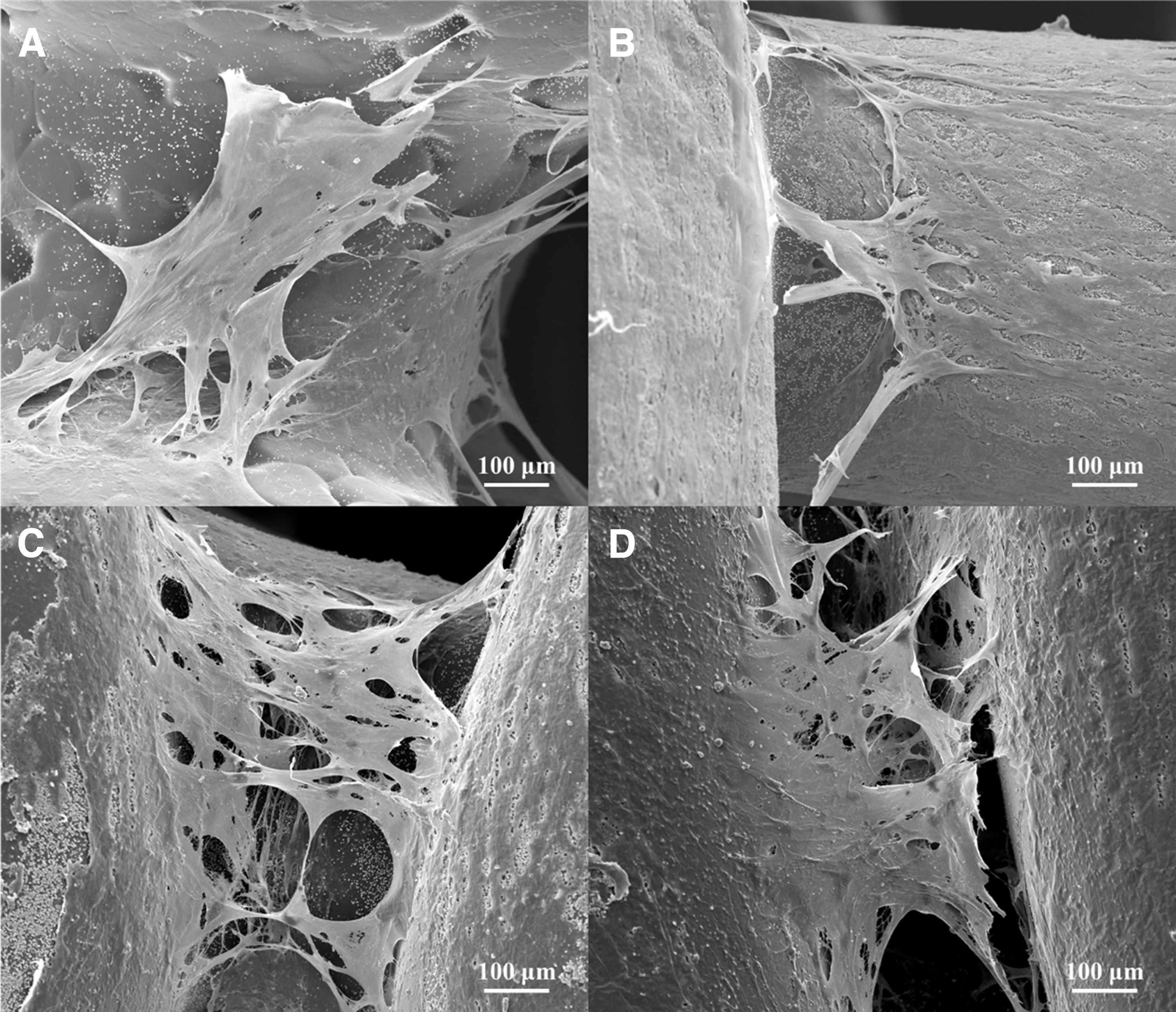
Figure 6 presents SEM images of cells attached and spreading on the surface and cross sections of the scaffolds after 14 days of osteogenic differentiation. Results show that cells are well spread over the scaffolds creating bridges between pore filaments, which are particularly significant in the case of PCL/bioglass scaffolds. Cell bridging is more evident on the inner regions of the scaffolds (low pore size) and this may compromise oxygen and nutrient supply to the cells that migrated to the internal regions of the scaffold.

SEM images of cells spreading on the scaffolds (top and cross section) after 14 days of cell differentiation.
Furthermore, this can confirm that gradient pore size can affect cell growth in a gradient manner. Moreover, high magnification images of differentiated cells bridging between neighboring filaments and scaffolds top surface show that cells are attaching and spreading with reduced kinetics on PCL, PCL/HA, and PCL/TCP scaffolds compared with PCL/bioglass scaffolds (Fig. 7). This can be explained due to the high bioactive nature and high mineralization properties of bioglass particles, implying the positive long-term impact on cells growth, proliferation, and differentiation.
High magnification SEM images of cells spreading between printed filaments and scaffolds top surface after 14 days of cell differentiation.
Figure 8 shows the confocal images of differentiated cells on the scaffolds on day 14, on two different regions (inner and outer region). Furthermore, Figure 8 demonstrates that cells are attaching and differentiate faster in the inner region (Fig. 8A, C, E, G) and slower in the outer region of the scaffolds due to larger pore size (Fig. 8B, D, F, H). Finally, it is possible to observe that all scaffolds are presenting a large number of cells attached on them with slightly higher of PCL/bioglass and PCL/HA scaffolds.
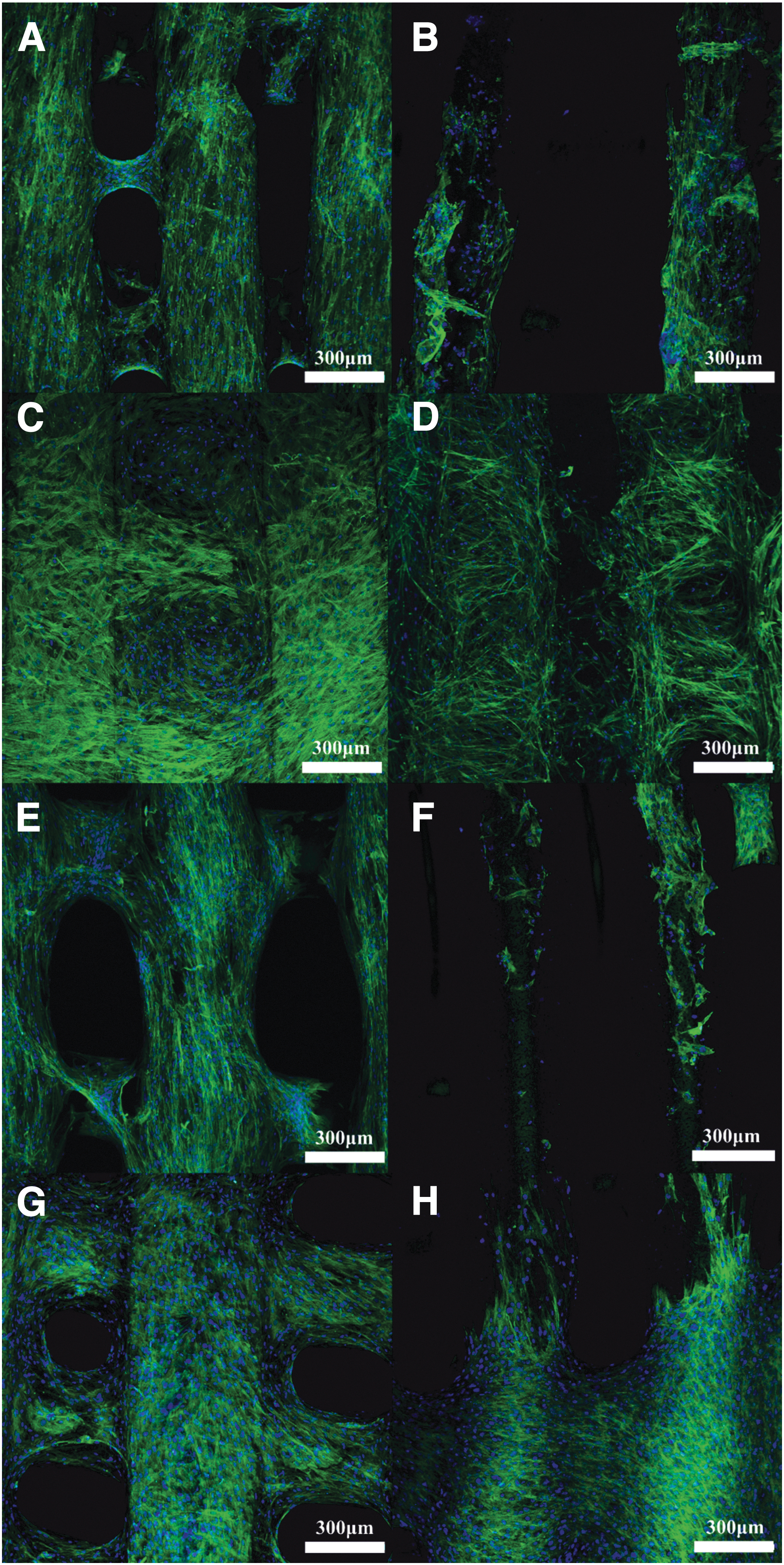
Confocal images of differentiated cells attached and spreading on day 14 of inner
Conclusions
This article investigates the in vitro biological performance of anatomically designed functionally graded pore size scaffolds based on PCL and containing relatively small amounts (20 wt%) of bioactive ceramic reinforcements (HA, TCP, and bioglass). Previously, we demonstrated that using a simple melt blending process to prepare the blends and a screw-assisted extrusion additive manufacturing process it was possible to produce scaffolds with mechanical properties suitable for both trabecular and cortical bone, with mechanical properties of 187 MPa for PCL, 239 MPa for PCL/HA, 247 MPa for PCL/TCP, and 343 MPa for PCL/bioglass.
Moreover, contrary to other studies, where the addition of these ceramic reinforcements were not able to improve the overall mechanical properties, our material preparation and fabrication approach allowed good interfacial adhesion between the polymer matrix and the reinforcement particles, and the addition of ceramic particles resulted in a significant increase on both compressive modulus and yield strength. This article shows that the adopted strategy does not hinder the osteogenic characteristics of the reinforcements and as a consequence PCL/bioglass scaffolds enable fast apatite layer formation on the surface of the scaffolds, high cell proliferation, and osteogenic differentiation. Results seem to indicate that among the scaffolds investigated, PCL/bioglass scaffolds present a high ability for rapid bone bonding and fast new bone formation being the most relevant scaffolds for future preclinical evaluation as combined mechanical properties suitable for load-bearing applications and osteoproductive and osteoconductive properties.
Footnotes
Acknowledgment
This article is a result of the study completed by Evangelos Daskalakis toward a PhD in mechanical engineering, granted by the University of Manchester in September 2022.
Authors' Contributions
Conceptualization and visualization by E.D. and P.B.; methodology by E.D., B.H., M.H.H., A.M.O., C.V., A.A.A., and A.F.; software by E.D. and A.F.; validation by E.D., B.H., M.H.H., A.M.O., C.V., A.A.A., A.F., and P.B.; formal analysis by E.D., B.H., and P.B.; investigation by E.D., B.H., M.H.H., A.M.O., C.V., A.F., and P.B.; resources, data curation, and writing—review and editing by all authors; writing—original draft preparation by E.D.; supervision by G.C., A.W., G.B., B.K., and P.B.; project administration and funding acquisition by P.B. All authors have read and agreed to the published version of the article.
Author Disclosure Statement
On behalf of all authors, the corresponding author states that there is no conflict of interest.
Funding Information
This project has been supported by the University of Manchester and the Engineering and Physical Sciences Research Council (EPSRC) of the United Kingdom, the Global Challenges Research Fund (GCRF), grant number EP/R01513/1 and EPSRC Doctoral Prize Fellowship EP/R513131/1.