
Abstract
Abstract
It is known that acupuncture is effective for lowering blood pressure and treating some cardiovascular diseases. Recent work and reports from practitioners confirm that electroacupuncture (EA) is a more effective therapy; however, the basis of this observation has not been explained in terms of Western medical science. One approach is the use of calcium channel blocker agents, particularly L type of channels. This ultimately relaxes the muscle in the walls of the arteries. The same effect can also be observed by the use of EA wherein low-frequency and low-current electrical pulses increase the gating voltage and block the calcium channels. This alteration of voltage across the Ca++ channels by EA is examined herein.
Introduction
The causes for hypertension are multiple, including age, long-term stress, obesity, smoking, high-salt diet, and a sedentary lifestyle. Generally, the recommended treatment is medications. For example, diuretics in combination with other medication are advised because they increase urine output and thus reduce blood volume. 2 β-Blockers are used to reduce the heart rate and the force of heart contraction. Angiotensin-converting enzyme inhibitors are recommended because they help to relax the blood vessels. Similarly, calcium channel blockers are used to relax the muscle of artery walls. 3 Thus, the problem of hypertension may need to be treated with several different approaches and a combination of medications is generally used.
According to traditional Chinese medicine, hypertension is due to an inharmonious condition of the whole system, which includes the improper function of the heart, liver, kidney, and some parts of the digestive system. 4 Acupuncture is based on long-term empirical observations and needs to be correlated with Western medical approaches. Such efforts have been carried out for specific cases such as pain, 5 particularly low back pain 6 and torticollis. 7 Western medicine considers hypertension a cardiovascular disease, including an artery disorder such as excessive contraction of smooth muscle cells. In this case, treatment is focused on the relaxation of smooth muscle cells. This article focuses on the latter aspect only.
One common treatment for controlling arterial hypertension is the use of drugs like amlodipine, valsatran, nifedipine, or similar pharmaceuticals. 8 The principal action of these drugs is to block the calcium channels. This inhibits the flow of Ca++ across the transmembrane of the cardiac muscle and the smooth muscle. This reduces the resistance of the vascular system, relaxes the muscles, and reduces the arterial blood pressure.
It has also been found that electroacupuncture (EA) can be useful for treating hypertension-related problems.9,10 This view has been strongly supported in Russian literature. 10 Research organized by the National Heart, Lung, and Blood Institute confirms that a low level of electrical stimulation at specific points on the front leg of rats reduces blood pressure by more than 40%. Cardiovascular depression can also be treated with acupuncture stimulation. In general, there are findings that show that acupuncture stimulation helps to reduce blood pressure.
Taking into account the experimental data, a systematic study of hypertension and blood circulation was carried out by stimulating at very low frequency (0.1 Hz). 9 There was an increase in the vascular smooth muscle contraction. If the stimulation frequency is slightly higher (10 Hz), the muscle blood flow and the lymphatic circulation are found to be enhanced. Moreover, the peripheral circulation is clearly improved together with changes in the temperature with EA. The combined effects proved beneficial for blood pressure–related problems.
Earlier work 4,10 in EA and reports from several practitioners suggest the need to explain these observations on the basis of Western medical science.
It is well known that one of the approaches to reduce hypertension is vasodilation, 11 which is achieved with the help of relaxation of the smooth muscle surrounding the blood vessels. The origin for the contraction of the smooth muscle is associated with the excess of intracellular calcium ion concentration; hence, blood pressure is controlled by blocking the flow of calcium channels with specific drugs.
Ea and Hypertension Treatment
An extensive investigation was done by Mayor 4 on the effectiveness of EA for hypertension. Williams et al 9 used well-established points, e.g., LR 3, ST 36, and LI 11. Depending on the cause of hypertension, other points like Zusanli (ST 16), Renying (ST 9), and Fengchi (GB 20) may be useful for lowering blood pressure due to their positions with respect to the arteries (Figure 1). It is clear that acupuncture points are closely spaced with the major artery pathways. For example, PC 3 is very close to the radial artery; similarly, ST 36 is close to the popliteal artery, and LV 3 is located on the plantar arch. Thus, all the points used in EA therapy should be close to or associated with the principal arteries (Figure 1). Low-frequency pulses of electrical current influence the arterial system or some part of it. This aspect has several significant consequences; one is the effect on the voltage-sensitive calcium channels located on the membrane of the smooth cells, which are sandwiched between the internal and external elastic lamina of the artery. This is illustrated with the help of a typical artery, smooth muscle cells, and the location of calcium ion channels in Figure 2. This illustrates the mechanisms and correlation of the EA and hypertension, but also the selection of the proper acupuncture points according to the symptoms and the requirement of the patients.
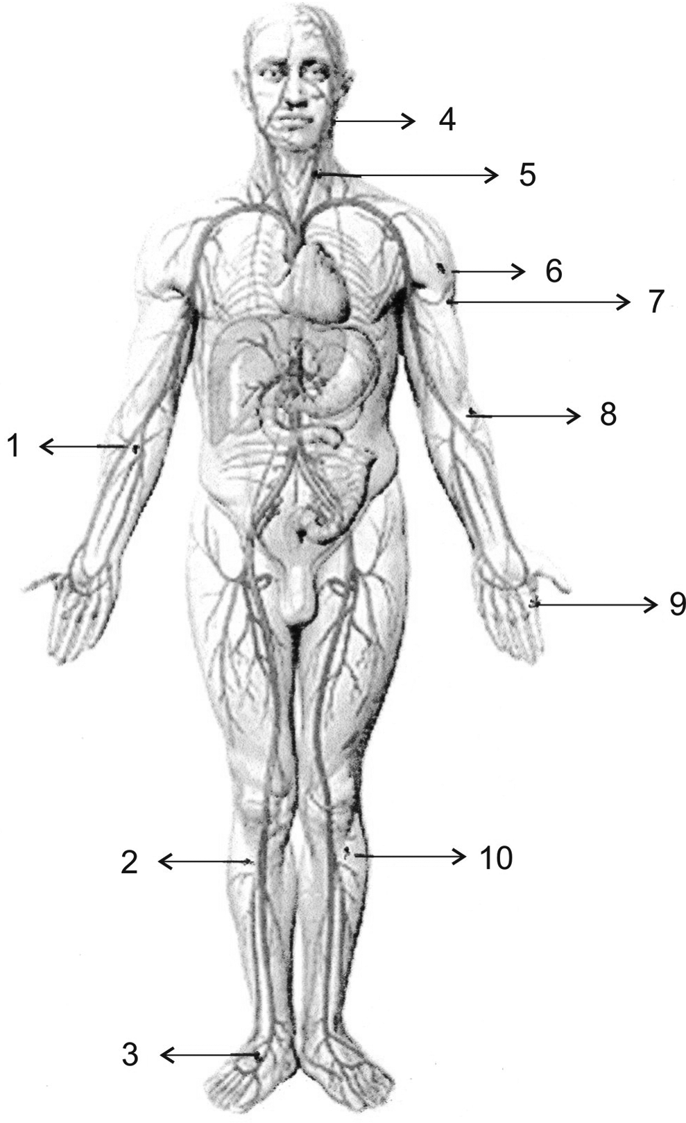
Acupuncture points used for hypertension treatment: (1) PC 3 (Quze), (2) ST 36 (Zusanli), (3) LR 3 (Taichong), (4) GB 20 (Fengchi), (5) ST 9 (Renying), (6) LI 14 (Binao), (7) LU 3 (Tianfu), (8) LI 11 (Quchi), (9) LI 3 (Sanjian), (10) GB 34 (Yanglingquan).
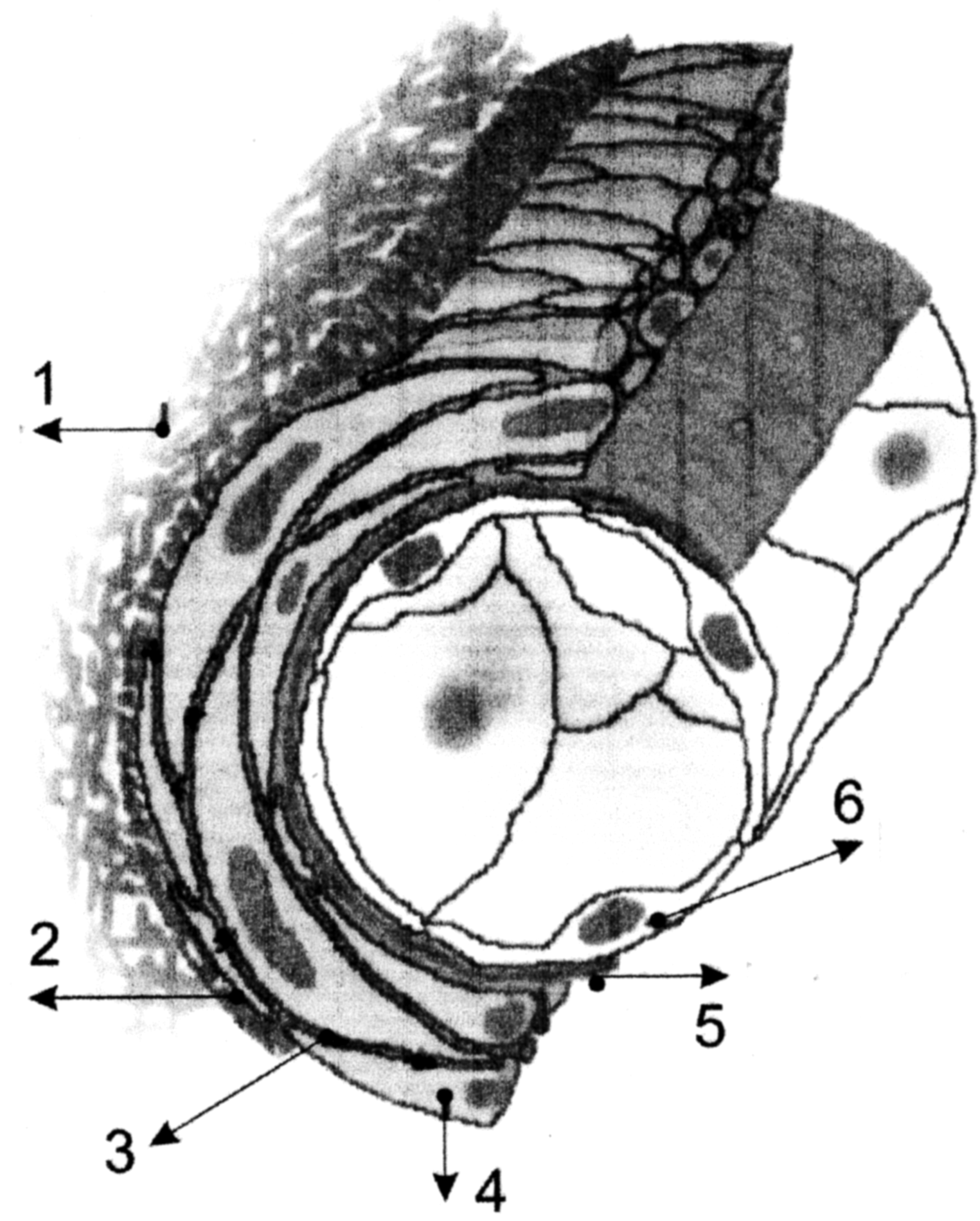
Structure of artery wall through which electrical pulse passes: (1) connecting tissue, (2) external elastic lamina, (3) calcium channel, (4) smooth muscle cell, (5) internal elastic lamina, (6) endothelium.
Morphology Of Artery
The typical structure of the artery wall indicating the details of tunica externa, tunica media, and intima is shown in Figure 2. Tunica media largely consists of vascular smooth muscle cells (VSMCs), which have the property of contraction and relaxation of the muscle. This quality is related to the concentration of calcium ions within the cells. 11 The membrane of the smooth cell consists of voltage-sensitive calcium channels, and the alteration of the voltage (though minor) has a dramatic effect on the opening or closing of the channels. This has important and direct consequences.
The trans membrane of voltage-gated (VTm) calcium channels constitute the main path for Ca ++ entry in VSMCs. 12 In conventional treatment, it is often blocked by special drugs, but the same goal may be achieved with the help of electrical pulses, which can modulate the gate voltage. The diameter and the impedance (resistance to electrical pulses) depend on the section of the artery under consideration. Impedance of the artery wall also varies from patient to patient.
Cellular Structure and Calcium Ion Channels
The voltage-sensitive calcium ion channels located on the thin membrane of the smooth muscle cells are shown in Figure 3. The difference between T (transient) and L (longlasting) types of channels is not made in the present figure and the corresponding discussion—as the general arguments are valid for both types of channels except that the magnitude of the voltage gate for which channels are opened is slightly different. The densities of L and T channels vary from cell to cell and from this point of view, the membrane is not uniform for electrical properties.

Voltage-gated calcium channel structure located at the membrane: (1) outside region, (2) membrane region, (3) cytoplasm (the numbers marked within the region of Ca++ channels corresponds to known subunit of the channel).
Discussion
The precise estimation of the electrical properties of the artery wall, particularly tunica adventitia, tunica media, and the membrane of the smooth muscle cell, is a complex issue. These elements are highly anisotropic because of the alignment of the biological tissues. The collagen external tunica (connective tissue with elastic lamina) have much lower resistivity in the longitudinal direction than in the radial direction. Therefore, part of the current is shunted along the longitudinal axis due to the high conductance. In the same way, the extracellular region has a high conductance and is also a pathway for the current. However, in EA treatment, the constant magnitude of the current (not the voltage) is maintained. In general, the membrane resistance and the capacitance have relatively high values in the radial direction as compared with the longitudinal direction; therefore, the current passes through a high conductivity path. Because of this, there is no substantial voltage drop across the external tunica.
The passage of the electrical current through the artery wall (axial direction), particularly the smooth muscle cells, is difficult to evaluate because the precise information about the resistance and other electrical properties of tunica adventitia, external elastic layer, smooth muscle cell, epithelium, etc., is not available. Moreover, these properties are anisotropic and therefore the data in a specific direction is a must. Hettrick et al 13 developed a finite element model on the basis of their experimental results. The resistance of a vascular smooth cell of 0.8 mm thickness is about 100 Ω-cm. This resistance mostly originated from the membrane of the smooth cell, as the inside region is much more conductive. This means that the resistance varies between approximately 6 Ω and 8 Ω. In fact, this is an approximation because it varies from person to person and the region of the artery under consideration. In an EA procedure, the current used is of the order of 10 mA or more; it means that the voltage drop across the membrane of a smooth cell is roughly between 60 mV and 80 mV. For this value, the calcium channels of both types (T and L) are blocked, 14 and the calcium flow of current from the outside to the inside of the cell is stopped.
All the biological membranes also have capacitive resistance, which depends on the applied frequency. Such measurements have been carried out for certain types of membranes and they are of the order of μF/cm 2 . From these values, the extrapolation for the micron region is not recommended for the complex system under consideration. All the components involved are nonuniform in thickness; they are heterogeneous in structure and in chemical composition, and their surface resistivity varies from one micron region to another. Because of these reasons, the charge storage capacity and the capacitance are different from one region (or tissue) to another or even one part of the membrane to another part. In addition to these obstacles, there is a theoretical limitation also. There is a charge flow at the boundary region of the capacitance, which is known as the edge effect. 15 Near the edge of the capacitor, the charge is not uniform and the field gradient is created. This is not significant when the dimensions of the capacitor are of the order of centimeters or more. When the dimensions of the capacitors are very small (submicron or less), the edge effect is stronger and the extrapolation of the values of capacitance may lead to incorrect values. Because of all these reasons, we have to rely on the experimental results from the smooth muscle cell itself.
Earlier results
16
show that the alternating electric fields (0-100 Hz) have an effect on the flow of calcium ions and on the concentration of the cytosolic calcium. This means that the impedance varies with the applied frequencies, indicating that there is a contribution from the capacitive resistance. Similar results are also observed in EA treatment and in some cases the frequency dependence is confirmed.
4
This is possible only if the capacitive resistance (or impedance) is of the same order of the membrane ohmic resistance. In this case, the resistance and the capacitance are in series and therefore the impedance of the system across the membrane of the smooth cell is given by
15
Here, R is the resistance of the smooth cell membrane in a radial direction, C is the capacitance of the membrane, and ω is the frequency. For this calculation, the membrane resistance, R, is 8 Ω. The value of 1/C is expected of the same order and the plot is examined for the values from 1 to 8. The frequency, ω, of the applied field used in EA is generally between 0 and 100 Hz. However, the interesting region for this set of values is found to lie between 0 and 5 Hz, and for higher values of ω, the variation in impedance is practically imperceptible. Figure 4 illustrates the plot up to 5 Hz. For a certain combination of R and 1/C, the frequency dependence shows a maximum, but the variation in the impedance is not so critical for hypertension-related disorders. The general trend of the plot remains the same.
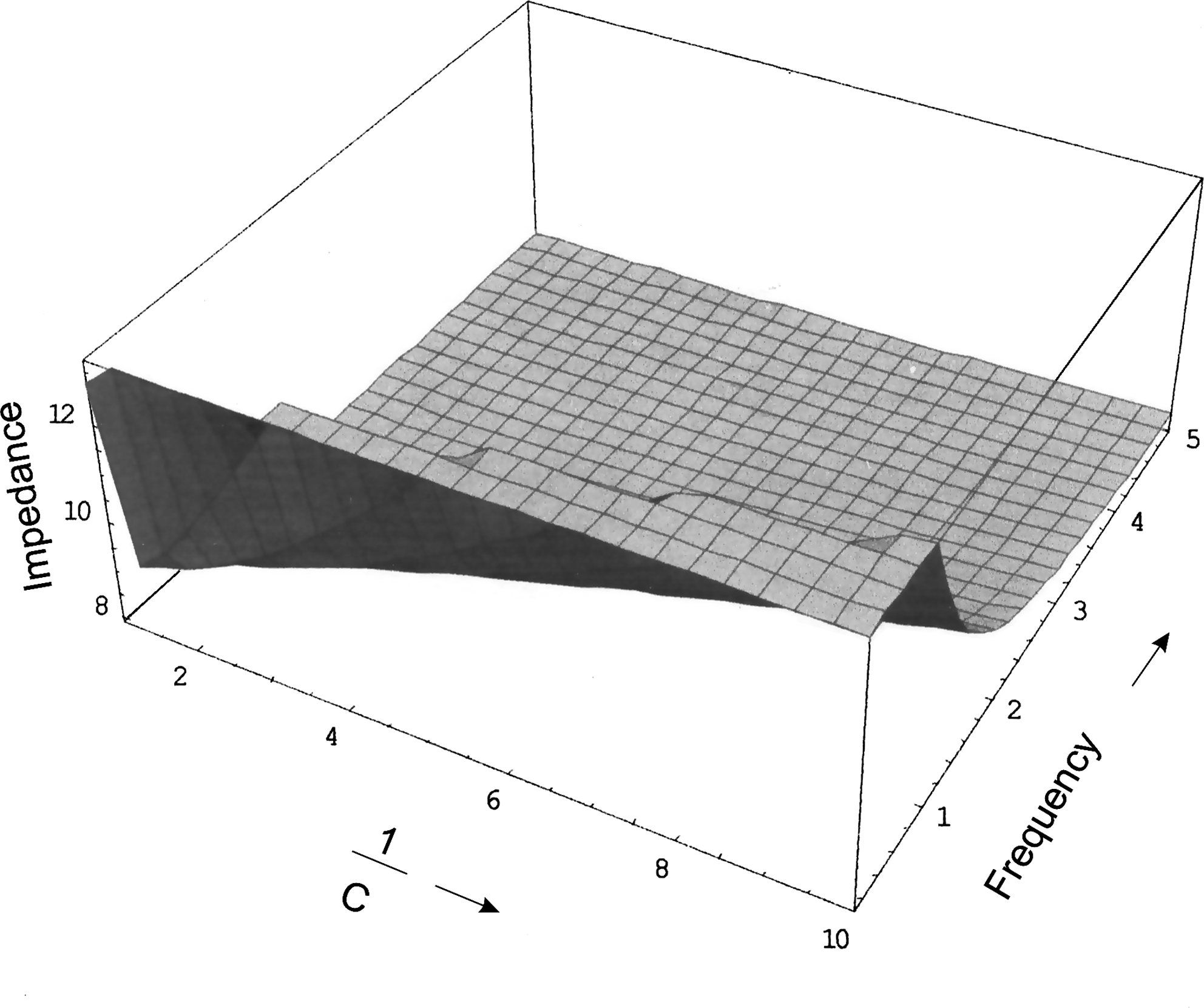
A plot of impedance against the resistance and capacitance of the membrane of the smooth cell. For certain combination of resistance the impedance varies for low frequency.
It is clear from this figure that a variation in the values of the capacitance does not change the impedance of the system substantially. This figure also indicates that for lower values of ω, the impedance increases; however, the increase is not substantial. Independent of the frequency used, the impedance varies from about 8 Ω to 12 Ω and the transmembrane voltage, VTM, is shifted toward +80 mV. For some patients, it is possible that the radial resistance of the membrane capacitance is a little less than 8 Ω and in that case, the magnitude of the current needs to be high. Fortunately, commercially available equipment can provide the current up to 40 X 10−3 A or even more. Therefore, the adjustment of the current to increase the transmembrane potential is not a problem.
There are several types of calcium channels such as L,N,P,Q, R, and T; some of them are ligand gated and others are voltage gated (L and T types). The ligand-gated channels are useful for pharmacological agents; their functions are beyond the scope of the present article. However, only 2 types of calcium channels, namely T type (transient, tiny) and L type (longlasting, large), play a predominant role in the flow of extracellular calcium into the smooth muscle cell. These channels (L and T types) coexist in the same patch and have different properties related to kinetics, permeability, and pharmacology. 17 Both are voltage gated but the magnitude of the voltage activation domain is different. The T types of channels are active between −60 mV and +10 mV while the L type channels have an active domain between −30 mV and +60 mV. 12 The latter channels are closely connected with the signaling and modulating processes of the strength of the contraction. It is generally accepted that the tension of the arterial muscle is sensitive to L type channels and they are abundant virtually in all excitable cells including cardiac and in artery smooth muscle cells, and it is confirmed that its contribution is substantial in controlling the arterial blood pressure. However, in certain cases (e.g., outer groove of the ear of the rabbit), 14 the contribution from the T type of channels is also appreciable. The blocking of both types of channels may help control the arterial blood pressure, which is possible to achieve just by monitoring the voltage across the membrane so that the calcium channels will be blocked.
The pharmacological properties of L and T types of channels are also quite different. The L type have specific ligands while T types do not. Therefore, medications based on calcium antagonists such as dihydropyridines and benzodiazepines act effectively only on L channels. However, by controlling the gate voltage, it is possible to block both types of channels and stop the flow of calcium ions. This can be accomplished by EA wherein pulses of low frequency (0 to 200 Hz) are applied to patients with hypertension. Pulses of a few mA are enough to create a potential difference across the membrane of the smooth muscle cell of the order of magnitude of 70 mV. This blocks both types of channels, stops the flow of Ca++, and relaxes the smooth muscle. EA could be more effective because drugs act only on L type of channels; meanwhile, the electrical field blocks L and T types of channels.
A complete mechanism of blocking or unblocking voltage-gated channels is not well understood. However, these channels are believed to be operated by the alteration in conformational changes in the protein structure originated from the variation in the membrane potential. Cell membrane depolarization exerts an electrical force on voltage sensors, which are integral to ion channels. Ion selectivity is also related to a specific change in a given protein. Therefore, these channels are selective for Ca++ and not for Na, K, and Mg ions so the side effects are minimized.
The electrical pulses used in the acupuncture process may block the calcium channels, relax the smooth muscle cells, and reduce arterial blood pressure. EA may be more efficient because it blocks both types of channels. The conformational change induced by EA could be more stable than the results achieved with conventional drugs, for which continuous treatment is recommended. Special research in this direction is needed.
Footnotes
Acknowledgments
We are thankful to Professor Luis Hernandez for his discussion and valuable comments.
Disclosure Statement
No competing financial interests exist.