




Abstract

A ROLE FOR SFRP5 IN ADIPOCYTE BIOLOGY AND OBESITY
Dissertation by Tyler C. Prestwich, Ph.D.
University of Michigan, 2008
Advisor: Ormond A. MacDougald
Research conducted over the last decade has established the Wnt/β-catenin-signaling pathway as an important regulator of adipocyte differentiation. It is now known that preadipocytes secrete various Wnt proteins that act through autocrine/paracrine mechanisms to inhibit preadipocyte differentiation. Further complexity arises through the regulated expression of endogenous inhibitors of Wnt/β-catenin signaling, including the family of secreted frizzled-related proteins (sFRPs). sFRPs are thought to prevent downstream Wnt signaling by binding to and sequestering Wnt molecules in the extracellular space, although recent reports have indicated sFRPs can function through mechanisms independent of Wnt inhibition.
Here we have sought to characterize a novel function for sFRP5 in adipocyte biology and obesity. We show that sFRP5 expression is strongly induced during adipocyte differentiation in vitro and in various models of obesity. Furthermore, sFRP5 expression is highly correlated with increasing adiposity and adipocyte size. Mice that lack functional sFRP5 resist diet-induced obesity as evidenced by lower total body weight and decreased fat mass. Detailed morphometric analysis revealed that sFRP5 Q27 stop mice challenged with a high- fat diet have fewer large adipocytes than wild-type mice. Utilizing a model of adipose tissue transplantation, we show that sFRP5 regulates adipocyte size during obesity in a tissue autonomous manner. In our studies to elucidate the mechanism of action of sFRP5, we found that sFRP5 regulates adipocyte clustering of 3T3-L1 cells, a similar result to that observed upon activation of integrin signaling in adipocytes. Indeed, we show that sFRP5 can functionally interact with integrins in a gel contraction assay, and that activation of the integrin/ERK-signaling pathway is altered in 2 distinct models of sFRP5-deficient adipocytes in culture. Thus we provide evidence that sFRP5 regulates adipocyte expansion during obesity, and that the integrin/ERK cascade may mediate these effects.
Finally, we describe additional preliminary results suggesting that sFRP5 may directly or indirectly regulate the seemingly distinct processes governing food intake under specific conditions, bone mass, and mitochondrial oxidative phosphorylation in adipocytes, thus expanding the scope of sFRP5 function and providing insight into the overall impact of this factor in vertebrate biology.
EFFECTS OF ADIPOGENESIS ON INSULIN SENSITIVITY: TWO TIME-COURSE STUDIES IN DIET-INDUCED OBESE MOUSE MODELS
Dissertation by Y. Loe, Ph.D.
University of California, Berkeley, 2008
Advisor: Marc K. Hellerstein
We investigated the role of adipogenesis in the development of obesity and insulin resistance. A heavy water (2H2O) labeling method was utilized for measuring proliferation rates of adipocytes in vivo, which were isolated with improved purity. The general objective of this work was to advance understandings of the dynamics of adipose tissue components, including cells, triglyceride content, and adipokines, and their relationship to insulin resistance. Two studies were carried out to test the effects of dietary induced insulin resistance and the effects of pharmacologic insulin sensitizers in this system.
The first study examined the role of adipocyte proliferation in the natural progression of diet-induced obesity and insulin resistance. Mice were placed on high fat diet for periods of different length and compared to control animals that were on standard low fat diet. Adipogenesis and adipocyte triglyceride synthesis rates were markedly increased in the high fat diet group. Adipocyte size still increased, however, and evidence of ectopic fat storage also appeared as indicated by an increase in estimated intramyocellular triglyceride content, concurrently with the development of tissue insulin resistance based on the deuterated glucose disposal test (2H-GDT) and impaired oral glucose tolerance. With these findings, we concluded that while adipogenesis is increased in response to diet-induced obesity, the degree of stimulation is not sufficient to maintain adequate fat storage in adipose tissues to prevent fat spillover to non-adipose tissue and subsequent insulin resistance.
In the second study, the time course of effects of an insulin sensitizer, rosiglitazone, was explored. Mice were first placed on a high fat diet to induce obesity and insulin resistance, then given rosiglitazone while staying on the high fat diet for varying periods of time. Compared to control mice that remained on high fat diet, treated mice showed improvement of insulin sensitivity after only 4 days, without any changes in adipocyte proliferation rates or adipocyte number. There was a beneficial redistribution of fat from epididymal (visceral) to inguinal (subcutaneous) as indicated by trends of adipocyte size shift (toward smaller size for epididymal adipocytes and toward larger size for inguinal) and a proportional increase of the inguinal fat depot, but no changes in estimated intramyocellular triglyceride content. Plasma adiponectin concentrations were increased by >2-fold after 4 days of treatment, and plasma free fatty acid levels were reduced after 40 days. The third study followed up on the second study to reexamined rosiglitazone’s early effects by repeating early time points (4, 8, and 12 days) and adding a time point of 0 day to rule out non-treatment-related inter- group differences. The results corroborated our findings in the second study. Improved IR appears as early as 4 days after starting rosiglitazone administration and does not require stimulation on adipogenesis. Other mechanisms, including increased plasma adiponectin levels, may have facilitated the early enhancement of insulin sensitivity, while increased storage of lipids in adipose tissues may have resulted in decreased plasma FFA and contributed to late improvements in IR. The results from both studies indicated that rosiglitazone may exert several actions that ameliorate insulin resistance and reduce the progression toward diabetes mellitus, but an increase in proliferation of fat cells (adipogenesis) is not one of these actions.
In summary, these studies demonstrated that adipogenesis responds to dietary induction, but is inadequate as a mechanism to increase adipose fat storage and prevent fat spillover to non-adipose tissues during periods of surplus energy intake. Pharmacologic insulin sensitizers improve insulin resistance, but this effect is not due to changes in adipogenesis. More effort needs to be invested in exploring ways to modulate adipogenesis, if it is to be used for therapeutic purposes.
Rosiglitazone

THE CIRCADIAN CLOCK AND NR2F2 (COUP-TF) REGULATION OF ADIPOGENESIS
Dissertation by Matthew Heckman, Ph.D.
University of Rochester, 2009
Advisor: David J. H. Wu
The Circadian Clock is being recognized as a significant part of the regulation of mammalian metabolism and energy balance. However, specific effects of clock dysfunction at the cell and tissue level remain to be characterized. I discovered an overweight phenotype in the Clock D19/D19 mutant mouse, and subsequently characterized changes in adipose tissue gene expression. I present data demonstrating the significant reduction of Nr2f2 expression in adipose tissue associated with the Clock D19/D19 mutation in vivo, with novel anomalies in transcript processing detected. To model the Circadian Clock defect of Clock D19/D19 mice in vitro, I used preadipocyte cell lines overexpressing Cry1 and Cry2 and found that the Circadian Clock of preadipocytes can cause enhanced lipid accumulation and adipogenesis in a manner consistent with the overweight phenotype I observed in the Clock D19/D19-mutant mouse. Changes in Nr2f2 expression were also detected in this in vitro model of Circadian Clock dysfunction, consistent with other evidence suggesting Nr2f2 as a candidate Circadian Clock-controlled gene. Finally, I demonstrate that the Nr2f1 (COUP-TF1) and Nr2f2 (COUP-TF2) paralogs code for proteins (COUP-TF) that act as redundant, negative regulators of adipogenesis, with relatively small changes in expression of these genes causing measurable changes in adipogenesis. Altogether my results support a mechanism whereby Circadian Clock dysfunction can act at the cellular level in adipose tissue, potentially by reducing levels of COUP-TF, to increase lipid accumulation and adipogenesis during development, contributing to an overweight phenotype.
FUNCTIONAL ANALYSES OF NRF1 AND NRF2 TRANSCRIPTION FACTORS
Dissertation by Laura Leung, Ph.D.
University of California, Irvine, 2008
Advisor: Jefferson Y. Chan
Nrf1 and Nrf2 are CNC-bZIP proteins that bind to the antioxidant response element (ARE) and mediate expression of several phase II metabolic enzymes. Despite significant structural homology between these 2 transcription factors, they exhibit different functions. Nrf2, although dispensable for development, is crucial for regulation of basal and induced antioxidant gene expression. In contrast, Nrf1 appears to play a minor role in mediation of the oxidative stress response but is essential for embryogenesis and normal hepatocyte function.
Our studies here delve further into the specific functions of Nrf1 and Nrf2. We first investigate whether Nrf1 and Nrf2 exhibit functional redundancy by analyzing mice that are deficient in both Nrf1 and Nrf2. These double knockout animals die during early gestation and exhibit extensive apoptosis that is inherent to the loss of Nrf1 and Nrf2 function. Apoptosis is attributable to exacerbation of decreased antioxidant gene expression already present in single knockout cells. Analysis of Nrf1 and Nrf2 function beyond mediation of antioxidant gene expression reveals distinct roles for both transcription factors. Nrf1 appears to be involved in ER homeostasis as loss of Nrf1 in mouse liver leads to activation of the PERK pathway in the unfolded protein response (UPR). Nrf1 knockout fibroblasts similarly show increased susceptibility to ER stressors, suggesting that the effect in liver is primary. Dysregulation of ER homeostasis in Nrf1LKO livers culminate in apoptosis mediated by CHOP, caspase 9, and caspase 3. Our investigations also reveal a novel function for Nrf2 as a regulator of adipogenesis. Nrf2 knockout mice are resistant to high fat diet-induced obesity. This defect appears to be intrinsic to adipocytes, as cells deficient in Nrf2 function exhibit decreased adipogenesis due to down-regulation of key regulators involved in adipocyte differentiation.
Altogether, we demonstrate that Nrf1 and Nrf2 not only have a collective function in mediating antioxidant response, but also individual roles in regulating ER homeostasis and adipogenesis, respectively.
INHIBITORY ROLES OF NRF2 AND AN OLEANOLIC TRITERPENOID ON ADIPOCYTE DIFFERENTIATION AND OBESITY
Dissertation by Soona Shin, Ph.D.
The Johns Hopkins University, 2009
Advisor: Thomas W. Kensler
Nuclear factor (erythroid-derived 2)-like 2 (Nrf2) and the aryl hydrocarbon receptor (AhR) are transcription factors that control expression of a wide range of enzymes involved in xenobiotic metabolism. This study first demonstrates that Nrf2 regulates expression of AhR and subsequently modulates transcription of downstream cytochrome P450s Cyp1a1 and Cyp1b1. Quantitative RT-PCR studies using Nrf2−/− mouse embryonic fibroblasts (MEFs) demonstrated that Nrf2 genotype affects constitutive mRNA levels of AhR. 1-[2-cyano-3,12-dioxooleana-1,9(11)-dien-28-oyl]imidazole (CDDO-Im), a pharmacologic activator of Nrf2 signaling, up-regulated mRNA expression of Cyp1a1 and Cyp1b1 in an Nrf2 genotype-dependent manner. Luciferase reporter assay and chromatin immunoprecipitation (ChIP) confirmed a direct binding of Nrf2 to one antioxidant response element (ARE) found in the −230 by region of the promoter of Ahr.
Recent studies have shown importance of the AhR-signaling pathway in negative regulation of adipocyte differentiation. Our hypothesis that Nrf2 inhibits adipocyte differentiation through cross-talk with AhR has been tested in MEFs undergoing adipocyte differentiation. Indeed, adipocyte differentiation was markedly accelerated in Nrf2−/− MEFs, while it was delayed in Keap1−/− MEFs compared to their congenic wild-type cells. Nrf2−/− MEFs were rescued from differentiation by ectopic expression of Ahr, confirming that Nrf2 inhibits adipocyte differentiation through interaction with AhR.
CDDO-Im
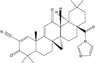
Having confirmed the negative role of Nrf2 in adipogenesis in MEFs, effects of CDDO-Im treatment on obesity and related diseases were evaluated in mouse models. CDDO-Im effectively prevented increases in body weight gain in mice fed a high fat, obesogenic diet but not a control diet. This effect was associated with a reduction of adipose mass and decreased lipid accumulation following CDDO-Im treatment in the mice fed a high fat diet. Furthermore, CDDO-Im treatment improved glucose tolerance in high fat-fed and streptozotocin-treated mice. Indirect calorimetry and quantitative RT-PCR confirmed that energy expenditure and fat oxidation were up-regulated while expression and activity of fatty acid synthesis enzymes were down-regulated following CDDO-Im treatment. Collectively, these results raise the possibility of the use of CDDO-Im for prevention of obesity, diabetes, and fatty liver.
ROLE OF BIOTRANSFORMATION IN 3-(3,5-DICHLOROPHENYL)-2,4-THIAZOLIDINEDIONE (DCPT)-INDUCED HEPATOTOXICITY IN RATS
Dissertation by Christine M. Crincoli, Ph.D.
University of the Sciences in Philadelphia, 2008
The compound 3-(3,5-dichlorophenyl)-2,4-thiazolidinedione (DCPT), which contains a 2,4-thiazolidinedione (TZD) ring, has been found to be heptatotoxic when administered to Fischer 344 (F344) rats. TZD rings are also found in a number of drugs that are used in the treatment of type II diabetes. The glitazone class of drugs includes troglitazone (TGZ), rosiglitazone (RGZ), and pioglitazone (PGZ). The use of these drugs in a clinical setting revealed idiosyncratic liver damage that ranged from mild to so severe that liver transplantations were required or death occurred. Toxicity seen with one member of this class in particular, TGZ, was so profound that it was withdrawn from the market 4 years after its initial introduction. While the exact mechanism behind the hepatotoxicity observed with these drugs is not understood, evidence in the literature is suggestive of the formation of reactive intermediates from biotransformation in the TZD ring.
A particular drawback to the exploration of glitazone-induced hepatotoxicity is the lack of a reliable and predictable animal model. In comparison to TGZ, however, DCPT produces liver damage that is consistently reproducible in a normal and common laboratory animal species. The structural similarity plus reproducibility of hepatotoxicity lead us to believe that DCPT may be a useful compound to study the potential involvement of the TZD ring in toxicity. In analogy to the glitazones, DCPT may also require biotransformation in order to produce toxicity, therefore a series of experiments were conducted to test this hypothesis.
DCPT

First, DCPT was synthesized and tested for its potential toxicity in cytochrome P450 (CYP) inhibitor-pretreated male Fischer 344 rats. The compound was administered in 2 previously determined heptatotoxic doses (0.6 and 1.0 mmol/kg, i.p. in corn oil) to CYP-inhibitor (1-aminobenzotriazole and troleandomycin)-pretreated rats. Liver and kidney functions and morphology were assessed 24-h post-dosing. The results from these experiments showed that CYP inhibitor-pretreated rats were protected from the liver damage observed in animals that did not receive the inhibitor pretreatment. Alternatively, male F344 rats that were pretreated with a CYP-inducer (dexamethasone) prior to a previously known non-toxic dose of DCPT (0.2 mmol/kg) exhibited elevated ALT levels and histology similar to that of animals that were dosed with a hepatotoxic dose.
To further explore the role of the CYP450 enzymes in the production of hepatotoxicity, female rats were administered the same doses of DCPT as male rats. The results of this experiment showed the lack of hepatic damage in female rats compared to the hepatotoxicity observed at matching doses in the male rats. One possible explanation for this gender difference could be the expression pattern of CYP enzymes. Male rats have predominantly CYP3A1/2 while female rats express mainly the CYP3A9 isoform of the enzyme. Similar to TGZ, DCPT may be extensively metabolized into a reactive species via the CYP3As.
A glutathione (GSH) conjugation trapping study was done to determine if putative reactive intermediates of DCPT could be detected in rat microsomal incubations and possibly elucidate a metabolic pathway of DCPT toxicity. Microsomes from corn oil-pretreated rats were incubated in the presence of GSH and appropriate cofactors. An isocyanate–GSH conjugate was found via reversed-phase HPLC. However, this conjugate could be derived via non CYP-mediated metabolism of DCPT. Further work is necessary to determine the mechanism of formation of the GSH conjugate.
In conclusion, the results of this study suggest that DCPT requires metabolism to exhibit its hepatotoxic effects. The exact mechanism of toxicity, however, remains to be elucidated, and further investigations are required.
THE ROLE OF STEAROYL-COA DESATURASE-1 IN MEDIATING WHOLE BODY ENERGY METABOLISM
Dissertation by Harini Sampath, Ph.D.
The University of Wisconsin, Madison, 2008
Advisor: James M. Ntambi
Stearoyl-CoA desaturase-1 is a delta-9 desaturase that catalyzes the conversion of saturated fatty acids to monounsaturated fatty acids. SCD1-deficient mice (SCD1−/−) are lean and protected from diet- and leptin deficiency-induced obesity and insulin resistance; they also have severe cutaneous abnormalities stemming from sebocyte hypoplasia in the skin. Given that dietary saturated fats are the substrates for SCD1-mediated desaturation, the first part of this thesis examined the role of SCD1 in mediating the pro-lipogenic effects of dietary saturated fat. SCD1−/− mice were largely protected from induction of de novo lipogenesis in response to dietary saturated fat. Instead, SCD1−/− mice catabolized cellular saturated fats by increasing fatty acid oxidation. This set of studies revealed that desaturation of dietary saturated fats by SCD1 is an essential aspect of their pro-lipogenic effects.
Since obesity increases the risk for insulin resistance, and given the role of SCD1 in the obesogenic pathway, the second set of studies in this thesis sought to determine the role of SCD1 in obesity-induced insulin resistance. Using 3 different mouse models of obesity, it was determined that SCD1 deficiency protects leptin-resistant Agouti obese mice and high fat-fed obese mice from obesity and insulin resistance. In contrast, leptin deficient ob/ob mice were not protected from insulin resistance due to SCD1 deficiency, despite significant reductions in adiposity.
Lastly, given the cutaneous abnormalities observed in SCD1−/− mice, it was of interest to determine the role of SCD1 in the skin. To address this, conditional knockouts of SCD1 lacking the SCD1 gene only in the skin (SKO mice) were generated. Deletion of SCD1 from the skin reconstituted the sebocyte hypoplasia and alopecia observed in SCD1−/− mice. In addition, skin-specific deletion of SCD1 also recapitulated the increased energy metabolism and protection from diet-induced obesity and insulin resistance observed in SCD1−/− mice secondary to increased fatty acid oxidation and uncoupling sebocyte hypoplasia, observed in global SCD1 deficiency. In summary, the results presented in this thesis indicate that SCD1 does indeed play a critical role in regulating whole body energy metabolism, hepatic lipogenesis, and insulin sensitivity as well as maintaining an intact skin barrier.
NON-ALCOHOLIC FATTY LIVER DISEASE ALTERS THE THREE STAGES OF HEPATIC DRUG MANAGEMENT
Dissertation by Craig D. Fisher, Ph.D.
The University of Arizona, 2008
Advisor: Nathan J. Cherrington
In pharmacotherapeutics, the term “correct dosing” is based on the concept that too high a systemic concentration will lead to drug toxicity, while drug levels that are too low may not produce the intended therapeutic effect. Often, the factors determining the ability of a patient to manage a given dose rely on their capacity to efficiently metabolize and eliminate drugs from the body. The liver plays a crucial role in the processing of many clinically relevant drugs via 3 stages of hepatic drug management. Drugs must first be taken into hepatocytes by uptake transporters. Drugs are then metabolized by phase I and phase II enzymes to make them more manageable. Finally, metabolites are removed from the hepatocyte by efflux transporters either into the bile for elimination or reintroduction to systemic blood. Alterations in one or more of the hepatic drug management stages increase the potential for adverse drug reactions (ADRs).
In the United States, ADRs account for between 3% and 12% of admissions to hospitals, and ∼5% of deaths each year. While <20% of these cases are due to genetic polymorphisms, the vast majority of ADRs are due to environmental factors including disease. Non-alcoholic fatty liver disease (NAFLD) comprises a spectrum of conditions progressing from steatosis to non-alcoholic steatohepatitis (NASH) and often leading to cirrhosis. Presently, NASH patients represent the greatest population of candidates for liver transplant, illustrating the severity as well as the incidence of this disease. Patients with NAFLD are typically treated for co-existing conditions of the metabolic syndrome (ie, hyperlipidemia or type II diabetes) and therefore represent a distinct population at risk for adverse drug reactions.
The following studies show that experimental NAFLD affects both the signal transduction pathways regulating hepatic drug management genes as well as the hepatic uptake transporter function. Additionally, patient livers diagnosed with progressive stages of NAFLD, display altered CYP activity and efflux transporter expression similar to those previously reported in experimental NAFLD. Given that changes observed in experimental NAFLD result in functional changes in hepatic drug management, similar changes observed in patients with this disease suggest an increased risk for ADRs.
INVESTIGATION OF THE MOLECULAR MECHANISMS OF ERV26P-DEPENDENT PROTEIN SORTING
Dissertation by Catherine A. Bue, Ph.D.
Dartmouth College, New Hampshire, 2009
Advisor: Charles K. Barlowe
Studies of the secretory pathway have revealed a multitude of secretory proteins that depend on the coat protein complex II (COPII) for export from the endoplasmic reticulum (ER) in transport vesicles. Some secretory proteins require protein-sorting adaptors to provide an indirect link to the COPII coat. Protein-sorting adaptors in ER-to-Golgi transport are generally thought to contain sorting signals that mediate binding to COPII as well as the retrograde-directed co-atomer protein (COPI) coat for bidirectional trafficking between the ER and Golgi. However, mechanisms of protein-sorting adaptor-dependent ER export remain unclear, including what fraction of secretory proteins require sorting adaptors, how many types of sorting adaptors are needed to accommodate the diversity of cargo, and the regulation of sorting adaptor-dependent transport. In this thesis, studies were performed on the uncharacterized Saccharomyces cerevisiae ER vesicle protein known as Erv26p, and indicate that it is a novel protein-sorting adaptor. Experimental evidence demonstrates that Erv26p is packaged efficiently into COPII vesicles, and strains lacking Erv26p show an ER accumulation of vacuolar pro-alkaline phosphatase (pro-ALP). pro-ALP interacts with Erv26p and is packaged into COPII vesicles in an Erv26p-dependent manner. To investigate the molecular mechanism of Erv26p-dependent sorting, mutations in conserved regions of Erv26p were made and analyzed. Erv26p interactions with both coat and cargo are required for efficient pro-ALP transport, and are independent of one another. Additionally, Erv26p homodimerization is important for pro-ALP interaction. The C-terminus of Erv26p is required for efficient COPII-dependent ER export, whereas the third luminal loop domain is involved in cargo recognition. These results have revealed mechanisms governing Erv26p-dependent transport, and future studies on this sorting adaptor–cargo pair should allow an elucidation of general mechanisms underlying receptor-mediated ER export.
MOLECULAR ARCHITECTURE OF THE YEAST DSL1P TETHERING COMPLEX
Dissertation by Yi Ren, Ph.D.
Princeton University, 2009
Advisor: Frederick M. Hughson
Multisubunit tethering complexes are essential for intracellular trafficking and have been proposed to mediate the initial interaction between a vesicle and its target membrane. The Dsl1p tethering complex, consisting of Tip20p, Dsl1p, and Sec39p, is required for trafficking from the Golgi apparatus to the ER. It interacts both with COPI vesicle coat proteins and with ER-resident SNAREs. Here, I have combined X-ray crystallography and negative stain EM to elucidate the architecture of the Dsl1p complex.
Three crystal structures are presented in this work: a Dsl1p fragment, a Tip20p–Dsl1p fusion protein, and a Dsl1p–Sec39p complex. Superimposing overlapping regions—among these structures and that of Tip20p—yielded a model of the entire Tip20p–Dsl1p–Sec39p complex. In this model, Dsl1p is located at the center of the complex, containing an internal disordered region capable of binding COPI-coated vesicles at a distance. Using its N- and C-terminal regions, Dsl1p binds to Tip20p and Sec39p, respectively. EM analysis was consistent with this model and, in addition, revealed significant flexibility at the center of the Dsl1p subunit. Recent biochemical studies in our lab establish that Tip20p and Sec39p interact with 2 different ER SNAREs, Sec20p and Use1p, respectively. These results provide a mechanism for vesicle tethering through the simultaneous recognition of vesicles, via direct interactions with the COPI coat, and the ER, via direct interactions with Sec20p and Use1p. The interactions with the COPI coat through Dsl1p could trigger conformational changes in Tip20p and/or Sec39p that are transmitted to Sec20p and Use1p, possibly facilitating SNARE complex assembly.
The crystal structures reported here establish that Dsl1p resembles Tip20p. Moreover, Dsl1p and Tip20p resemble known subunits of the exocyst tethering complex, which functions in Golgi-to-plasma membrane trafficking, but not the TRAPPI tethering complex, which functions in ER-to-Golgi trafficking. This finding thereby indicates a structural connection among several of the known multisubunit tethering complexes and strongly implies a potential mechanistic unity among them. The structural studies in this work, and future mechanistic investigation of the Dsl1p complex, may therefore provide significant insights into the fundamental common mechanisms underlying the operation of these tethering complexes.
STRUCTURE/FUNCTION ANALYSIS OF THE DIPHTHERIA TOXIN T1 MOTIF IN THE CATALYTIC DOMAIN ENTRY PROCESS
Dissertation by Carolina Trujillo, Ph.D.
Boston University, 2009
Advisor: John R. Murphy
The translocation of the catalytic domain of diphtheria toxin (DT) from the lumen of early endosomes into the cytosol of eukaryotic cells is an essential step in the intoxication process. Previous studies have shown that the translocation process requires target cell cytosolic proteins including b-COP. This study was focused on gaining a better understanding of the interactions between the transmembrane domain of DT and cytosolic proteins and how this domain influences the translocation of the catalytic domain. We reported the identification by BLAST analysis of a motif in DT transmembrane helix 1 (T1 motif) also found in other bacterial toxins. Results from pull-down experiments using the fusion protein GST-DT140–271 containing the T1 motif demonstrated binding of several COPI subunits including b-COP in a T1 motif-dependent fashion. We characterized a translocation-deficient, non-toxic single point mutant DAB 389 (L221E) IL-2. Because the L221E mutation falls within the T1 motif, we postulated a potential role for the T1 motif region in the formation of specific protein–protein interactions required in the catalytic domain translocation process. The interactions between the COPI complex and the T1 motif region were further characterized. We demonstrate by a COPI precipitation assay that synthetic peptides, which carry the T1 motif or mutant sequences, are predictive of interactions with the COPI complex, and that these interactions are mediated by specific dibasic signatures. It was also demonstrated that substitution of lysine to alanine in the T1 motif region of the fusion protein toxin DAB 389 IL-2 results in mutant proteins that are either non-toxic or have reduced toxicity relative to the wild-type parental protein. Furthermore, a mutant toxin in which the entire T1 motif was replaced with the conserved p23 COPI adaptor cytosolic tail region resulted in a substitution mutant that is as toxic as the unmodified DAB 389 IL-2. Thus, interactions between the T1 motif and COPI complex proteins are both essential for the efficient delivery of the DT catalytic domain into the target cell cytosol and are mediated by distinct lysine signatures that mimic the profiles presented to COPI by endogenous adaptor molecules.
REGULATION OF PEROXISOME PROLIFERATOR-ACTIVATED RECEPTOR ALPHA BY RIBOSOMAL PROTEIN L11, MURINE DOUBLE MINUTE 2, AND E6-ASSOCIATED PROTEIN
Dissertation by Lakshmi Gopinathan, Ph.D.
The Pennsylvania State University, 2008
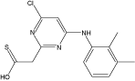
The peroxisome proliferator-activated receptors (PPARs) regulate genes involved in lipid and glucose metabolism, adipocyte differentiation, fatty acid oxidation, and inflammation. Regulation of these diverse responses requires a series of highly coordinated cellular events that include binding of ligand, association with a heterodimeric partner, recruitment of transcriptional machinery, and multiple protein–protein interactions. This thesis will examine a subset of these protein–protein interactions and detail the effects of the association on PPARα transcription activity. The ribosomal protein L11 (rpL11) is a PPARα-interacting protein that inhibits receptor transcriptional activity. Ablation of rpL11 expression by RNAi resulted in increased ligand-induced mRNA levels of PPARα target genes, and increased PPARα transactivation in PPRE-dependent reporter assays. Growth inhibitory conditions such as serum deprivation and treatment with low concentrations of actinomycin D facilitate increases in the level of non-ribosomal L11; these conditions decreased transcriptional activity of PPARα. Ribosomal protein L11 also acts as a negative regulator of murine double minute 2 (MDM2), mediating p53 stabilization. Whether interactions with rpL11 resulted in a convergence of PPARα and p53 pathways in response to peroxisome proliferators (PPs) was examined. Although treatment of Hepa-1 cells with the PPARα ligand Wy-14,643 decreased transcriptional activity of p53, this effect could not be attributed to rpL11. Increased p53 activity mediated by rpL11 remained unaffected by Wy-14,643 or coexpression of PPARα. Thus, rpL11 regulates both p53 and PPARα pathways, although the effects of peroxisome proliferators on p53 activity cannot be attributed to rpL11 associating with PPARα.
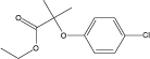
The identification of rpL11 as an MDM2-interacting protein and regulator of the MDM2/p53 pathway prompted further studies investigating the role of MDM2 in regulation of PPARα. MDM2 interacted with the A/B domain of PPARα and regulated the transcriptional activity of the receptor. Knockdown of MDM2 by siRNA in FaO hepatoma cells decreased ligand-induced mRNA levels of several PPARα target genes involved in lipid metabolism, indicating that MDM2 enhances the transcriptional activity of PPARα. MDM2 associated with PPARα on target gene promoters, and this association increased in the presence of Wy-14,643. Interestingly, treatment of mice with clofibrate resulted in decreased protein expression of MDM2 in wild type, but not PPARα null mice, suggesting that PPARα controls MDM2 through a negative feedback mechanism. MDM2 coexpression increased ubiquitination of PPARα; the E3 ubiquitin ligase activity of MDM2 regulated PPARα protein expression and transcriptional activity. PPARα protein expression was decreased in FaO cells transfected with MDM2 siRNA indicating that MDM2 stabilized PPARα protein levels. In support, MDM2 null mouse embryonic fibroblasts (MEFs) expressed lower levels of PPARα protein and responded to Wy-14,643 only upon reintroduction of MDM2. These studies identify MDM2 as a component of PPRE-associated coregulator complex that increases PPARα transcriptional activity in response to ligand, and that is in turn regulated by PPARα through a negative feedback loop.
Wy-14643
Although MDM2 ubiquitinates PPARα, it is not the only E3 ligase that is capable of regulating this nuclear receptor. The E6-associated protein (E6-AP), previously identified as a nuclear receptor coactivator, was examined as a potential PPARα-interacting protein and ubiquitin E3 ligase. E6-AP is a PPARα-interacting protein that when overexpressed resulted in ligand-independent inhibition of PPARα transcriptional activity. This effect required the ubiquitin ligase activity of E6-AP. Treatment of mice with clofibrate resulted in an increase in E6-AP expression in wild type, but not PPARα null mice, suggesting that E6-AP acts as a feedback regulator of PPARα.
Emerging studies suggest a role for MDM2 in insulin signaling via interactions with the insulin receptor substrate 1 (IRS1) and glycogen synthase kinase 3 (GSK3). Understanding the role of the PPARα–MDM2 interaction in insulin signaling and manipulating this interaction may have important implications in developing improved PPARα-based therapeutics. The significance of the MDM2–rpL11 interaction in tumorigenesis is highlighted by reports of cancer-associated mutations in MDM2 that disrupt its interaction with ribosomal proteins such as rpL11. The studies presented in this thesis also provide a basis for examining the role of PPARα in regulating the MDM2–rpL11 association during cancer development.
Clofibrate
A GENETIC SCREEN FOR MODULATORS OF THE NOTCH PATHWAY IN Drosophila melanogaster IDENTIFIES NOT1 AS A POSITIVE REGULATOR OF NOTCH SIGNALING
Dissertation by E. Morreale, Ph.D.
Boston College, 2009
Advisor: Marc A.T. Muskavitch
The Notch pathway is an evolutionarily conserved mechanism of intercellular signaling that plays a central role in the development of metazoans. Here I summarize 2 genetic screens that utilize a rough eye phenotype created by Delta overexpression in the Drosophila eye to identify modulators of Notch pathway-signaling activity. Among the many “hits” obtained from both screens, I have mapped to the Not1 gene a single complementation group that exhibits strong genetic interactions with Notch pathway mutants. NOT1 is a component of the CCR4–NOT complex, a global regulator of gene expression that exerts its effects through a variety of mechanisms, including mRNA deadenylation and direct transcriptional repression. I have conducted a series of genetic and molecular experiments in an effort to obtain more insight into the relationship between the CCR4–NOT complex and the Notch pathway. Both Not1 EMS mutations and RNAi-mediated knockdown of NOT1 expression produce phenotypes that mimic those of Notch loss-of-function pathway mutants. Knockdown of NOT1 in the developing bristle organ disrupts Notch-mediated inhibition of neuronal specification, resulting in supernumerary neurons and aberrant sheath cell specification. Knockdown of NOT1 within the developing wing margin disrupts expression of the Notch target genes Cut and Wingless, as well as the Notch ligand Delta. Phenotypic rescue experiments imply that Not1 functions downstream of Notch signal activation and acts directly on Notch target gene expression. These results suggest that NOT1 is required for Notch signal transmission in certain developmental contexts and implicate the CCR4–NOT complex as a positive regulator of the Notch pathway.
NOTCH SIGNALING IN MOUSE EMBRYONIC NEURAL STEM CELL REGULATION AND CANCER
Dissertation by L. Dang, Ph.D.
The Johns Hopkins University, 2009
Advisor: Nicholas Gaiano
The processes of neural development and cancer share common regulatory molecules, such as those in the Notch pathway. Activated Notch signaling maintains neural stem cell fate in the developing nervous system, and pathway dysregulation causes cancer in various organ systems, including the nervous system.
Of the Notch receptors expressed in the developing mouse telencephalon (Notch1–3), Notch1 has been most heavily studied. Because Notch3 can oppose Notch1 in some situations, a retrovirus expressing activated Notch3 was injected into the forebrain vesicle near the onset of neurogenesis to carry out a gain of function analysis. Activated Notch3 promoted the formation of radial glia embryonically and astrocytes postnatally, similar to previous findings with Notch1. Interestingly, Notch3 activation also caused the formation of choroid plexus tumors and eye tumors, but similar experiments with Notch1 did not.
In order to study endogenous levels of Notch signaling, the Gaiano Lab developed a mouse line containing a transgene with the CBF1-responsive element driving green fluorescent protein (GFP) expression. It was found that the ventricular zone of the developing brain had clusters of GFP-high and GFP-low cells, an indication of differential CBF1/Notch signaling among progenitors. GFP-high cells possessed characteristics of neural stem cells, while GFP-low cells had characteristics of more differentiated intermediate neural progenitors. Prospective isolation of GFP-high and GFP-low cells followed by in vivo cell transplantation revealed that the GFP-high cells gave rise to neurons, oligodendrocytes, and astrocytes, while the GFP-low cells gave rise to mostly neurons and oligodendrocytes.
To explore what molecules cause this differential CBF1/Notch signaling, the Deltex proteins, known regulators of the Notch-signaling pathway, were examined in the developing telencephalon. When expressed in vivo by in utero electroporation, Deltex1 and Deltex4 caused cells to cluster in the subventricular zone, a proliferative region in the embryonic forebrain. However, Deltex1 expression in vivo could not block CBF1-dependent Notch signaling.
This work builds upon evidence that Notch signaling can be oncogenic, shows that differences between Notch1 and Notch3 are context dependent, unveils a novel mechanism of neural progenitor diversification, and suggests that Deltex proteins can affect cell fate specification in the developing brain.
THE BRAIN-SPECIFIC PROTEIN, DELTA-CATENIN, IS CRITICAL FOR THE MAINTENANCE OF CORTICAL NEURONAL STRUCTURE AND FUNCTION IN VIVO
Dissertation by C. Matter, Ph.D.
University of California, Los Angeles, 2009
Advisor: Xin Liu, Joshua Trachtenberg
Delta (d)-catenin is a brain-specific member of the adherens junction complex that is heavily expressed in neurons and frequently deleted in the severe mental retardation syndrome, Cri-du-Chat (CDCS). Identified through its interaction with presenilin-1 (PS1), the most commonly mutated protein in familial Alzheimer’s disease (FAD); d-catenin localizes within the post-synaptic and dendritic compartments, and is implicated to play a role in dendritic outgrowth via modulation of the actin cytoskeleton. Mice with a targeted disruption of d-catenin were shown to have severe impairments in hippocampal dependent learning. The mechanisms underlying these deficits are not clearly defined. In vitro, d-catenin mediates neuronal morphogenesis implicating alterations in neuronal connectivity in contributing to the cognitive deficits in the d-catenin (−/−) mice. This work addresses the role of d-catenin in vivo in the maintenance of cortical neuronal structure and function. We found that loss of d-catenin in vivo results in a progressive loss of dendritic arbor length and decreased dendritic spine stability resulting in a significant reduction in spine density. Accompanying the progressive structural decline, we observe progressive reductions in cortical responsiveness and functional organization to physiological stimuli. Direct electrode stimulation confirmed the presence of a cortical component in the decline in responsiveness. Our data indicate that d-catenin is critical for the maintenance of cortical neuronal structure and function in vivo and may further our understanding of the pathology associated with the mental retardation syndrome Cri-du-Chat and neurodegenerative diseases such as Alzheimer’s disease.
STRUCTURAL ANALYSIS OF TWO FAMILIES OF INTRAMEMBRANE PROTEASES: RHOMBOID AND SITE-TWO PROTEASE (S2P)
Dissertation by Zhuoru Wu, Ph.D.
Princeton University, 2008
Regulated intramembrane proteolysis (RIP) plays important roles in activating cellular signaling pathway in both prokaryotes and eukaryotes. The realization that transmembrane proteins can be cleaved in their membrane spans in the lipid bilayer inspired the extensive studies of the intramembrane proteases responsible for these cleavages. There are 4 known families of intramembrane proteases: rhomboid, site-2 protease, presenilin, and signal peptide peptidase. Due to the unique feature of RIP occurring in a hydrophobic environment, it is intriguing to investigate the detailed molecular mechanism of the intramembrane proteolytic reactions.
In this dissertation, I will present 2 three-dimensional protein structures of the transmembrane core domains from 2 intramembrane protease families: GlpG, the Escherichia coli homolog of rhomboid; and mjS2P, the Methanococcus jannaschii homolog of S2P.
Structure analysis helped shed light on how these proteases carry out their functions in intramembrane proteolysis. First, water molecules, which are essential for the scission of peptide bonds, appear to gain access to the active sites of GlpG and mjS2P via a similar mechanism. GlpG contains an open cavity full of water that converges on the active site residue Ser201 whereas mjS2P has a polar channel that allows water entry to the catalytic zinc atom in the closed conformation. More importantly, the 2 proteases share significant similarity in substrate entry as well, which is proposed to be a gating mechanism regulated by transmembrane helices. This mechanism involves the lateral movements of transmembrane helices within the lipid bilayer to allow substrate access the active site of intramembrane proteases. In GlpG, bending of the C-terminal half of the transmembrane helix a5 was proposed to open the gate for substrate entry. Likewise, in mjS2P, the rotation and translocation of transmembrane domain (TM) 1 as well as the translocation of TM6/TM5 appear to allow substrate entry.
CURCUMIN INHIBITION OF THE MAMMALIAN TARGET OF RAPAMYCIN SIGNALING PATHWAY
Dissertation by Christopher S. Beevers, Ph.D.
Louisiana State University Health Sciences Center–Shreveport, 2008
The goal of our present study was to investigate the potential anticancer effects of curcumin against rhabdomyosarcoma (RMS). The work presented in this dissertation demonstrates that curcumin significantly inhibited the proliferation/growth, motility, and survival of the Rh1 and the Rh30 RMS cell lines. Investigations into the molecular mechanisms of curcumin revealed that the compound potently inhibited the mammalian target of rapamycin (mTOR) complex 1 (mTORC1)-mediated phosphorylation of p70-S6 Kinase 1 (S6K1) and eukaryotic initiation factor 4E (eIF4E)-binding protein 1 (4E-BP1), the phosphorylation of mTOR, and the mTOR complex 2 (mTORC2)- and the phosphatidylinositol-dependent kinase 1 (PDK1)-mediated phosphorylation of Akt/protein kinase B (Akt/PKB). We observed this phenomenon also in human breast (MCF-7), prostate (DU145), cervical (HeLa), and colon (HT29) cancer cell lines.
We discovered that curcumin inhibits mTOR signaling independently of protein phosphatase type 2A (PP2A) and the tuberous sclerosis complex 2/AMP-activated protein kinase (TSC2/AMPK) network using analysis of phosphorylation and methylation states, inhibitors, and dominant-negative and inactive forms of the proteins, small hairpin ribonucleic acid (shRNA) down-regulation, and cell lines containing genomic knockouts of these various factors.
Curcumin
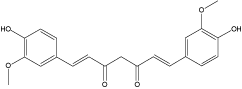
Focusing on mTOR itself as the potential target for curcumin, we found that the compound blocks both insulin-like growth factor type 1 (IGF-1)- and phosphatidic acid (PA)-mediated stimulation of wild-type mTOR, but the expression of a rapamycin-resistant form of mTOR suppressed this inhibitory action of curcumin to a certain degree. Curcumin was able to block the kinase activity of the mTORC1 and the mTORC2 by disrupting mTOR-raptor and mTOR-rictor interaction.
N-acetyl-L-Cysteine (NAC)

Phenylarsine oxide (PAO)

However, we discovered that both N-acetyl-
GENETIC AND BIOCHEMICAL CHARACTERIZATION OF THE TSC-RHEB-TOR SIGNALING PATHWAY IN DROSOPHILA
Dissertation by Y. Zhang, Ph.D.
The Johns Hopkins University, 2009
Advisor: Duojia Pan
The insulin/target of rapamycin (TOR)-signaling pathway regulates cell growth, cell metabolism, and cell survival; it also plays pivotal roles in the genesis of human diseases including cancer, diabetes, and aging. Previous studies from our lab have shown that tuberous sclerosis (Tsc) tumor suppressors negatively regulate insulin signaling in cell growth and act in a parallel pathway (Gao and Pan, 2001). We have also shown that Tsc1 and Tsc2 antagonize amino acid-TOR-signaling pathway (Gao et al., 2002; Gao and Pan, 2001). Tsc2 possesses a domain that shares homology with the GTPase-activating protein (GAP) domain of Rap1–GAP2, suggesting that a GTPase might be the physiological target of Tsc2. Here, we show that the small GTPase Ras homolog enriched in brain (Rheb) is a direct target of Tsc2 GAP activity both in vivo and in vitro. Point mutations in the GAP domain of Tsc2 disrupted its ability to regulate Rheb without affecting its ability to form a complex with Tsc1. Our studies identify Rheb as a molecular target of the Tsc tumor suppressors.
The 280-kDa TOR protein functions as the catalytic component of 2 large multiprotein complexes and consists of an N-terminal HEAT-repeat domain and a C-terminal Ser/Thr kinase domain. To gain insights into the functions of and interactions between different domains of TOR proteins, we conducted an EMS mutagenesis screen and recovered a series of mutations in the Drosophila Tor gene. We have shown that combinations of mutations in the HEAT and kinase domains of TOR displayed the rare genetic phenomenon of intragenic complementation, in which 2 or more defective proteins assemble to form a functional multimer. We present biochemical evidence that TOR self-associates into multimer in vivo and show that this multimerization is unaffected by positive or negative signals upstream of TOR. Consistent with multimerization of TOR, recessive mutations in the HEAT and kinase domains can dominantly interfere with wild-type TOR function in cells lacking Tsc1 or Tsc2. Our studies demonstrate that Drosophila target of rapamycin kinase functions as a multimer.
TOR complex 1 (TORC1) and TOR complex 2 (TORC2) are 2 distinct complexes that have been identified recently (Bhaskar and Hay, 2007). Previous studies involving TOR complexes were mostly performed in cell culture, so the roles of TORC1 and TORC2 in animal development remain elusive. We investigated the roles of TORC1 and TORC2 in Drosophila development by generating dTORC mutant flies and characterizing them side by side. We show that dTORC1 is required early in development, while dTORC2 mutants are viable and only show minor effects on cell growth. Flies ablating dLST8, a shared component of dTORC1 and dTORC2, are also viable but show severe growth defects. In the fat body, dLST8 mutants recapitulate the growth inhibition-phenotype of dTor mutants, but other dTORC mutants show no effects. Furthermore, dLST8 mutant flies show enhanced resistance to oxidative stress treatment. Our results indicate that dLST8 carries novel functions beyond its role in dTORC2.
THE ROLE OF THE AKT/TSC/MTOR SIGNALING PATHWAY IN KAPOSI’S SARCOMA-ASSOCIATED VIRUS G-PROTEIN-COUPLED RECEPTOR NEOPLASIA
Dissertation by Risa Chaisuparat, Ph.D.
University of Maryland, Baltimore, 2009
Advisor: Silvia Montaner
Kaposi’s sarcoma (KS), a multifocal vascular neoplasm, is the most frequent cancer arising in HIV-infected individuals and in immunosuppressed patients. KS is caused by the KS-associated herpesvirus (KSHV). A single lytic gene of KSHV genes, G-protein-coupled receptor (vGPCR), is sufficient to induce Kaposi-like sarcomas in mice. This vGPCR has been shown to promote the activation of PI3K/Akt, involved in transformation of endothelial cells. However, the Akt downstream effectors required for vGPCR to promote Kaposi’s sarcomagenesis are still unknown.
Here, we have found that vGPCR induces the phosphorylation and inactivation of tuberin (TSC2), promoting the activation of mTOR. Moreover, overactivation of TSC/mTOR is sufficient to render endothelial cells oncogenic. Treatment with rapamycin (mTOR inhibitor) efficiently prevented the growth of vGPCR tumors in vivo. Collectively, these results implicate the mTOR-signaling route in Kaposi’s sarcomagenesis and provide experimental evidence demonstrating that drugs targeting mTOR may represent an effective mechanism-based therapy for the treatment of KS.
We have observed that although rapamycin has been shown to be an efficient therapy for patients with iatrogenic or classic KS, the activity of Akt was transiently increased in vGPCR-expressing endothelial cells (EC-vGPCR). We therefore investigated the efficacy of PI-103, a novel dual PI3Ka/mTOR inhibitor, in preventing vGPCR transformation. PI-103 treatment effectively and independently blocked the activation of both PI3K and mTOR in EC-vGPCR. This resulted in the effective inhibition of endothelial cell proliferation and survival in vitro, and tumor growth in vivo, suggesting that PI-103 may be an effective therapeutic option for the treatment of KS.
We also found that the angiogenic growth factors secreted by EC-vGPCR can induce the activity of mTOR in endothelial cells, suggesting that vGPCR regulates mTOR through both direct and indirect (paracrine) mechanisms. In addition, we found that the effect of rapamycin on vGPCR sarcomagenesis is not dependent on the direct activation of mTOR in vGPCR-expressing cells. Rather, the profound sensitivity of these tumors to rapamycin treatment may be due, in part, to the inhibition of the paracrine activation of mTOR in neighboring (bystander) cells by the angiogenic factors elaborated by EC-vGPCR. Collectively, these results support the key role of paracrine-transforming mechanisms in vGPCR sarcomagenesis.
PI-103

IDENTIFICATION OF TARGETED THERAPEUTICS FOR MALIGNANT PERIPHERAL NERVE SHEATH TUMORS
Dissertation by Gunnar L. Johansson, Ph.D.
University of Cincinnati, 2008
Advisor: Nancy Ratner
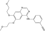
Neurofibromatosis type 1 (NF1) is one of the most common autosomal dominant disorders, affecting 1 in 3,500 worldwide. The hallmark of NF1 is the expression of benign tumors of the peripheral nerve (neurofibromas). In addition, patients have a 5%–13% life time risk of developing malignant peripheral nerve sheath tumors (MPNST), a soft tissue sarcoma with poor prognosis. NF1 functions as a negative regulator of active RAS; and elevated levels have been observed in both the neurofibromas and MPNST. The levels of epidermal growth factor receptor (EGFR) and mammalian target of rapamycin (mTOR) signaling are also increased in MPNST. In an attempt to define treatment options, we have treated MPNST cell lines grown as xenografts in nude mice, with the mTOR inhibitor RAD001 and the EGFR tyrosine kinase inhibitor erlotinib. When treatment was initiated prior to the formation of the tumors, RAD001 prevented the growth of the tumors and erlotinib reduced tumor growth by 35%. In already established tumors, erlotinib had no effect. RAD001 significantly, but transiently, delayed tumor growth, and decreased vessel permeability within xenografts. To find additional drugs that can target NF1-related tumors, we have recently screened NF1-related and sporadic MPNST cell lines using a high-throughput screening approach. The only known difference between these tumors is the absence of NF1, and the up-regulation of active RAS levels. Drugs that show selectivity against the NF1-related MPNSTs are likely to target pathways directly affected by NF1 and could potentially be used in both neurofibromas and MPNST.
RAD001 (everolimus)

RAD001 (everolimus)

Erlotinib
THE IMMUNOSUPPRESSIVE EFFECTS OF TRIPTOLIDE AND RAPAMYCIN ON MOUSE MODEL OF CARDIAC TRANSPLANTATION
Dissertation by Yan Liu, Ph.D.
University of Hong Kong, 2008
Current immunosuppressive strategies for transplantation have failed to achieve long-term graft survival. Triptolide (TPT) is a component of the Chinese herb Triptergium wilfordii and has potent immunosuppressive and anti-inflammatory effects. In this study, I have investigated the effects of TPT alone and in combination with rapamycin (Rapa) on graft survival together with the changes induced in pathology and immunological responses. Since dendritic cells (DCs) play a critical role in the initiation of alloimmune responses to foreign grafts, this study was designed to further evaluate the effects of TPT in the functional phenotype of bone marrow (BM)-derived DCs and to determine the function of DC-SIGN on DCs in transplant immunology.
Heterotopic heart transplantation model was established using MHC fully mismatched mice (C57BL/6 and BALB/c) as donors and recipients, respectively. TPT and Rapa were administered either alone or in combination. Complete cessation of the cardiac beat was used to determine the survival time of the allografts. In vitro experiments, BM-derived DCs were cultured with or without TPT. For evaluation of DC-SIGN function, DCs were treated with DC-SIGN soluble protein or DC-SIGN expression was inhibited. DC maturation, T-cell stimulatory properties, and cytokine secretion were examined.
Triptolide (TPT)
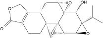
The mean survival time (MST) of the heart allografts in animals receiving TPT alone or combined with Rapa was significantly prolonged compared to the no treatment group (23.5 ± 5.3 days at 3 mg/kg of TPT and 93.5 ± 6.7 days in combination with Rapa vs. 7.7 ± 0.8 days in untreated group). TPT and/or Rapa treatment inhibited LPS-induced DC maturation. This was accompanied by up-regulated synthesis of IL-10 and down-regulated CCL19, CCR5, CCR7, IFN-γ, and IL-12, facilitating the expansion of regulatory T (Treg) cells. Furthermore, I observed that TPT increased cell surface expression of DC-SIGN and that treated DCs with DC-SIGN soluble protein enhanced their ability to secrete IL-10. However, DC-SIGN protein had no inhibitory effects T-cell-mediated responses.
Taken together, current results demonstrate that the combination of TPT and Rapa can significantly prolong allograft survival in a mouse model of cardiac transplantation. These compounds attenuated graft rejection by inhibition of DCs maturation, conditioning DCs to adopt tolerogenic phenotype, and the expansion of Treg cells. Furthermore, up-regulation of DC-SIGN by TPT may contribute to the ability of TPT to maintain DC homeostasis in immune responses. Whether DC-SIGN has any effects in graft rejection still needs to be further investigate. Present studies add weight to the application of combination therapy in transplantation.
INVESTIGATIONS INTO NEURONAL CILIA UTILIZING MOUSE MODELS OF BARDET-BIEDL SYNDROME
Dissertation by Nicolas F. Berbari, Ph.D.
The Ohio State University, 2008
Advisor: Kirk Mykytyn
Cilia are hairlike microtubule-based cellular appendages that extend 5–30 µm from the surface of most vertebrate cells. Since their initial discovery over a 100 years ago, cilia have been of interest to microbiologists and others studying the dynamics and physiological relevance of their motility. The more recent realization that immotile or primary cilia dysfunction is the basis of several human genetic disorders and diseases has brought the efforts of the biomedical research establishment to bear on this long overlooked and underappreciated organelle.
Several human genetic disorders caused by cilia defects have been identified, and include Bardet-Biedl syndrome, Joubert syndrome, Meckel-Gruber syndrome, Alstrom syndrome, and orofaciodigital syndrome. One theme of these disorders is their multitude of clinical features such as blindness, cystic kidneys, cognitive deficits, and obesity. The fact that many of these cilia disorders present with several features may be due to the ubiquitous nature of the primary cilium and their unrecognized roles in most tissues and cell types.
The lack of known function for most primary cilia is no more apparent than in the central nervous system. While it has been known for sometime that neurons throughout the brain have primary cilia, their functions remain unknown. Research on neuronal cilia has suffered from a paucity of tools to study them. To this end, some of this work describes the development of the first in vitro neuronal culture system where primary cilia are present (Chapter 2).
It is apparent that the functions of cilia, at least in part, are defined by the specific proteins that localize within and on the organelle. Indeed access of proteins and signaling modules into and out of the ciliary compartment appears to be tightly regulated. In order to better understand the physiological roles of cilia throughout the body, an understanding of the signaling proteins that localize to the cilium and the mechanisms behind cilia localization is needed. To these ends, some of this work describes novel ciliary signaling proteins such as the localization of adenylyl cyclase III to primary cilia throughout the adult mouse brain, and the localization of the G-protein-coupled receptor (GPCR) melanin concentrating hormone receptor 1 to certain neuronal cilia (Chapters 3 and 4). We also have identified a specific GPCR sequence involved in the localization of GPCRs to the ciliary compartment (Chapter 4). This sequence will aid in determining the mechanisms that target proteins to the cilium.
The development of these tools has allowed for the opportunity to test the hypothesis that certain clinical features of the ciliary disorder Bardet-Biedl syndrome (BBS) are due to neuronal cilia dysfunction. Indeed, we have shown that mouse models of BBS fail to localize specific receptors to their neuronal cilia (Chapter 5). These data have set the ground work for more extensive investigations into neuronal cilia function. Further, this work suggests neuronal cilia dysfunction may contribute not only to the cognitive defects associated with ciliary disorders, but may also underlie the obesity observed in these disorders.
THE PSEUDOSUBSTRATE AND C1B DOMAINS OF PKC-EPSILON ARE NECESSARY AND SUFFICIENT FOR ACCUMULATION OF PKC-EPSILON AT THE PHAGOSOMAL MEMBRANE DURING Fcgamma R-MEDIATED PHAGOCYTOSIS: EVIDENCE FOR A NOVEL MEMBRANE TARGETING MECHANISM
Dissertation by K. Cheeseman, Ph.D.
Albany Medical College of Union University, 2008
Advisor: Michelle Lennartz
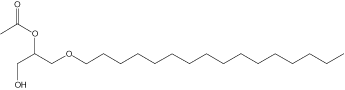
Binding of antibody-opsonized particles to macrophage Fcγ receptors (FcγR) transduces intracellular signals resulting in phagocytosis, and the production and secretion of proinflammatory compounds. Protein kinase C-ε (PKC-ε) accumulates at IgG-phagosomes and modulates the rate of FcgR-dependent phagocytosis, defining its mechanism of translocation is important for understanding its mechanism of action. Using confocal microscopy, deletion mutants, and a point mutant of GFP-conjugated PKC-ε, we found that deletion of the εC1 and εC1B domains or mutation of amino acid 259 within εC1B-domain (C [arrow right]G) decreased PKC-ε accumulation at phagosomes during phagocytosis and at the plasma membrane in response to exogenous diacylglycerol (DAG). PKC-ε translocation to phagosomes and FcγR-mediated phagocytosis was inhibited by the DAG antagonist, 1-hexadecyl-2-acetyl glycerol (EI-150). The role of phospholipase D (PLD) and phosphatidylinositol-specific phospholipase C (PI- PLC) in PKC-ε accumulation was assessed. Although GFP-PLD2 localized to phagosomes and enhanced phagocytosis, its inhibition with 2,3-diphos-
Endogenous PI-PLC-γ1 and GFP-PI-PLC-γ1 and PKC-ε accumulated at phagosomes in macrophages from PI-PLC-γ2−/− mice. PI-PLC-γ1 was transiently phosphorylated on tyrosine 783 in nascent phagosomes. These results indicate that εC1B is necessary for PKC-e translocation to membranes and this is dependent on DAG and PI-PLC-γ1.
Chimeras of PKC-δ containing eC1B [δ (εC1B)] and PKC-e containing δC1B [ε(δ C1B)] were used to determine if εC1B was sufficient for phagosome targeting. δ (εC1B) did not localize to phagosomes whereas ε(δ C1B) did. Truncation and deletion mutants revealed that removal of the pseudosubstrate domain (εPS) prevented PKC-ε accumulation. The δ (εPSC1B) chimera localized to phagosomes, whereas d (εPS) did not, indicating that εPS and εC1B are necessary and sufficient for phagosomal targeting. To identify the pseudosubstrate-binding partner at the phagosome, gluthatione-S-transferase (GST)-conjugated PS mutants were constructed and used in lipid-overlay assays. GST-εPS bound to phosphatidylinositols: PI(3)P ≥ PI(5)P > PI(4)P > PI(4,5)P ≥ PI(3,4,5)P. The PI (3)P reporter, 2xFYVE, accumulated at the phagosomes and colocalized with PKC-ε. Inhibition of PI(3)P production with Wortmannin prevented PKC-ε localization and decreased the rate of phagocytosis. These data are consistent with an εPS-PI(3)P interaction at the phagosome that would result in activation of the PKC-ε by removal of εPS from the active site.
EI-150
DESIGN AND SYNTHESIS OF PROTEIN KINASE C MODULATORS AS THERAPEUTIC LEADS
Dissertation by Jung-Min Kee, Ph.D.
Stanford University, 2008
Advisor: Paul A. Wender
Synthetic chemistry has played a central role in many branches of science. By enabling access to desired molecular entities, either natural or designed, it has provided scientists with invaluable tools to understand and even intervene nature at the molecular level. Natural product chemistry and drug discovery are examples of such areas where synthetic chemistry played critical roles. Although natural products with exquisite biological activities have been excellent leads in drug discovery, these natural leads are not optimized for therapeutic use in humans, and many of them often suffer from scarce availability. Therefore, synthesis of the natural products and their designed analogs is of high significance in improving the function and supply of these therapeutic leads. With this regard, this thesis describes the progress on the development of the synthetic strategies to daphnane and tigliane diterpenes and their designed analogs. In particular, studies on kirkinine, a daphnane diterpene with potent neurotrophic activity, and prostratin, a tigliane diterpene with promising anti-HIV activities, are presented. Progress in the development of staurosporine analogs as protein kinase C (PKC) inhibitors is also reported in this dissertation.
Prostratin

A common theme in the function of these compounds is the modulation of protein kinase C (PKC) activity. In Chapter 1, therefore, a general review on PKC is presented, with emphasis on its implication in human diseases. PKC is a serine/ threonine kinase that is activated by calcium or diacylglycerol (DAG), which are secondary messengers in cell signaling. Since PKC plays a central role in cellular signaling pathways, anomaly in its activity is implicated in a variety of human diseases such as heart disease, cancer, Alzheimer’s disease, and immunological disorders. The roles of PKC and its individual isoforms in these health problems are discussed in this chapter.
In Chapter 2, advances in the synthesis of tigliane and daphnane diterpenes are reviewed. Total syntheses and formal syntheses of phorbol, an archetypical PKC-activating tigliane diterpene, are summarized and compared with each other. Total synthesis of resiniferatoxin (RTX), a daphnane diterpene with potent vanilloid receptor activation, is also described. Summary of these syntheses is to serve as a primer for Chapters 3 and 4, which discuss the advances in the synthesis of kirkinine and its analogs. In addition, important reactions of tigliane diterpenes are also reviewed in Chapter 2 as a prelude to Chapter 6, which describes a semisynthesis of prostratin and its analogs starting from phorbol, a readily available tigliane diterpene.
Chapters 3 and 4 discuss the development of 2 synthetic strategies toward kirkinine and its analogs. The first-generation strategy starting from
Chapter 5 presents a general semisynthesis strategy to 12-deoxyphorbol derivatives, which is exemplified by the synthesis of prostratin, a tigliane diterpene with anti-HIV activities, and its analogs. The first-generation synthesis of prostratin and its analogs was achieved in 9 steps from phorbol, an abundantly and renewably available tigliane diterpene. More significantly, a second-generation strategy without the use of protecting groups was developed to afford prostratin and its analogs in only 5 steps, providing a practical solution to the supply problem of the natural therapeutic lead. Synthesis and biological evaluation of the prostratin analogs are also reported, opening the door to the future development of superior clinical agents based on prostratin.
Finally, Chapter 6 discusses the development of staurosporine analogs as PKC inhibitors. By consecutive use of a rhodium-catalyzed intermolecular [5+2] addition and an intermolecular [4+2] cycloaddition, the pentacyclic core structure of staurosporine analogs was synthesized in a highly convergent and efficient manner. A late-stage diversification strategy to access various staurosporine analogs as well as the biological evaluation of these analogs are presented in this chapter. The lessons learned in this study were valuable in the development of the second-generation PKC inhibitors with improved potencies.
THREE DIMENSIONAL STRUCTURAL ANALYSIS OF POROUS MATERIALS VIA MICRO-TOMOGRAPHY IMAGE PROCESSING
Dissertation by Yves Defrenne, Ph.D.
University of Minnesota, 2008
Advisor: Shri Ramaswamy
Porous materials such as paper present a very intricate structure that is difficult to understand and analyze, particularly when the goal is to characterize it to allow for accurate modeling and prediction of mechanical or optical or transport properties.
The past 2 decades have given way to a non-intrusive and non-destructive method, X-ray microtomography, as an extremely powerful tool to obtain the internal rendering of porous media. The complete 3-dimensional understanding of the fiber matrix, combined with powerful image analysis tools, has shown great potential in providing accurate results, and a promising replacement technique for standardized bulk measurements that can only give crudes averages for the overall of the media being studied.
Recently, access to high-resolution imaging facilities such as the European synchrotron has opened the door to very high-resolution scans of porous materials, allowing for much higher accuracy in visualizing the structural features. The major drawback was the amount of noise in the data collected preventing further analysis.
A new set of fully automated image analysis tools was developed to denoise the raw tomographic data and accurately identify the various components of the porous media. Thus obtained higher quality images were then analyzed for structural parameters such as porosity, interfacial area, pore size distribution for samples of varying structure, fiber sources, and process treatments and compared with experimental data from conventional techniques. The results indicate interesting differences between the high resolution and low-resolution images as well as the different mechanical treatment of fibers.
An improved method for estimating transport properties of the porous media using random walk simulations in actual 3-dimensional images of the samples is also presented. In addition to single phase transport, a method for simultaneous multiphase transport through the porous media is also presented and results for varying sample structures are compared with experimental data. This provides a unique tool for directly predicting transport behavior of porous materials.
REFINING GEL-BASED PROTEOMICS: DEVELOPING AND APPLYING OPTIMIZED METHODS OF ANALYSIS
Dissertation by Hussain R. Butt, Ph.D.
University of Calgary, Canada, 2008
The study of the proteome provides fundamental insights into the molecular world as a discovery directed means of identifying potentially critical components of underlying molecular mechanism and (patho)physiology. Most current comparative proteomic analyses utilize the robust and reliable methods of 2-dimensional gel electrophoresis (2DE) coupled with automated image analysis and mass spectrometry to resolve, detect, quantify, compare, and identify proteomes. Yet, it is recognized that there are limitations to these gel-based proteomic analyses; there remain concerns that the technique may not provide the optimal method of analysis of the native biological complexity of the sample. Here some of the known and suspected limitations of 2DE are systematically investigated. Routine and reliable methods of preparing and resolving membrane proteomes are described. In this vein, it is demonstrated that sample preparation is critically important for 2DE and novel methods of automated frozen disruption of biomedically relevant tissues are described. Simultaneously optimal methods for coupled proteomic and expression analyses by concomitant 2DE/RNA microarray analysis were developed. Novel “post-fractionation” strategies were developed for improving the resolution of specific proteins that are inherently poorly resolved, or even occluded by hyperabundant components of the analyte, in traditional 2DE. Additionally, the first detailed characterization of infrared fluorescence detection of Coomassie blue stained gels is provided, demonstrating that this dye can perform at the cutting edge of in-gel protein detection, and beyond, at a fraction of the cost of other fluorescent methods. Finally, although technical developments are the focus of this thesis, the overall objective of this work is to provide analyses of (patho)physiology. Thus, optimal methods of analysis developed herein are applied in an initial investigation of human preterm labor, one of the last major obstacles in obstetrics and gynecology, accounting for the majority of perinatal morbidity and mortality, and an already large and growing healthcare concern. This thesis includes the first reported gel-based proteomic analysis of preterm labor, identifying 11 potentially critical molecules that may contribute to this condition, and laying the groundwork for substantial further investigation of labor (patho)physiology using this gel-based proteomics approach.