Xie J, Zhang H, Yea K, Lerner RA: Autocrine signaling based selection of combinatorial antibodies that transdifferentiate human stem cells.Proc Natl Acad Sci USA2013;110:8099–8104.
Abstract: We report here the generation of antibody agonists from intracellular combinatorial libraries that transdifferentiate human stem cells. Antibodies that are agonists for the granulocyte colony stimulating factor receptor were selected from intracellular libraries on the basis of their ability to activate signaling pathways in reporter cells. We used a specialized “near neighbor” approach in which the entire antibody library and its target receptor are cointegrated into the plasma membranes of a population of reporter cells. This format favors unusual interactions between receptors and their protein ligands and ensures that the antibody acts in an autocrine manner on the cells that produce it. Unlike the natural granulocyte colony-stimulating factor that activates cells to differentiate along a predetermined pathway, the isolated agonist antibodies transdifferentiated human myeloid lineage CD34+ bone marrow cells into neural progenitors. This transdifferentiation by agonist antibodies is different from more commonly used methods because initiation is agenetic. Antibodies that act at the plasma membrane may have therapeutic potential as agents that transdifferentiate autologous cells.
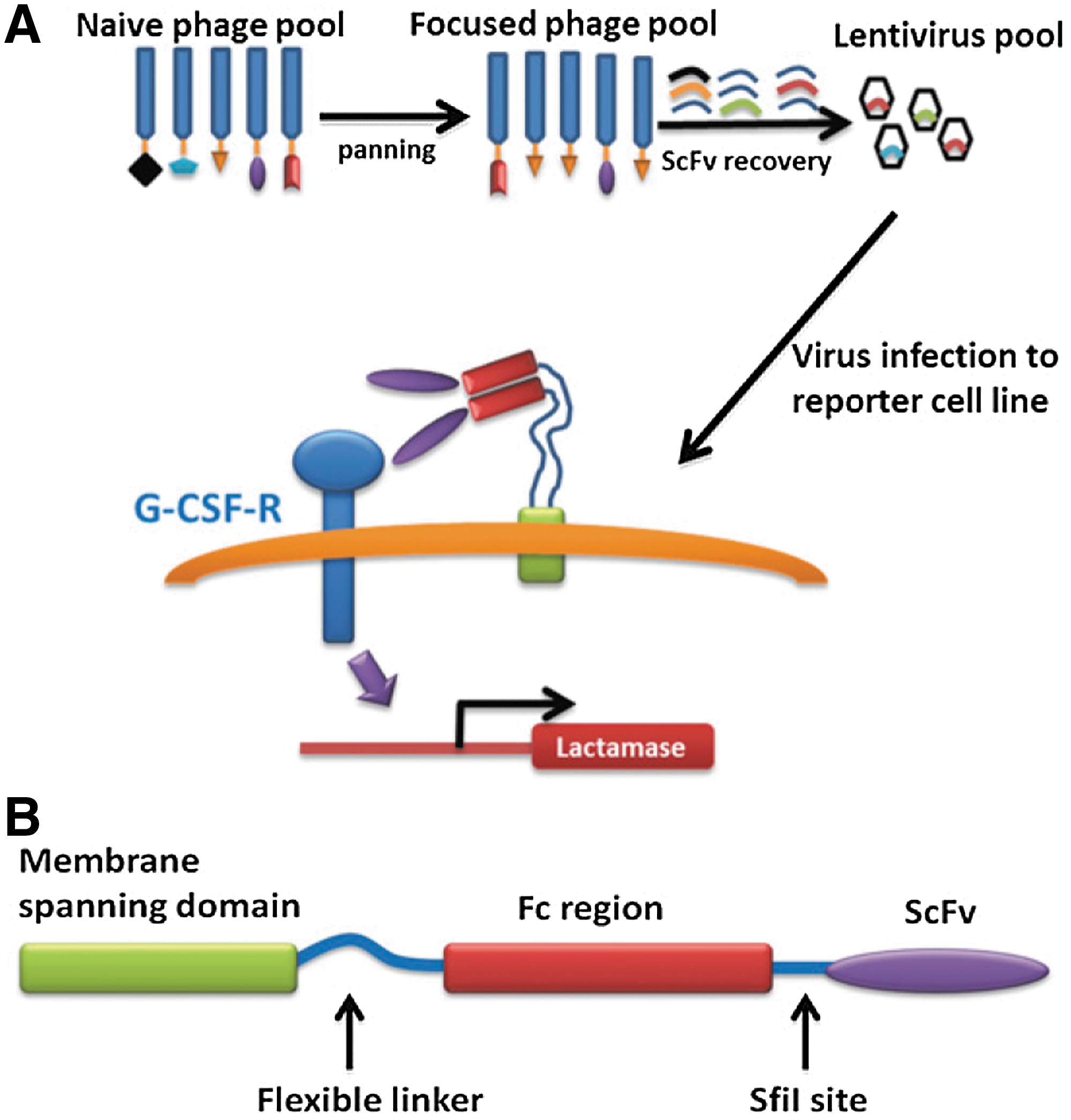
Commentary:Building upon their previously reported method in which combinatorial antibody libraries were expressed in eukaryotic cells through a lentivirus infection, members of the Lerner laboratory now extend the platform to discover antibodies with unique transdifferentiating properties. By coexpressing antibodies, along with their intended target, on the surfaces of cells, a direct selection of agonists that regulate cellular phenotypes is possible. In the present work, combinatorial antibody libraries were expressed along with the granulocyte colony-stimulating factor receptor (G-CSFR). A platelet-derived growth factor receptor (PDGFR) membrane-spanning domain served as an anchor for single chain ScFv dimerized by the appended Fc domains, effectively displaying them as dimers into the plasma membrane. The authors termed these “near neighbor libraries” wherein the coupling of constrained interaction geometries to a very high effective molarity for the interacting pairs, along with the target being present in its natural environment, could result in the identification of new antibodies acting by unusual mechanisms that would otherwise be inaccessible through the traditional solution phase selection process (see
figure
). The process of selection was further aided by the ability to screen for the desired outcome through the use of high-throughput fluorescence-activated cell sorting, allowing one to test approximately 2 million events per hour. Indeed, using this strategy, the team isolated an agonist antibody to the G-CSFR that was later shown to induce human CD34+ stem cells, isolated from five individuals, to form neural progenitor cells, representing a transdifferentiation event from a myeloid to neural lineage. Because CD34+ stem cells are of the myeloid lineage, this rare antibody appears to induce a transdifferentiation process. The implications of this discovery and of the platform in general can be far-reaching because antibodies that transdifferentiate autologous stem cells can potentially be used to produce differentiated cells from patients and in turn those differentiated cells can find applications in a variety of therapeutic scenarios such as rebuilding damaged organs. Contributed by Anton Simeonov.
(A) Schematic illustration of the strategy for selecting the granulocyte colony-stimulating factor receptor (G-CSFR) agonist antibody. A naïve phage library was used to pan against the purified receptor ectodomain. This pool that was enriched for antibodies that bound to the G-CSFR was converted to a membrane bound version in lentivirus. A reporter cell line was infected with these lentiviruses and individual cells were selected for β-lactamase gene expression. (B) Topology of the membrane-tethered antibody (MTA) fusion protein.
TARGET FISHING NOW CATCH AND RELEASE
Brigham JL, Perera BGK, Maly DJ: A hexylchloride-based catch-and-release system for chemical proteomic applications.ACS Chem Biol2013;691–699.
Abstract: Bioorthogonal ligation methods that allow the selective conjugation of fluorophores or biotin to proteins and small molecule probes that contain inert chemical handles are an important component of many chemical proteomic strategies. Here, we present a new catch-and-release enrichment strategy that utilizes a hexylchloride group as a bioorthogonal chemical handle. Proteins and small molecules that contain a hexylchloride tag can be efficiently captured by an immobilized version of the self-labeling protein HaloTag. Furthermore, by using a HaloTag fusion protein that contains a protease cleavage site, captured proteins can be selectively eluted under mild conditions. We demonstrate the utility of the hexylchloride-based catch-and-release strategy by enriching protein kinases that are covalently and noncovalently bound to ATP-binding site-directed probes from mammalian cell lysates. Our catch-and-release system creates new possibilities for profiling enzyme families and for the identification of the cellular targets of bioactive small molecules.
Commentary:Bioorthogonal chemistry approaches aim to label biological components without affecting their function. Small chemical moieties capable of undergoing alkyne-azide cycloaddition reactions (“click chemistry”) are examples of bioorthogonal conjugation chemistry, which has become popular for tagging proteins. Certain enzymes that undergo self-labeling reactions can also be used as bioorthogonal reagents and can be used as traps to pull down interacting proteins when fused to a protein of interest. For example, a mutant of O6-alkylguanine-DNA alkyltransferase (known as AGT, also called SNAP-tag) can react with benzylguanine analogs to form covalent linkages, and an engineered dehalogenase (HaloTag) can be covalently linked to hexylchloride-linked (HLP) proteins. This study used both SNAP-tag and HaloTag proteins linked via a protease recognition site to construct a “catch and release” system for proteomic analysis (see
figure
). The SNAP-tag can be linked onto solid supports without loss of functional activity by using analogs of benzylguanine. Initially a TEV protease site was placed between the SNAP-tag and the HaloTag enzyme but it was found that cleavage by TEV was inefficient. Therefore, a SUMO sequence, which can be cleaved by Ulp1 protease, was substituted. This showed more efficient cleavage but with a high degree of background cleavage by endogenous proteases in the cell lysate. Therefore, a mutated form of SUMO was used that could be recognized by engineered forms of Ulp1. The mutated SUMO/protease system eliminated background cleavage. The final SNAP-tag(AGT)/SUMO*/HaloTag (ASH*) fusion protein was found to be stable to detergents and high salt washes; however, high concentrations of detergent or denaturants inhibited the cleavage reaction, a result likely due to the cleavage site being dependent on the fold of SUMO instead of a simple linear peptide sequence. The ASH* system was then shown to enrich for HLP proteins in HeLa cell lysates (see
figure
). The authors then used the ASH* system to examine protein kinases. In this case an ATP competitive inhibitor of SRC kinase was modified to contain a photoactivable moiety (benzophenone) and a hexylchloride tag. The modified inhibitor retained potency showing an IC50 of 49 nM against SRC kinase. Examining COS7 lysates spiked with 50 nM SRC showed that 11% of the total SRC was labeled after UV irradiation. After ASH* addition and immobilization on affinity resin, all the labeled SRC was captured and could be released from the resin following cleavage by Ulp1*. Selectivity was demonstrated by using unmodified ATP competitive inhibitors to SRC that blocked this reaction. This approach was then applied to potent Type II (allosteric) inhibitors of LCK. Type II protein kinase inhibitors often have slow on/off rates making them difficult to use as affinity probes because they cannot be easily competed off the enzyme. However the ASH* system was able to enrich for 13 protein kinases, some of which were not previously known to bind to the kinase inhibitor used in this study. This study demonstrates the use of self-labeling proteins and bioorthogonal chemistry approaches to enable chemical proteomic studies and should be useful to identify the targets of small molecules arising from phenotypic screens. Contributed by Doug Auld.
Selective catch-and-release of a hexylchloride-labeled protein in HeLa lysate. (A) Schematic for the selective catch-and-release of hexylchloride-labeled protein (HLPs). ASH* is immobilized on resin and then incubated with mammalian lysate supplemented with a protein, HLP, which contains a single hexylchloride tag (shown in pink). HaloTag selectively captures the HLP, and the SUMO protease Ulp1 releases HaloTag (and any proteins that HaloTag has captured) from the beads. (B) Western blot analysis (anti-His6) of the catch-and-release experiment described in (A). (C) Silver stain analysis of the catch-and-release experiment described in (A). SNAP-tag, mutant of O6-alkylguanine-DNA alkyltransferase; ASH*, SNAP-tag(AGT)/SUMO*/HaloTag.
Abstract: Hydrogen sulfide (H2S) is a reactive small molecule generated in the body that can be beneficial or toxic owing to its potent redox activity. In living systems, disentangling the pathways responsible for H2S production and their physiological and pathological consequences remains a challenge in part due to a lack of methods for monitoring changes in endogenous H2S fluxes. The development of fluorescent probes with appropriate selectivity and sensitivity for monitoring production of H2S at biologically relevant signaling levels offers opportunities to explore its roles in a variety of systems. Here we report the design, synthesis, and application of a family of azide-based fluorescent H2S indicators, sulfidefluor-4, sulfidefluor-5 acetoxymethyl ester, and sulfidefluor-7 acetoxymethyl ester, which offer the unique capability to image H2S generated at physiological signaling levels. These probes are optimized for cellular imaging and feature enhanced sensitivity and cellular retention compared with our previously reported molecules. In particular, sulfidefluor-7 acetoxymethyl ester allows for direct, real-time visualization of endogenous H2S produced in live human umbilical vein endothelial cells upon stimulation with vascular endothelial growth factor (VEGF). Moreover, we show that H2S production is dependent on NADPH oxidase–derived hydrogen peroxide (H2O2), which attenuates VEGF receptor 2 phosphorylation and establishes a link for H2S/H2O2 crosstalk.
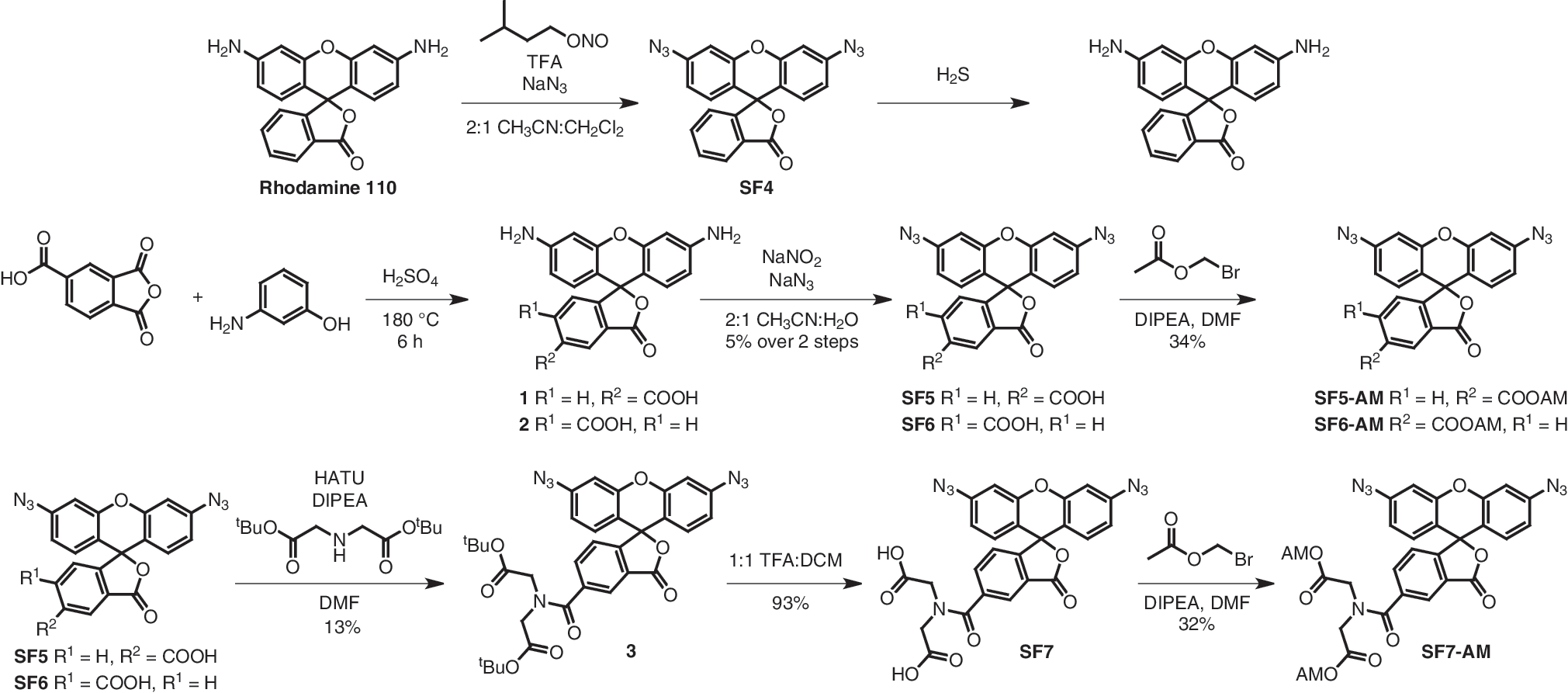
Commentary:H2S is increasingly being recognized as a ubiquitous signaling molecule, akin to nitric oxide (NO); however, streamlined methods for direct H2S monitoring in cells have been lacking. Here, a series of imaging probes, sulfidefluor- (SF)4, SF5 acetoxymethyl ester (-AM), and SF7-AM are presented (
first figure
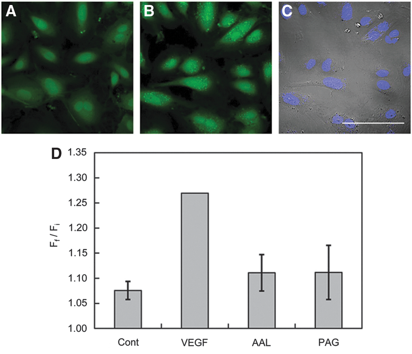
). These operate through chemoselective H2S-mediated reduction of biocompatible azides to amines, displaying up to 40-fold fluorescent increases. Furthermore, they were shown to be selective for H2S relative to the more abundant cellular thiols, reactive sulfur species, reactive nitrogen species, and reactive oxygen species. The best probe from the series, SF7-AM, was used to visualize VEGF-triggered H2S production in live human umbilical vein endothelial cells (HUVECs) through time-lapse imaging (
second figure
). The studies demonstrated that production of H2S was dependent on VEGF and cystathionine gamma-lyase (CSE) activation. Furthermore, it was discovered that the VEGF activation (through vascular endothelial growth factor receptor 2 [VEGFR2] phosphorylation) was dependent on H2O2 produced by NADPH oxidase. The successful validation of the new fluorescent probe, along with the discovery of a crosstalk between the H2O2 and H2S signaling pathways, should spur the use of this imaging agent to further advance our understanding of the fascinating roles of this small molecule. Contributed by Anton Simeonov.
Design and synthesis of trappable probes for H2S imaging. TFA, trifluoroacetic acid; SF, sulfidefluor-; HATU, O-(7-azabenzoltriazol-1-yl)-N,N,N′,N′-tetramethyluronium hexafluorophosphate; DIPEA, N,N-diisopropylethylamine; DMF, dimethylformamide; DCM, dichloromethane.
Confocal images of endogenous H2S detection in live HUVECs using SF7-AM. (A) HUVECs incubated with 5 μM SF7-AM for 30 min at 37°C, washed, and then imaged. (B) The same field of HUVEC in A was treated on stage with 40 ng/mL VEGF for 30 min at 37°C and then imaged. (C) Brightfield images of the same field of cells in B overlaid with images of 1 μM Hoechst stain at 37°C. Images in A and B are the maximum intensity projections of 8 μm×2 μm z-stacks. Scale bar, 100 μm. (D) Quantification of confocal fluorescence images of H2S signaling in live HUVECs using SF7-AM. HUVECs were incubated with 2.5 μM SF7-AM, washed, and imaged before treatment with 0.1% bovine serum albumin (BSA) in H2O as a vehicle control (Cont; n=8), 40 ng/mL total VEGF stimulation (VEGF; n=14), 30 μM AAL-993 for 40 min before VEGF stimulation (n=3), or 100 μM PAG for 10 min before treatment with 40 ng/mL VEGF (n=3). Data were normalized to VEGF-stimulated positive control. Data are expressed as a ratio of final mean fluorescence intensity (Ff) to the initial mean fluorescence intensity (Fi), and error bars are ±SEM. HUVEC, human umbilical vein endothelial cell; VEGF, vascular endothelial growth factor; AAL-993, 2-[(4-pyridyl)methyl]amino-N-[3-(trifluoromethyl)phenyl]benzamide; PAG, dl-propargylglycine.
NEW COMBINATION OF TARGETS SHOWS PROMISE FOR LEUKEMIA
Dai Y, Chen S, Kmieciak M, Zhou L, Lin H, Pei X, Grant S: The novel Chk1 inhibitor MK-8776 sensitizes human leukemia cells to HDAC inhibitors by targeting the intra-S checkpoint and DNA replication and repair.Mol Cancer Ther2013;12:878–889.
Abstract: Interactions between the novel Chk1 inhibitor MK-8776 and the HDAC inhibitor (HDACI) vorinostat were examined in human leukemia cells harboring wild-type (wt) or deficient p53. MK-8776 synergistically potentiated vorinostat-mediated apoptosis in various p53-wild type (wt) or -deficient leukemia cell lines, while p53 knock-down by shRNA sensitized p53-wt cells to lethality of this regimen. Leukemia cell lines carrying FLT3-ITD were also sensitive to the MK-8776/vorinostat regimen. Synergistic interactions were associated with inhibition of Chk1 activity, interference with the intra-S phase checkpoint, disruption of DNA replication, and down-regulation of proteins involved in DNA replication (e.g., CDT1) and repair (e.g., CtIP and BRCA1), resulting in sharp increases in DNA damage, reflected by enhanced γH2A.X formation, and apoptosis. Moreover, leukemia cells expressing kinase-dead Chk1 (D130A) or Chk1 shRNA were significantly more sensitive to HDACIs compared to their wild-type counterparts, and displayed down-regulation of CtIP and BRCA1 phosphorylation following HDACI exposure. Finally, the MK-8776/vorinostat regimen was active in primary AML blasts, particularly against the CD34+/CD38−/CD123+ population enriched for leukemia-initiating cells. In contrast, identical regimens were relatively sparing toward normal cord blood CD34+ cells. Together, these findings indicate that the novel Chk1 inhibitor MK-8776 markedly potentiates HDACI lethality in leukemia cells displaying various genetic backgrounds through mechanisms involving disruption of the intra-S checkpoint, DNA replication, and DNA repair. They also argue that leukemic cells, including those bearing oncogenic mutations associated with poor prognosis (e.g., p53 deletion/mutation or FLT3-ITD) may also be susceptible to this strategy.
Commentary:Acute myeloid leukemia (AML) has been the subject of intense research efforts in recent years with clinical trials ongoing. A recent focus for this disease has been in combination therapies. The authors here looked at the combination of a CHK1 (checkpoint kinase) inhibitor and an HDAC inhibitor (histone deacetylase) for their effect on AML cells with various genetic backgrounds (wild-type or mutant p53 and wild-type FLT3 or FLT3-ITD). Defects in p53 are much more common in relapsed/refractory AML than at initial AML presentation, and FLT3-ITD AML carries a worse prognosis than many other types of AML. CHK1 acts at various checkpoints including the intra-S phase checkpoint as shown with flow cytometry. Checkpoints are important components of the DNA-damage response in cells, and causing DNA-damage while targeting the cells' ability to cope with the insult is one strategy for the development of cancer therapeutics. The CHK1 inhibitor studied here, MK-8776, is currently in clinical trials in combination with DNA-damaging agents, such as cytarabine, for the treatment of AML. CHK1 inhibitors have also been previously shown to target cancer stem cells in non–small-cell lung cancer and pancreatic cancer. Cells with p53 mutations had a greater susceptibility to MK-8776. The HDAC inhibitor investigated here was vorinostat, and HDAC inhibitors are important for modifying chromatin structure, which impacts gene expression. HDAC inhibitors are also known to lead to DNA damage. The synergy between MK-8776 and vorinostat in a variety of leukemia cell lines was tested by looking at apoptosis and colony-forming assays (see
figure
). Co-administration of the two compounds led to large increases in caspase-3 and caspase-9 cleavage as well as PARP degradation. Synergy was observed in the AML cells with these two inhibitors regardless of the genetic background of the cells, and there was a sharp increase in the DNA damage (enhanced γH2A.X). The synergy observed could also be replicated with kinase-dead CHK1 or CHK1 shRNA and the HDAC inhibitor. The combination was also active in primary AML blasts but was relatively less toxic to normal CD34+ hematopoietic cells. Interestingly, the combination also had an effect on CHK1 mRNA levels. This particular combination is intriguing, and it will be interesting to see whether the synergy occurs in vivo as well. Contributed by Mindy I. Davis.
MK-8776 synergistically interacts with HDAC inhibitors (HDACIs) to induce apoptosis in leukemia cell lines with various genetic backgrounds. (A) U937, MV-4-11, OCI-AML-3, and MOLM-13 cells were characterized by immunoblotting analysis (upper panels). Dose responses to MK-8776 were then examined in these lines by Annexin-V-FITC/PI staining and flow-cytometry (lower panel). (B) Cells were exposed (48 h for OCI-AML-3; 24 h for others) to MK-8776 ± 1.5 μM vorinostat, apoptosis was measured. (C) Alternatively, immunoblotting was performed to detect cleavage of caspase-9 and PARP. (D) After 24-h exposure to 500 nM MK-8776 ± HDACIs, a soft-agar assay was performed to assess the colony-forming capacity of U937 cells. CF, cleaved fragment.
MSD-SET
Estep P, Reid F, Nauman C, Liu Y, Sun T, Sun J, Xu Y: High throughput solution-based measurement of antibody-antigen affinity and epitope binning.mAbs2013;5:270–278.
Abstract: Advances in human antibody discovery have allowed for the selection of hundreds of high affinity antibodies against many therapeutically relevant targets. This has necessitated the development of reproducible, high throughput analytical techniques to characterize the output from these selections. Among these characterizations, epitopic coverage and affinity are among the most critical properties for lead identification. Biolayer interferometry (BLI) is an attractive technique for epitope binning due to its speed and low antigen consumption. While surface-based methods such as BLI and surface plasmon resonance (SPR) are commonly used for affinity determinations, sensor chemistry and surface related artifacts can limit the accuracy of high affinity measurements. When comparing BLI and solution equilibrium based kinetic exclusion assays, significant differences in measured affinity (10-fold and above) were observed. KinExA direct association (ka) rate constant measurements suggest that this is mainly caused by inaccurate ka measurements associated with BLI related surface phenomena. Based on the kinetic exclusion assay principle used for KinExA, we developed a high throughput 96-well plate format assay, using a Meso Scale Discovery (MSD) instrument, to measure solution equilibrium affinity. This improved method combines the accuracy of solution-based methods with the throughput formerly only achievable with surface-based methods.
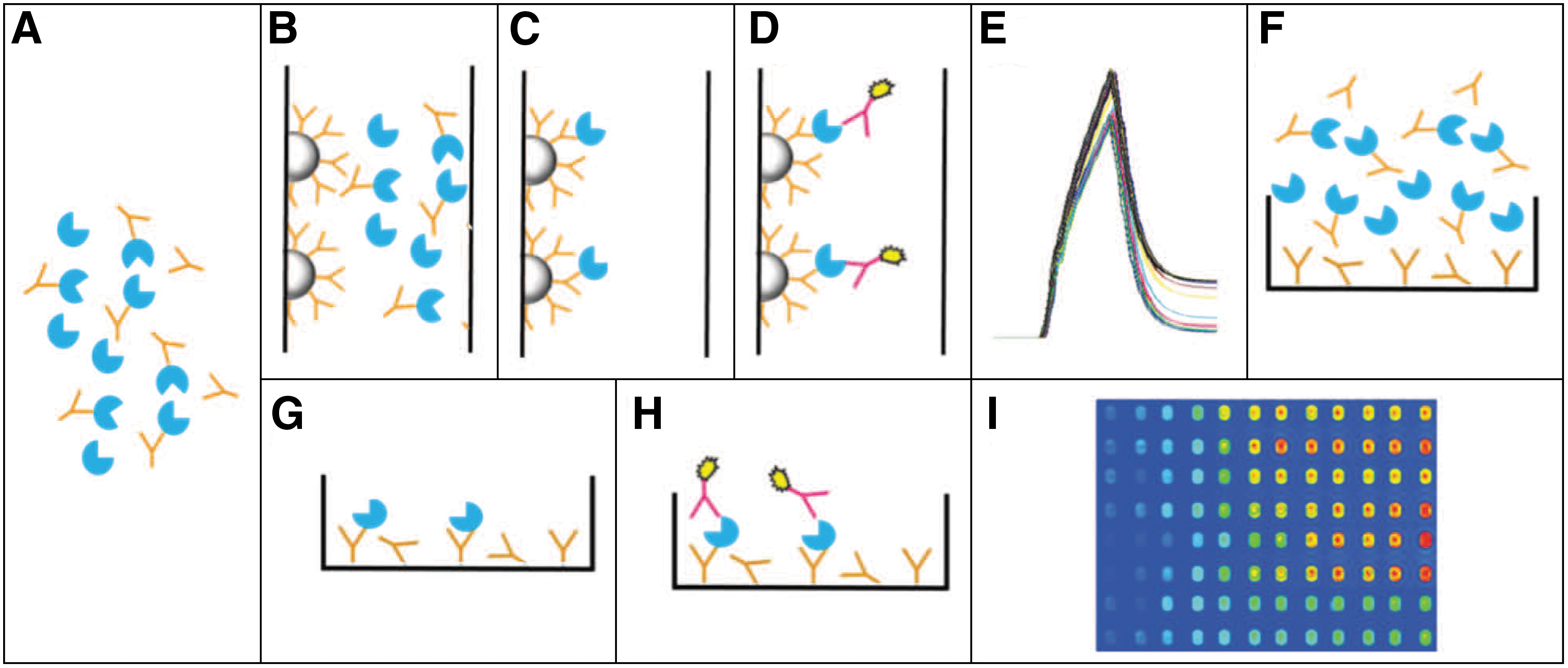
Commentary:For the advancement of antibody discovery, various label-free biosensors have been utilized to set up epitope binning assays and antibody–antigen affinity determination. These platforms can be broadly categorized into solution-based and surface-based methods, and the latter category could be further differentiated based on its surface immobilization orientation (antigen or antibody) (Abdiche et al., J Immunol Methods 2012;382:101–116). A few surface-based approaches, such as BLI and SPR, are appealing and can be considered as primary investigative platforms due to their availability in high-throughput format. On the other hand, to obtain reliable binding parameters, some intrinsic limitations that come with these methods (e.g., ligand loading density) cannot be overlooked. BLI and SPR formats have been adopted in industry for preliminary selection of high-affinity antibodies against a large number of therapeutically relevant targets. In the article by Estep and coworkers, the authors describe a primary screening platform based on an MSD-based enzyme-linked immunosorbent assay that combines the advantages of solution-based equilibrium titration (MSD-SET) and high-throughput properties. Specifically, following a similar principle as demonstrated in KinExA (see Darling et al., Assay Drug Dev Technol 2004;2:647–657), a second antibody is applied to a sandwich antigen that was previously captured by a surface-coated antibody and is not competitive with the first antibody (see
figure
, panelsG–I). An electrochemically activatable tag previously conjugated to the second antibody is then used as the assay readout. A few key parameters were identified that affected the affinity measurement, such as antigen contact time and coating density, and these were further optimized to balance signal strength and assay sensitivity. It was subsequently demonstrated that affinity values derived from this MSD-SET assay were within twofold of those obtained from solution-based KinExA platform. Furthermore, the authors provided valuable affinity data for performance comparison between BLI and KinExA, illustrating that sensor-related artifacts could be causes of a stronger affinity deviation. Overall, the good agreement in its affinity determination compared to those obtained through other method(s), high-throughput nature, and its reproducibility represent important attributes for this MSD-SET approach. MSD-SET could be considered as an alternative/orthogonal assay for epitope binning and identification of antigen heterogeneity. Contributed by Wendy Lea.
Solution equilibrium titrations (SET), KinExA, and Meso Scale Discovery (MSD) experimental design. (A) SET. A SET is prepared with antigen (blue) held constant and antibody (orange) titrated. The solution is allowed to come to equilibrium. (B) KinExA. A column is packed with antibody-conjugated beads. The SET sample is passed over the column at a fixed flow rate (typically 0.25 mL/min). (C) Only free antigen is captured on the antibody-conjugated beads. (D) The captured antigen is detected with a fluorophore-labeled antibody (pink) that binds in a noncompetitive manner on the antigen. (E) The fluorophore's signal results in the trace shown, where the end points of a raw signal trace can be used to plot signal as a function of antibody concentration and fit to a quadratic equation to extract a KD. (F) MSD. Antibody is coated to a standard-bind MSD plate. The SET sample is added by a liquid handling robot to the antibody-coated plate. (G) After 150 sec, free antigen is captured on the plate, while the remaining SET solution is washed off the plate. (H) The captured antigen is then detected by a sulfotag-conjugated antibody that binds in a non-competitive manner on the antigen. (I) The sulfotag is electrochemically activated to provide signal that is measured by a CCD camera. The raw signal can then be plotted as a function of antibody concentration and fit to a quadratic equation to extract a KD.
CPP-LINKED PKIS
van Wandelen LTM, van Ameijde J, Ismail-Ali AF, Quarles van Ufford HC, Vijftigschild LWA, Beekman JM, Martin NI, Ruijtenbeek R, Liskamp RMJ: Cell-penetrating bisubstrate-based protein kinase C inhibitors.ACS Chem Biol2013;8:1479–1487.
Abstract: Although protein kinase inhibitors present excellent pharmaceutical opportunities, lack of selectivity and associated therapeutic side effects are common. Bisubstrate-based inhibitors targeting both the high-selectivity peptide substrate binding groove and the high-affinity ATP pocket address this. However, they are typically large and polar, hampering cellular uptake. This paper describes a modular development approach for bisubstrate-based kinase inhibitors furnished with cell-penetrating moieties and demonstrates their cellular uptake and intracellular activity against protein kinase C (PKC). This enzyme family is a longstanding pharmaceutical target involved in cancer, immunological disorders, and neurodegenerative diseases. However, selectivity is particularly difficult to achieve because of homology among family members and with several related kinases, making PKC an excellent proving ground for bisubstrate-based inhibitors. Besides the pharmacological potential of the novel cell penetrating constructs, the modular strategy described here may be used for discovering selective, cell-penetrating kinase inhibitors against any kinase and may increase adoption and therapeutic application of this promising inhibitor class.
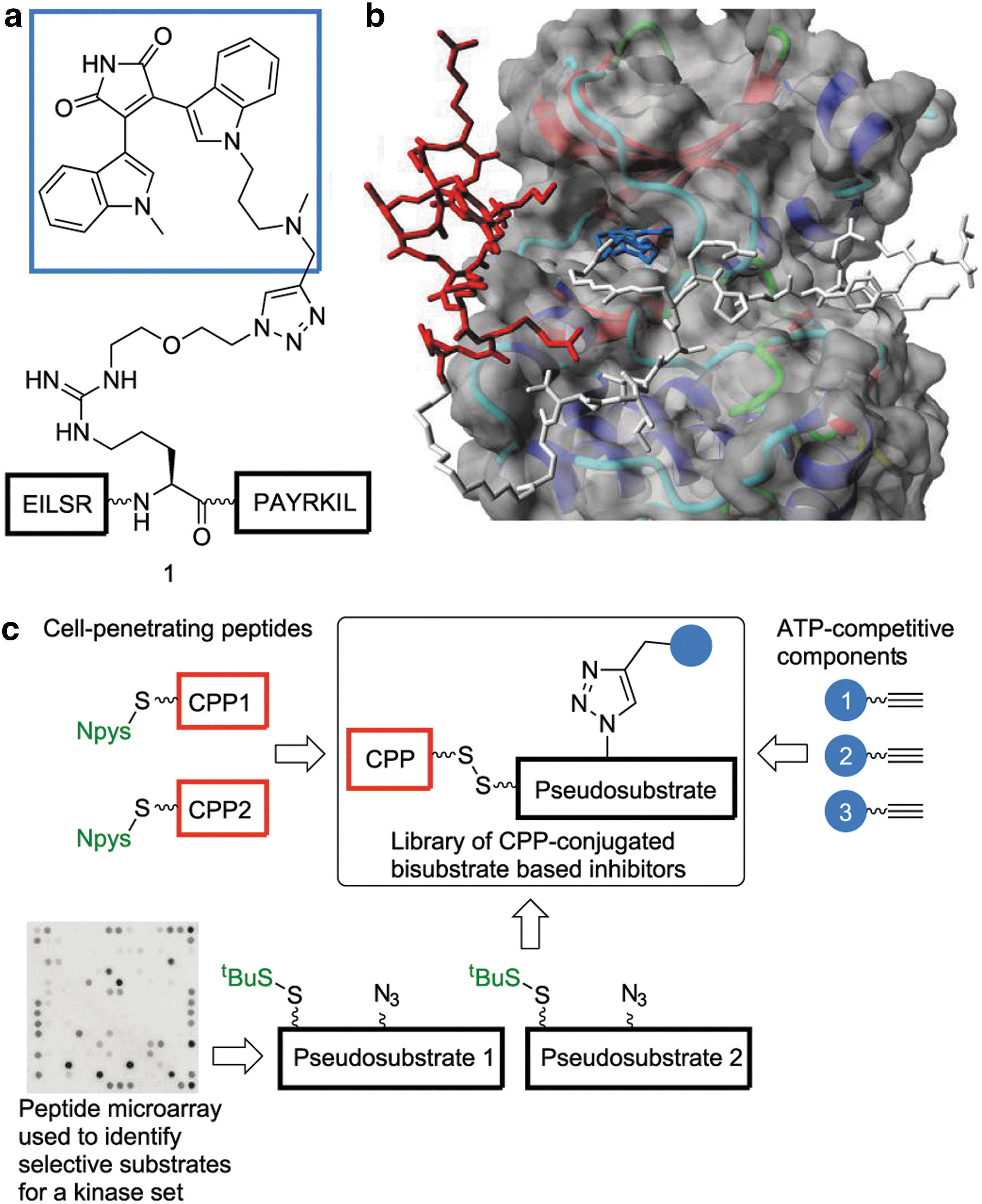
Commentary:Protein kinases bind two substrates, adenosine triphosphate (ATP) and a peptide, and bisubstrate inhibitors designed to interact with both sites can offer high potency and selectivity by combining the binding affinity from contacts in both ATP and peptide pockets. However, such inhibitors are often large and hydrophilic and therefore are not cell penetrant. This article describes a solution to this problem by incorporating cell penetrating peptides (CPPs) to bisubstrate inhibitors. Short peptide sequences derived from proteins such as the trans-activating transcriptional activator (Tat) from human immunodeficiency virus 1 and penetratin (a protein transduction domain derived from the homeoprotein Antennapedia) can act as CPPs to promote cell penetration of molecules into cells. To test this idea, a library targeting PKC was constructed. The library contained PKC pseudosubstrates that were linked to CPPs as well as an ATP-competitive inhibitor. Bisubstrate inhibitors were constructed by linking the pseudosubstrates to an ATP-competitive inhibitor using the alkyne-azido click chemistry reaction. The bisubstrate inhibitors were constructed to incorporate a CPP either by using a disulfide linkage (allowing release of the bisubstrate inhibitor upon cell penetration) or through addition of the CPP to the N-terminus of the substrate (see
figure
). Initially, fluorescein-labeled constructs were used to measure cellular uptake of the CPP-bisubstrate inhibitors. Approximately 85% of the viable cells showed uptake but the linearly, N-terminally linked CPPs showed a diffuse staining pattern, whereas the disulfide linker led to punctate staining suggesting endosomal localization. A cell-based enzyme-linked immunosorbent assay in HeLa cells treated with phorbol-12-myristate-13-acetate was then used to gage the potency of these bisubstrate inhibitors in cells. Although some differences between the linkage type and CPP employed were noted, many of the constructs showed activity in the micromolar range in cells. A lipophilic analog of compound 1 (see
figure
) showed a potency in cells that was similar to that measured in a cell-free assay suggesting that the lipophilic analog readily penetrates cells. The selectivity was evaluated by microarray analysis, which supported selective inhibition by the compounds. This approach could enable the application of potent and selective inhibitors that span both ATP and peptide substrate pockets to selectively target protein kinases in cells. Contributed by Doug Auld.
(a) Bisubstrate-based inhibitor 1. (b) Model of conjugate 7a with PKCθ (CPP in red, ATP-competitive part in blue, and pseudosubstrate in white). (c) Modular discovery strategy of CPP-conjugated bisubstrate-based inhibitors. PKC, protein kinase C; CPP, cell penetrating peptide; ATP, adenosine triphosphate.