
Abstract
Despite all the research aiming to treat ocular diseases, age-related macular degeneration (AMD) remains one of the serious diseases worldwide, which needs to be treated. Neovascularization is a key factor in AMD and thus antiangiogenic therapy is beneficial in reducing the development of new abnormal blood vessels. Axitinib, multireceptor tyrosine kinase inhibitor, is a small molecule that works by blocking vascular endothelial growth factor receptors (VEGFR) and platelet-derived growth factor receptors (PDGFR) responsible for developing neovascularization. The goal of this study is to develop a sustained release formulation of axitinib-loaded poly(lactic-co-glycolic acid) (PLGA) nanoparticles to minimize frequent administration of the drug by intravitreal injection. The nanoparticles were characterized for particle size and zeta potential, as well as using differential scanning calorimetry, transmission electrode microscope, and in vitro drug release profile. The cytotoxicity of the formulation was evaluated on human retinal pigmented epithelium ARPE19 cells by MTT [3-(4,5-dimethylthiazol-2-yl)-2,5-diphenyltetrazolium bromide salt] assay. The cellular uptake, antimigration assay, and vascular endothelial growth factor (VEGF) expression levels were found out in vitro using cells. The optimized formulation was 131.33 ± 31.20 nm in size with −4.63 ± 0.76 mV zeta potential. Entrapment efficiency was found to be 87.9% ± 2.7%. The cytotoxicity of ARPE19 cells was <12% for nanoparticles suggesting the in vitro compatibility at 10 μM concentration of drug. Cellular uptake, antimigration assay, and VEGF expression levels for the nanoparticles suggested greater uptake, significant antiangiogenic potential, and inhibition of VEGF activity. The results showed successful development of axitinib-loaded PLGA nanoparticles as an alternative potential treatment for AMD.
Introduction
Modern research work in the field of medicine and health focuses on better ways for drug delivery system to ensure improved therapeutic effects and reduced side effects. Poly(lactic-co-glycolic acid) (PLGA) has proved to be useful as a carrier for drug delivery and tissue engineering.
1
PLGA is a FDA-approved polymer that is efficient as a drug delivery vehicle due to its physical stability, biodegradability, and biocompatibility. It has been intensively scrutinized as a vehicle to assist the delivery of drugs, proteins, nucleotides, and peptides. The popularity of the PLGA nano/micro particles is also due to its ability to enhance a sustained drug delivery process, favorable deterioration abilities, and the experience in the clinical field.
2
Again, through control of various parameters, the physical properties of a polymer–drug complex can be adjusted to suit the appropriate dosage and release interval considering the type of the drug being dealt with. However, the toxicity associated with these require a critical analysis to enhance safety.
3
PLGA is a copolymer of two compounds; the polylactic acid (PLA) and the polyglycolic acid (PGA). PLGA is a racemic
Age-related macular degeneration (AMD), one of the major causes for loss of vision affects many elderly patients worldwide. 5 Among two types of AMD, wet AMD is the foremost cause of vision loss and is linked to choroidal neovascularization. The acellular debris accumulates, which leads to damage to macular site in retinal epithelium resulting in blurring of the vision. 6 In response to damage in the epithelial layer, vascular endothelial growth factor (VEGF) secretion begins. Additionally, due to dysfunction of ion channel and abnormal metabolism of lipids, oxidative damage to cells take place. 7 To pay for damages for the reduced blood supply at the site, new blood vessel will start to form, which will in turn increase inflammation. 8,9 Angiogenesis is an essential phenomenon of formation of new vessels in the vasculature and one of their important regulators are tyrosine kinases. Anti-VEGF therapy has become well known to reduce angiogenesis in cancer. Even for treatment of wet AMD, anti-VEGF therapy has gained a lot of interest to reduce neovascularization. Current therapy for AMD involves intravitreal administration of antiangiogenic agents, such as Lucentis™ (Ranibizumab), Macugen® (Pegaptanib), and Avastin® (Bevacizumab).
Axitinib is a tyrosine kinase inhibitor and works by blocking the vascular endothelial growth factor receptors (VEGFR), which in turn inhibit the activity of the endothelial growth factors. It also plays a crucial function in inhibiting the function of platelet-derived growth factor receptors (PDGFR), thereby decreasing the development of neovascularization. 10 Administering axitinib intravitreally has proved to be useful in managing wet AMD therapy, which reduces neovascularization and macular thickness due to AMD. 11
However, the disadvantages of the current therapies for AMD have rapid clearance of these agents from the site of action requiring frequent intravitreal instillations, which increase the risk such as detachment of retina, endophthalmitis, infections, vitreous hemorrhage, and increased intraocular pressure, 12 as well as reduces patient compliance. Thus, there is requirement for such a delivery system, which can minimize intravitreal instillation frequency of antiangiogenic agents for efficient therapy of AMD. In the present research, axitinib-loaded PLGA nanoparticles (AX-NPs) were developed and evaluated for size, zeta potential, surface morphology, and in vitro drug release along with its in vitro cellular uptake and cytotoxicity. Its antiangiogenic potential to inhibit cell migration and VEGF expression was also studied in vitro.
Materials and Methods
Materials
Axitinib was bought from the Selleck Chemicals, Houston, TX. PLGA (molecular weight [Mw] 15,000 Da) containing lactide/glycolide at a ratio of 50:50 was acquired from the Acros Organics. Polyvinyl alcohol, Mw 100,000, was purchased from the Fisher Scientific. Both the Coumarin 6 dye and the Dialysis membrane, Mw 10,000, was outsourced from Sigma-Aldrich (St. Louis, MO). Fetal bovine serum (FBS) and penicillin/streptomycin (10,000 U/mL) were purchased from Gibco Thermo Fisher Scientific. Both 3-(4,5-dimethylthiazol-2-yl)-2,5-diphenyltetrazolium bromide salt (MTT reagent) and nucleus stain DAPI (4′,6-diamidino-2-phenylindole) were outsourced from Tocris Bioscience. CellMask™ deep red plasma stain was obtained from Thermo Fisher Scientific. The BCA Protein Assay Kit and Invitrogen™ eBioscience™ Human VEGF-A Platinum Enzyme-Linked Immunosorbent Assay (ELISA) Kit were purchased from the Fisher Scientific branch in United States. ARPE-19, a human retinal pigment epithelial cell line (ATCC® CRL2302™) and Dulbecco's modified Eagle's medium F 12 (DMEM F12) was obtained from American Type Culture Collection (ATCC). Cell culture phosphate-buffered saline (PBS; 1 × ) was obtained from Corning Cellgro (Manassas, VA). Trypsin (0.05%) was purchased from Thermo Fisher (Lansing, MI). All the analytical reagents used were analytical grade and hence no further purification was needed.
Cell Culture
All the cell line studies were performed on the human retinal epithelial cell line, ARPE-19 (ATCC CRL2302). DMEM F12 supplemented with 10% v/v FBS and 1% 10,000 u/mL penicillin/streptomycin antibiotics were used for the maintenance of cells. The cells were incubated in a humidified atmosphere at 5% CO2 at 37°C.
Preparation of Axitinib-Loaded PLGA NPs
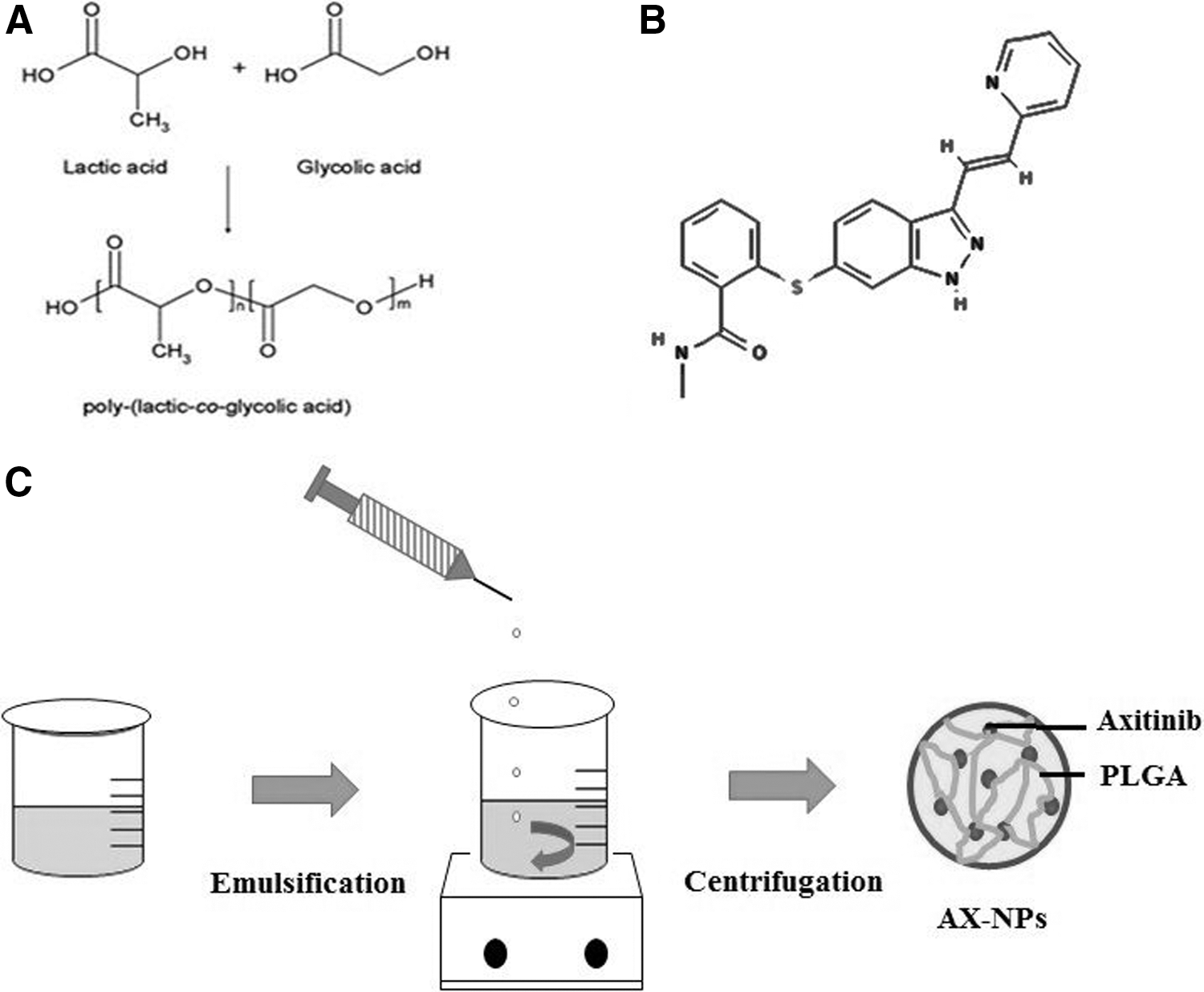
AX-NPs were prepared by the o/w solvent evaporation method. 2 The drug and the polymer were dissolved in a 2 mL of dimethyl sulfoxide (DMSO):acetone (1:9 v/v) at a weight ratio of 1:7 at room temperature (RT). The resultant solution was then slowly added into 1% w/v PVA solution using 23-G syringe with constant stirring at 700 rpm at RT. After that, the organic solvent was allowed to evaporate at RT, and the resulting volume of the aqueous dispersion was collected. The suspension was then centrifuged at 5,000 rpm for 5 min at RT so that the unentrapped drug can be removed. Furthermore, the supernatant was centrifuged at 18,000 rpm at 4°C for about 2 h to allow nanoparticles to settle down. After being centrifuged, pelleted nanoparticles were washed using distilled water and collected. 2 In the same manner, blank nanoparticles excluding the drug and Coumarin 6-loaded nanoparticles were formulated. Figure 1 shows the graphical presentation of the preparation method of AX-NPs.
Differential Scanning Calorimetry
The analyses of the sample using the differential scanning calorimetry (DSC) were conducted on the DSC-Q 20 (Q series Q20-2288-DSC software; TA Instruments, New Castle, DE). Blank aluminium pan was used as a reference. 13 About 4°mg of the sample powders was then heated at a temperature of about 30°C–300°C at a rate of 10°C min−1 under the nitrogen purge (50 mL/min). 14 The procedure was carried out in both the thermal analysis involving pure axitinib drug, the physical mixture of axitinib and the PLGA nanoparticles, polymer, and lastly on the AX-NPs.
Particle Size and the Zeta Potential
Through the aid of a laser dynamic light scattering (DLS), the mean particle sizes of AX-NPs were determined. This investigation was carried out within a measuring angle of about 90° at a temperature of ∼25°C using a sample, adequately diluted with filtered distilled water. 15 For every sample analyzed, the mean diameter and the standard deviation (SD) of five readings were determined through the application of the multimodal analysis. The outcome of the analysis was then reported in triplicates. The results of zeta potential were obtained along with polydispersity index (PDI).
Zeta potential of nanoparticles was determined based on Smoluchowski equation that considers electrophoretic mobility of the nanoparticles and their backscatter at 90°. 16 The analysis was carried out in triplicate after 10 times dilution of nanoparticles using double-distilled water using zeta cuvette and Zetasizer Nano ZS 90 (Zetasizer Software Ver. 7.10; Malvern Instruments Ltd.).
Transmission Electron Microscopy
Transmission Electron Microscope (TEM; JEOL JEM 1400 electron microscope with Gaton camera; Peabody), was used to study the morphology, size, and shape of AX-NPs. This involved placing the sample on the Electron Microscopy Sciences Formvar Support Film Square Grid, 200 Cu. It was then allowed to air dry approximately for 5–10 min. It was further treated with 2% w/v phosphotungstic acid for negative staining. The investigation was conducted at an accelerating voltage of about 120 kV with 40,000 magnification.
Entrapment Efficiency
AX-NPs were first centrifuged at 5,000 rpm for 10 min to remove the unentrapped drug from the suspension. Then the collected supernatant containing NPs was further centrifuged at 18,000 rpm for 20 min to collect AX-NP pellet. The pellet was washed thrice with deionized water to separate the traces of unentrapped drug from the surface and collected. Analysis was done using ultraviolet (UV) spectrophotometer at a wavelength of 260 nm (λmax) in methanol as a solvent. The contents of the AX drug were then determined from the UV curve produced. Hence, the AX entrapment efficiency and the loading efficiency were calculated using the following equations:
In Vitro Drug Release Study
The in vitro drug release study of axitinib solution in DMSO and AX-NPs was carried out using dialysis bag (molecular weight cut-off 10,000 Da) diffusion technique. The dialysis tube was immersed in 20 mL of release medium (PBS with pH 7.4) containing 0.1% (v/v) Tween 80. Using a magnetic stirrer, the contents were continuously stirred at 150 rpm and the temperature was adjusted to 37°C. At predetermined intervals of 0.25, 0.5, 1, 2, 3, 4, 5, 6, 12, 24, 48, 72, 96, 120, 144, and 168 h, 1 mL of sample was removed and replaced with the fresh release media. Drug concentration was determined by using the UV spectrophotometry at a wavelength of 260 nm after dilution of the sample with methanol.
Cytotoxicity Study
The MTT assay was initiated to help determine the level of cytotoxicity associated with the AX-NPs comparing it with the pure drug solution, in ARPE-19 cell line (human retinal pigment epithelial cells). The cells were cultured at a suitable density of 5,000 cells/well in a 96-well plate in 200 μL of DMEM supplemented with 10% FBS. The incubation was carried out at a temperature of about 37°C with access to 5% atmosphere CO2 for 24 h to enable proper attachment and assist in the growth of the cells. 17 Both the formulations involving the nanoparticles and the pure drug solution were subjected to dilution using as serum-free DMEM F12 medium to come up with varied concentrations of axitinib drug. After 24 h culture, the cells were treated with prepared formulations and the treatment was kept for 4 h. At the end of the incubation period, the treatment was removed, and cells were washed once using sterile 1 × PBS. The procedure was then followed by the addition of fresh complete DMEM F12 containing 10% FBS for a period of 24 − 48 h. After a specified time, 100 μL of MTT reagent (1 mg/mL) solution was added to each well and cells were incubated in the same condition as previous for 4 h. The medium in each plate was then replaced with 100 μL of dimethyl sulfoxide and intensity of the color of the dissolved formazan crystals was measured using the microtiter plate reader at wavelength 595 nm. Cells treated with DMEM F12 acted as the negative control, whereas the ones treated with 0.1% Triton X were the positive control in the experiment. Cell viability was given relative to that of the negative control. 15,18
Cellular Uptake
To determine the cell uptake potential of the AX-NPs, confocal microscopy was carried out. Formulations, such as blank nanoformulation, drug formulation, and coumarin formulations, were prepared. The cells were seeded at a density of 2 × 10⁵ per well in a six-well plate. The plate was incubated to achieve 70% confluency. The cells was washed thrice with PBS. The wells were then treated with the prepared coumarin-loaded nanoparticles and blank nanoparticle as a reference. The six-well plate was then incubated with all the prepared nanoformulations for the period of 4 h. The plate is then subsequently washed thrice with PBS and then fixed with 4% paraformaldehyde. DAPI was used as a nuclear staining followed by CellMask deep red staining for membrane staining. This is then examined under confocal microscope FV1200 (Olympus, Tokyo, Japan) at 60 × magnification.
Wound Scratch Assay
Wound healing assay procedure was carried out to assist in the analysis of the inhibitory effect of axitinib and its formulation on VEGF-induced angiogenesis. Human retinal pigment epithelial cells were grown in 24- well plates and allowed to reach 80% confluency. The wounds were then developed with much care using a pipette tip. The average size of the wounds developed was measured and found to be around 300 μm. For the sake of the study, wound width within 5% variation was considered. The wounds were then cleaned with sterile PBS two times to eliminate partly adhered cells on the plates due to the wound. These wells were then subjected to treatment using the serial dilution of drug formulation and nanoparticulate formulation in incomplete media at 1 and 10 μM concentrations.
Cell migration was analyzed under the microscope and quantified through obtaining the measurements of the area covered by the cells that had migrated from the wound edges using Image software. To inspect the effect of VEGF on cell migration, two wells were also kept at high two concentrations and treated with VEGF simultaneously. Thus, one well consisted of a complete medium, whereas the other one was treated with VEFG to act as reference and controls in that order. Incubation was done still at the average body temperature of about 37°C in a CO2 incubator (5%) for 48 h. The treatments were removed after incubation, and the cells were washed with PBS three times, fixed using 70% ethanol. Images were then obtained, and the wound width was measured using images captured by the microscope. The width of the untreated group at 0 h was considered as 100%, and the relative percent recovery of the wound was compared. 19
Anti-VEGF ELISA
Human retinal pigment epithelial cells were cultured in 24-well plates at the density of 5 × 10⁴ cells/mL and allowed to become confluent. On the day of the examination, the culture media were replaced with incomplete media and subjected to treatment. The treatment group consisted of a free drug solution and axitinib-NPs, each at 10 μM concentration and were further incubated for a total period of 72 h. 20 Quantification of the secreted VEGF in the culture media was done by ELISA method using the Human VEGF-A Platinum ELISA Kit following the manufacturer's instructions. The protein content in the cells was estimated using the Pierce BCA Protein Assay Kit after collecting cell lysate and by normalizing VEGF secretion to total protein. Samples were read using ELISA plate reader at 450 nm absorbance, and 550 nm and the difference were recorded, followed by calculating inhibition of VEGF secretion using standard curve.
Statistical Analysis
The results obtained were presented as mean and SD, and the investigation was conducted with the aid of student's t-test. The variance in the lipid profile was investigated statistically using one-way analysis of variance followed by Turkey's test. The probability values of p < 0.05 were considered to be statistically significant.
Results and Discussion
Differential Scanning Calorimetry
Pure AX drug, physical mixture of PLGA and AX, as well as AX-NPs were studied for physicochemical characterization using DSC technique. Figure 2 demonstrates DSC thermal analysis of AX, physical mixture of AX and PLGA and AX-NPs. 13,15,21 The physical state of drug molecule in NPs affects their release and solubility in external medium and so DSC thermograms is helpful in identifying the nature or physical state of the drug alone and while being loaded in NPs. 22 AX showed sharp endothermic peak at around 212°C indicating its specific melting point. This endothermic peak was not present in DSC spectra of AX-NPs approving the complete loading of AX in NPs and confirming amorphous form of loaded AX. Incompatibility between the polymer and drug can also be determined from DSC thermograms from the alterations in the transition temperature of individual components that generates added peaks in thermogram. 23 Thus, DSC study also disclosed the compatibility of the AX and PLGA.
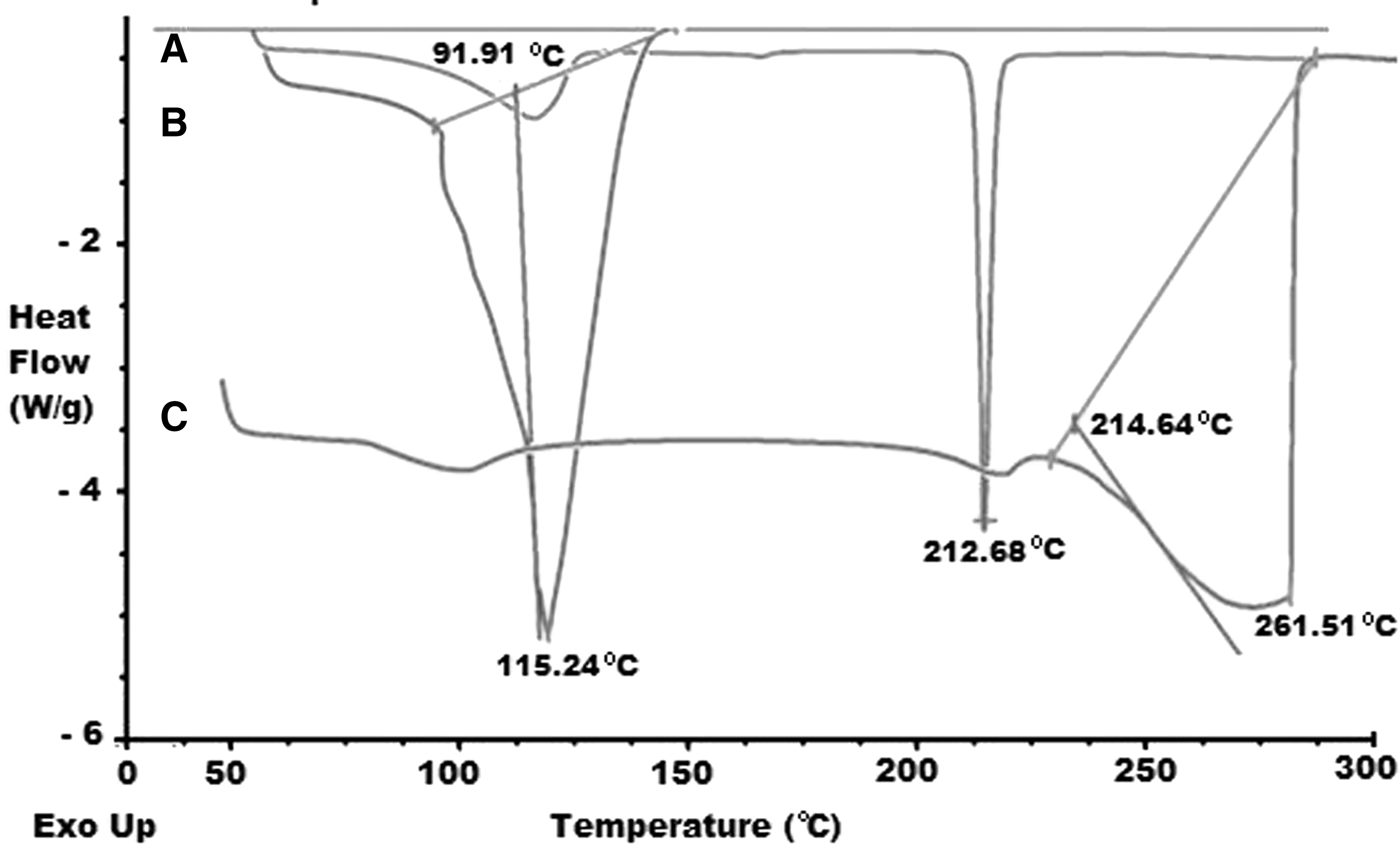
DSC spectra of
Particle Size and Zeta Potential
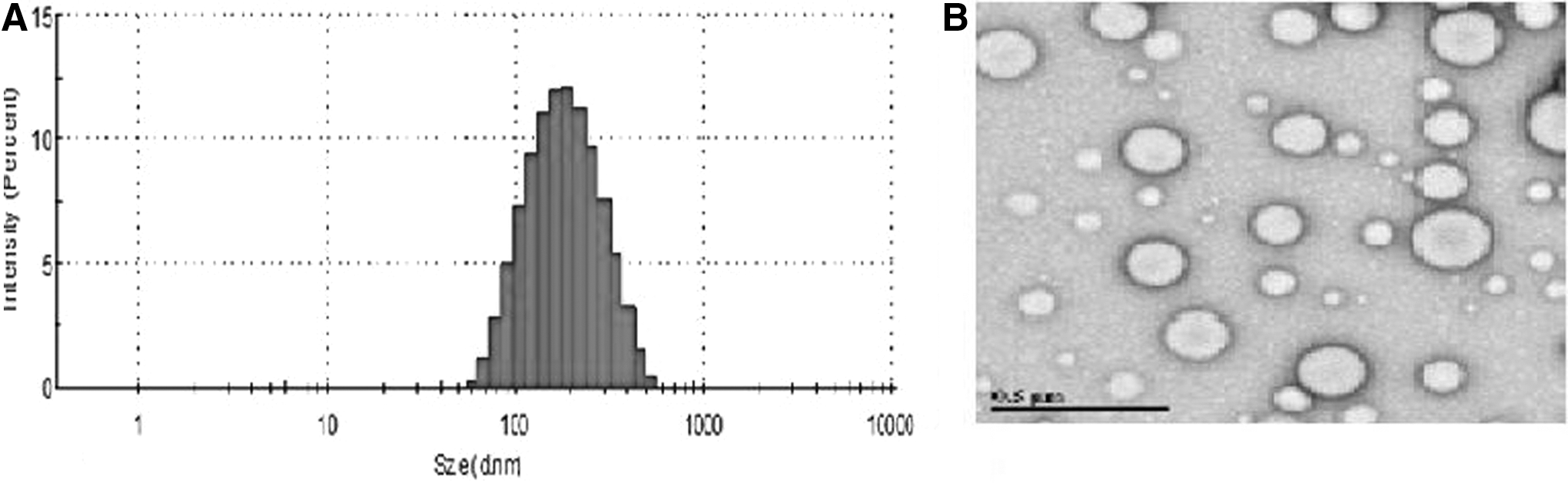
The size of AX-NPs was measured using DLS technique and was found to be 131.33 ± 31.20 nm with PDI of 0.108 ± 0.005. The mean zeta potential of AX-NPs was determined using a Malvern Zetasizer Nano ZS90 and was found to be −4.63 ± 0.76 mV. Figure 3A shows Z-average for the particle size for AX-NPs. 24 It has been observed that with increased concentration of PLGA a drug to PLGA weight ratio of 1:10, the axitinib entrapment was not increased significantly. However, an increase in the PLGA concentration resulted in increased viscosity of the organic phase. Thus the diffusional resistance of drug particles from the organic phases to the aqueous phase was increased. This led to increase in the particle sizes (Table 1). Thus, 1:7 weight ratio was optimized for preparation of AX-NPs. The zeta potential affects the stability of the nanoparticles at both extremes significantly, either in the positive side or the negative side, leading to higher repulsive forces being witnessed. The repulsive forces exist between similar electric charge blocks and hence they enhance the ease of dispersion. For the combined dynamic and steric stabilization, minimum zeta potential is recommended. Formulation involving the use of nonionic surfactants exhibited average zeta potential, whereas the formulations without the use of surfactant demonstrated a higher zeta potential. This scenario depicted could be due to the existence of carbonyl groups within the extremes of the polymeric chains of pure PLGA nanoparticles. 25
Effect of Various Parameters on Percent Entrapment Efficiency and Particle Size of Axitinib-Loaded PLGA Nanoparticles
Bold values are of optimized formulation.
% EE, entrapment efficiency; PDI, polydispersity index; PLGA, poly(lactic-co-glycolic acid); DL, drug loading.
Transmission Electron Microscopy
TEM revealed that the particle size of AX-NPs was around 110 nm. Although this was in disagreement with that of particle size obtained by DLS, such difference can be easily realized as DLS reports the hydrodynamic diameter of the particles, whereas in TEM analysis the size obtained was of the particle fixed in grid. 26 TEM images (Fig. 3B) confirmed nanoparticles of uniform size and were in concurrence with the DLS results and were spherical with smooth surface. TEM was used to confirm the information that the nanoparticles were spherical and was not in aggregate.
Entrapment Efficiency
Encapsulation efficiency was calculated by centrifuging the NP solution and resuspending the NP pellet in methanol. Entrapment efficiency was found 87.9% ± 2.7%. The percentage of drug loading was found to be around 11%. UV spectroscopy was used to determine the encapsulation efficiency of AX by comparing the absorbance in methanol to standard dilutions of AX in methanol (r 2 = 0.9994) at λmax of 260 nm. Three weight ratios (1:5, 1:7, 1:10) of AX:PLGA were studied to see the effect of polymer concentration on entrapment and loading of the drug in NPs. The drug to polymer weight ratio of 1:7 was optimized, as significant increase in entrapment efficiency was observed compared with 1:5. For drug to polymer weight ratio 1:10 there was no further increase in entrapment efficiency observed but the particle size was significantly higher as compared with 1:7. Thus, drug to polymer weight ratio of 1:7 was optimized. All the batches with different drug to polymer ratio for the AX-NPs were evaluated for the particle size (nm) and entrapment efficiency (% EE) as shown in the Table 1. All the variables were obtained to get the desired optimized formulation, which gives lower particle size and high percentage of the drug entrapment and drug loading. Particle size of the prepared batches was found to be in the range of 118.3–307.0 nm. Particle size of the optimized batch (1:7) was 131.3 ± 31.2 nm with PDI 0.108 ± 0.005 and drug loading 11%. From Table 1, it was observed that with increase in time, significant increase in the entrapment efficiency and drug loading was observed with decrease in the particle size, and hence 24 h time was optimized for stirring. While with change in stirring speed from 350 rpm to 1,100 rpm, initially the low speed resulted in very low entrapment. The 700 rpm was optimized as it resulted in AX-NPs with highest % EE and lesser size. With increase in speed from 700 rpm to 1,100, no further improvement in % EE and size was observed.
In Vitro Drug Release Study
The in-vitro release study of AX from drug solution and AX-NPs were carried out by dialysis method. To mimic the in vivo eye posterior area environment, PBS having pH of 7.4 was used as the release medium and the study was performed at 37°C. Tween-80 (0.1% v/v) was added to the release medium to improve the solubility of drug in the release medium as well as to maintain the sink condition. The release profile of AX from solution as well as NPs have been shown in Figure 4 for a period of 7 days. The release of AX from solution was rapid and almost 100% of AX was released within 6 h in release medium. While the AX-NPs initially demonstrated burst release of 18.4% ± 2.2% of AX within the first hour, may be because of the adhered drug to the outer surface of NPs. But, after that AX-NPs exhibited sustained release profile of AX with 23.7% ± 1.4% release at the end of 24 h followed by 84.2% ± 4.1% at the end of 7 days. 27 For release kinetics, AX drug solution followed first-order drug release with R 2 of 0.9762; however, AX-NPs followed Higuchi model with R 2 of 0.9869.
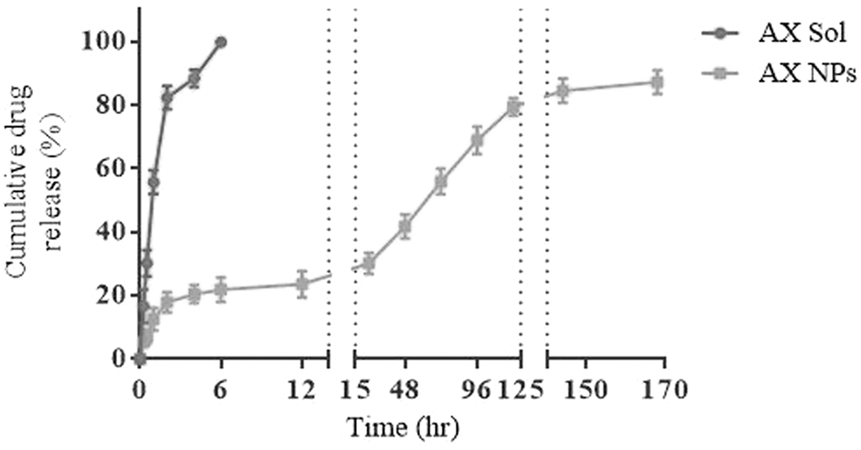
Cumulative drug release of AX from AX solution and AX-NPs up to 7 days at 37°C in phosphate-buffered saline at pH 7.4 (mean ± SD, n = 3). SD, standard deviation.
Cytotoxic Study
The cytotoxicity of the AX drug solution and AX-NPs was studied using the MTT assay in the human retinal pigmented epithelium cell line ARPE-19. Cells were treated in serum-free medium with various concentrations (0.001, 0.01, 0.1, 1, 10, and 20 μM) of AX in solution as well as NPs in triplicates for 24 and 48 h. To study the cytotoxic potential of polymer alone, blank PLGA NPs were also evaluated for MTT assay. The resulting cell viability was compared with that of the control untreated cells, which was established as 100%. Results specified that the viability of cells was higher than 90% for AX-NPs at 10 and 20 μM concentration tested for both 24 and 48 h time points. On the other hand, for drug solution viability at 20 μM was found to be 78% and 63%, respectively, for 24 and 48 h. Thus, the nanoparticles demonstrated nontoxic nature as compared with such a drug solution at 20 μM concentration of the drug (Fig. 5). The biocompatible and biodegradable nature of PLGA has been demonstrated so far and thus FDA has approved the use of PLGA based on its safety profile. 28 Data from MTT assay also supported its biocompatible nature with ARPE-19 cells.
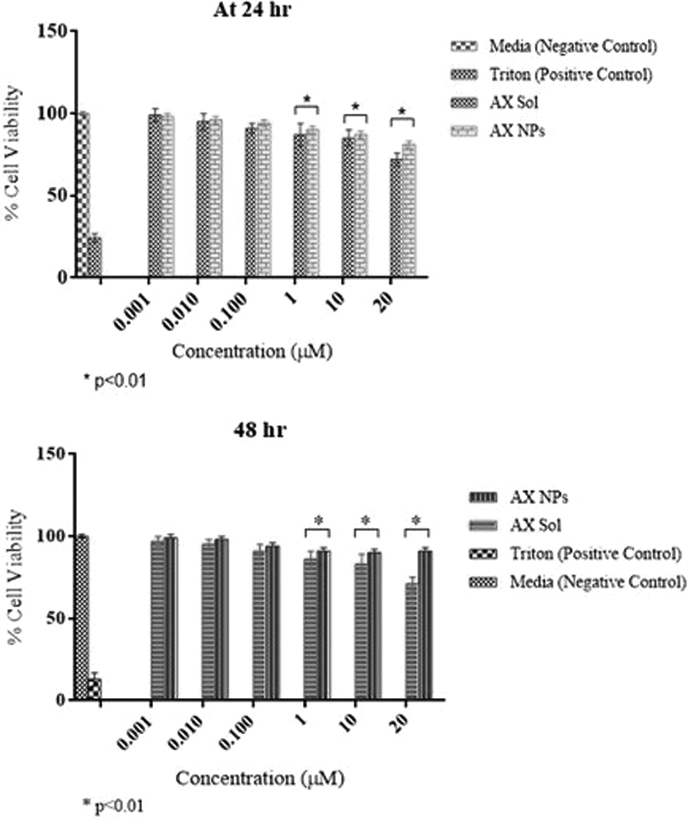
Percent cell viability at different concentrations of AX drug solution and AX-NPs at
Cellular Uptake
Coumarin-6-loaded PLGA NPs were evaluated for cellular uptake upon incubation with ARPE-19 cells at 37°C. Coumarin-6 is a hydrophobic fluorescent dye which mimics the hydrophobic nature of AX and was used to locate the nanoparticles and their uptake into the cells. Figure 6 shows confocal images of demonstration of the uptake of coumarin 6-loaded PLGA NPs into ARPE-19 cells at various time points at 37°C. The left first panel shows nucleus stained with nucleus staining dye DAPI with blue fluorescence. The next panel represents presence of cells as red fluorescence due to stained cell membrane with CellMask Deep Red stain. In the third panel from the left, uptake of coumarin-6 loaded NPs was observed and showing green fluorescence in the cytoplasm of the cells which confirms presence of NPs in cell cytoplasm. The last panel demonstrates the merged images of all. The blank in the figure is untreated cells which were kept as control. The intensity of green fluorescence depicting uptake of NPs in cells has been observed to increase with increase in treatment time. This may be because the uptake of NPs follows the receptor-mediated endocytosis process and with increase in the treatment time, more NPs may bind with the VEGF receptors present on the ARPE-19 cells which enhances cellular uptake as seen in Figure 6.
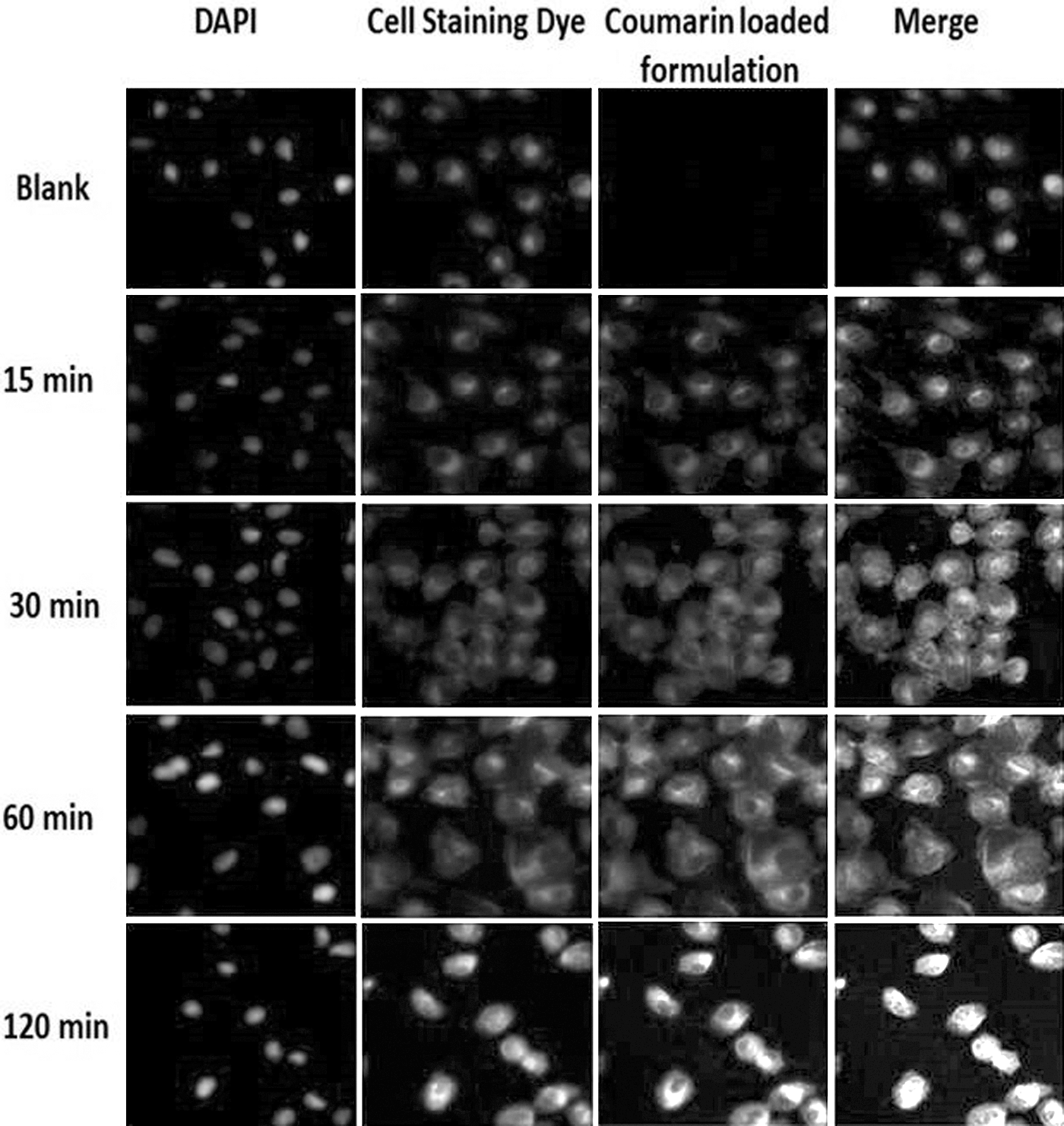
Cellular uptake of coumarin 6-loaded PLGA NPs in ARPE-19 cells at various time points at 37°C.
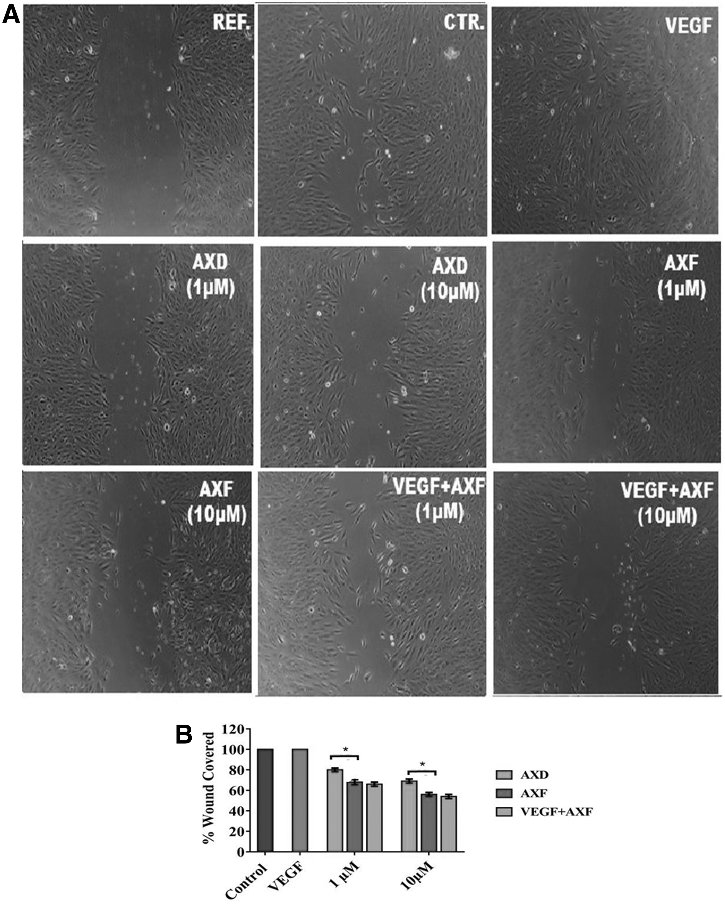
Wound Scratch Assay
Wound scratch assay was performed to study migration of the cell, which is one of the primary stages that leads to angiogenesis. The evaluation of the cell locomotion avails a significant estimation of inhibition of cell growth through conducting appropriate measurement on wound recovery under microscopic evaluation. 21 The cells were imaged immediately after making a wound served as a reference while the untreated group demonstrating covering of wound naturally served as a control. Figure 7A demonstrates microscopic images showing recovery of wound after treatment of cells with AX solution and AX-NPs in the presence and absence of angiogenesis-promoting agent (100 nM of VEGF165, rhVEGF; R&D Systems). Figure 7B shows the percent wound recovery for respective images in quantifying the graphical plot. Concentration-dependent inhibition of wound recovery was observed (p < 0.01); with least inhibition observed for the group with maximum concentration of AX in NPs (10 μM). The 100 nM of VEGF165 (rhVEGF; R&D Systems) increased the migration of cells to greatest extent and hence wound recovery as seen in Figure 7A. However, cotreatment of AX-NP formulation with VEGF165 could resist the action of VEGF165 as well as inhibit VEGF-induced angiogenic manifestations in ARPE-19 cells and did not significantly improve the cell migration-based wound recovery. These results indicate that AX-NPs efficiently and selectively inhibits VEGF-induced angiogenesis in ARPE-19 cells.
Anti-VEGF ELISA
The effect of the AX drug solution and AX-NPs on secretion of VEGF was studied using Anti-VEGF ELISA in ARPE-19 cells. Samples were collected after 12, 24, 48, and 72 h of treatment and the amount of VEGF-A was measured using the Human VEGF-A Elisa Kit. The percentage of VEGF secretion values are demonstrated using the control group value as 100% (Fig. 8). VEGF secretion was noticeably reduced by AX solution through all treatment groups. AX-NPs have similar effects on VEGF-A secretion. ELISA results showed significantly (p < 0.05) decreased VEGF protein levels after exposure to AX-NPs (10 μM equivalent of AX) in ARPE19 at 24, 48, and 72 h in comparison with control cells and cells treated with drug solution.
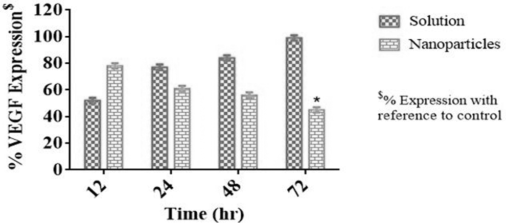
Percentage of VEGF expression in ARPE-19 cells treated with AX drug solution and AX-NPs for different time points determined using ELISA assay method (mean ± SD, n = 3). ELISA, enzyme-linked immunosorbent assay. *p < 0.05.
The observation depicted could be as a result of an initial high level of free drug available within the cells in case of employment of the solution form of the medicine, since the correlation of the scenario with the release profile of the formulation is hard to be determined. However, in case of nanoparticle formulation, there was an initial low level of VEGF control, which at later time points became stronger as the drug released from the matrix over a longer duration. 22,29 Therefore, sustained inhibitory effect of nanoparticle formulation as compared with drug solution was observed.
Conclusion
The AX-NPs were successfully developed, characterized, and tested in human retinal epithelial ARPE-19 cells manifesting its effectiveness in the wet AMD. This formulation may present an important approach of treating wet AMD due to its capability in ensuring a sustained release of drug and potential in inhibiting VEGF expression. It is therefore considered superior as compared with other oral formulations that portray low efficacy with more side effects. The intravitreal installation of the prepared nanoparticles can be conducted for efficient local delivery of the drug into the posterior segment of the eye, hence a sustained release formulation may be of great importance that reduces the frequent administration of intravitreal injection. Thus, this is an attractive approach that can be further investigated in vivo and in clinical setting to demonstrate its application for wet AMD therapy.
Footnotes
Acknowledgments
The authors would like to acknowledge the Lisa Muma Weitz Laboratory for Advanced Microscopy & Cell Imaging, USF Health, University of South Florida, Tampa, FL, for providing facility for microscopy and imaging and Department of Chemistry, College of Arts and Science, University of South Florida, for providing facility for DSC study.
Disclosure Statement
There is no conflict of interest.