
Abstract
Macrophages would engulf circulating oxidized (ox)- low-density lipoprotein and form lipid droplet-laden foam cells. Macrophage foam cells are considered an important therapeutic target of atherosclerosis. The aim of the study was to investigate a hypoxic foam cell model for anti-atherosclerotic drug screening using the chemical hypoxia-mimicking agent cobalt chloride (CoCl2). The oil red O stating results showed that treatment with CoCl2 could induce lipid accumulation and lead to cell transformation to spindle-shaped and lipid-rich foam cells in RAW 264.7 macrophages. Incubation with 150 μM CoCl2 for 24 h significantly increased the area of intracellular lipid droplets in macrophages, compared with the control group. Our findings indicate that CoCl2-triggered macrophage foam cells should be a potential in vitro hypoxia model for atherosclerosis drug discovery.
Introduction
Macrophages, a kind of phagocytes, are myeloid immune cells and involved in inflammatory processes of atherosclerosis. 1 Accumulation of cholesterol ester and low-density lipoprotein (LDL) in the intima of arteries would induce monocyte recruitment and differentiation through penetration of dysfunctional endothelium. 2 The monocytes differentiate into macrophages and express scavenger receptors that mediate the internalization of chylomicron remnant and modified LDL, and consequently become cytoplasmic lipid droplet-laden foam cells. 2 –4 In addition, macropinocytosis in macrophages is the other pathway to modulate uptake of LDL and foam cell formation in atherosclerosis. 5
Oxidized (Ox)- and minimally modified (mm)-LDLs play an important role in macrophage foam cell formation and subsequent fatty streak formation derived from accumulation of lipid and foam cell debris. 6 Activation of inflammatory signaling pathways in macrophage foam cells lead to more cell recruitment and LDL oxidation, and promote the progression of the atherosclerotic plaque. 2
Macrophage foam cells are reported as a therapeutic target in preventing cardiovascular diseases. 7 Inhibiting lipid droplet formation in macrophage and inducing removal of foam cells could reduce inflammatory responses and ameliorate atherosclerosis. 7,8 Recent study demonstrates that macrophage foam cell-targeting immunization improved atherosclerotic lesion in the whole aorta and the aortic root with enhanced lesion stability in apolipoprotein E-deficient (ApoE −/−) mice. 8 Regulating macrophage foam cell formation should be related to the progress of atherosclerotic lesions.
Hypoxia, presented in atherosclerotic plaques in humans and animal models, is considered related to the pathogenesis of atherosclerosis. 9,10 Hypoxia would induce the formation of lipid droplets in macrophages and increased local inflammation, and trigger atherosclerotic lesion progression. 9 It is also demonstrated that chronic hypoxia would accelerate the development of atherosclerosis in ApoE −/− mice. 11 The hypoxic macrophage foam cells should play a crucial role in the development of atherosclerosis.
Cobalt chloride (CoCl2) is one of the most commonly used hypoxia models in cell culture, through inducting expression of hypoxia-inducible factors (HIFs)-1α and 2α under normoxic conditions. 12 However, the effect of CoCl2 on lipid accumulation in macrophages remained unclear. The purpose of this study was to perform a hypoxic macrophage foam cell model for atherosclerosis drug discovery by using the chemical hypoxia agent CoCl2. We hypothesized that CoCl2 would induce lipid droplet and foam cell formation in macrophages, as well as in response to low oxygen condition.
Materials and Methods
Cell Culture
The murine macrophage cell line, RAW 264.7, was purchased from the American Type Culture Collection (Manassas, VA) and cultured as described in our earlier study. 13 RAW 264.7 macrophages were maintained in cell culture medium composed of Dulbecco's modified Eagle medium (Invitrogen Life Technologies, Carlsbad, CA), 10% fetal bovine serum (Invitrogen), 100 unit/mL of penicillin (Invitrogen), and 100 μg/mL of streptomycin (Invitrogen) at 37°C in a humidified incubator with 5% CO2.
Oil Red O Staining
The protocol of oil Red O staining was described in our earlier study.
14
In brief, oil red O powder (Sigma-Aldrich, St. Louis, MO) was dissolved in 2-propanol (Kanto Chemical Corporation, Tokyo, Japan) as 0.5% solution, and then diluted to 0.3% solution with distilled H2O. The oil red O solution was filtered through a 0.22 μm filter before use. RAW 264.7 macrophages were seeded on poly-
Quantification of Lipid Droplets
Quantification of cytoplasmic lipid droplets was measured based on a free and open source software ImageJ (Wayen Rasband, U.S. National Institutes of Health; Bethesda, MD;
Statistical Analysis
The data are presented as mean ± standard deviation. The statistical significance of differences between groups was analyzed using two-tailed unpaired Student's t-test by Microsoft Office Excel software (Microsoft Corp., Redmond, WA). The differences with a p < 0.05 were considered significant.
Results
CoCl2 Induced Lipid Droplet Formation in RAW264.7 Macrophages
To investigate the effect of CoCl2 on foam cell formation in macrophages, RAW264.7 macrophages were incubated with 300 μM CoCl2 for 24 h and then stained with oil red O. The results showed that treatment with CoCl2 led to cell transformation to spindle-shaped and accumulation of oil red O-labeled lipid droplets in macrophages (Fig. 1). The results of oil immersion microscopy confirmed that incubation with CoCl2 increased cytoplasmic lipid droplets surrounding the nucleus of macrophages (Fig. 2). Treatment with 150 and 300 μM CoCl2 for 24 h both markedly increased the density of intracellular lipid droplets in macrophages (Fig. 3). Our findings suggest that CoCl2 could induce lipid droplet and foam cell formation in macrophages.
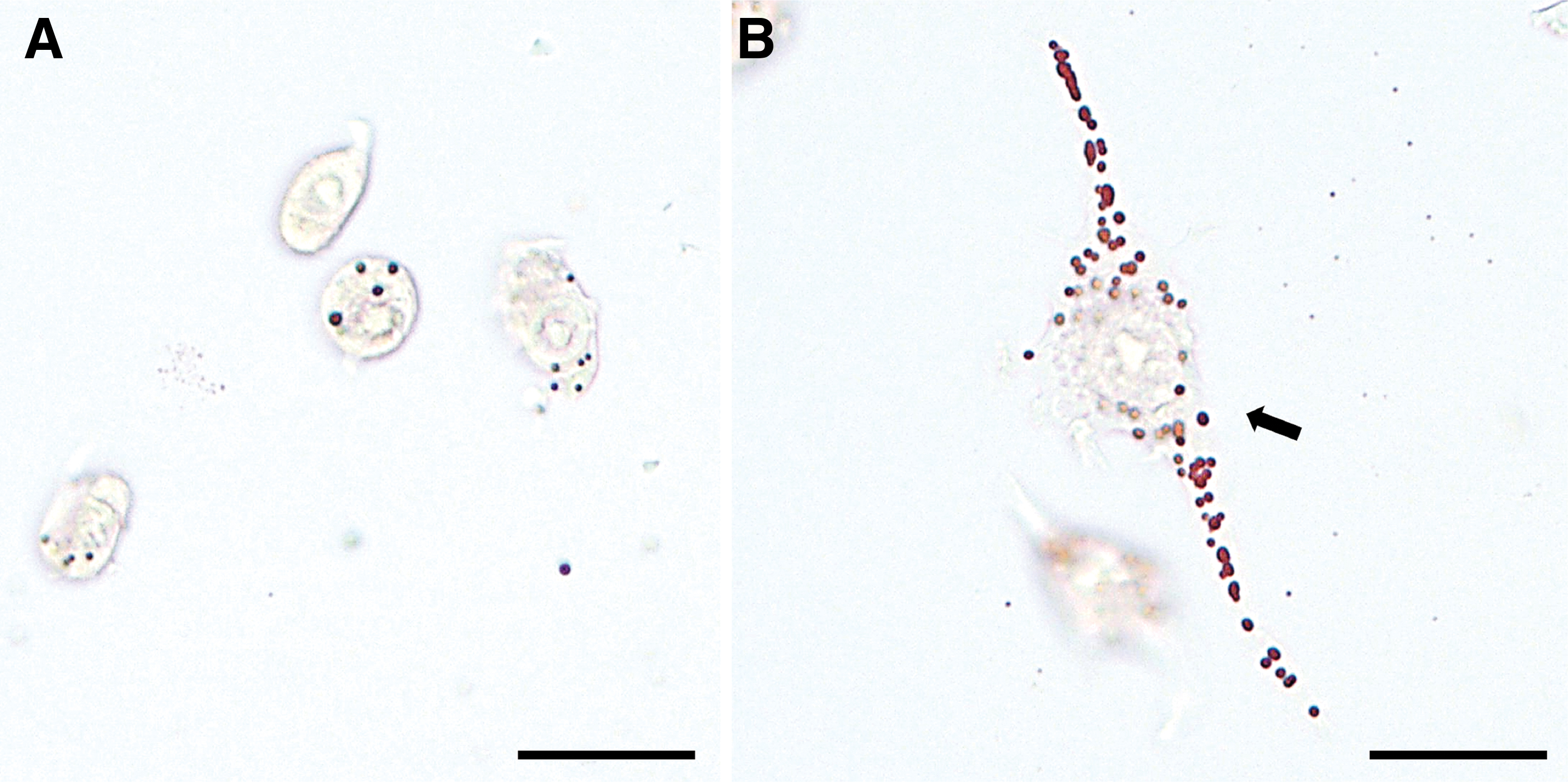
CoCl2 induced lipid accumulation and cell transformation in RAW264.7 macrophages. RAW264.7 macrophages were treated with 300 μM CoCl2 for 24 h and stained with oil red O.
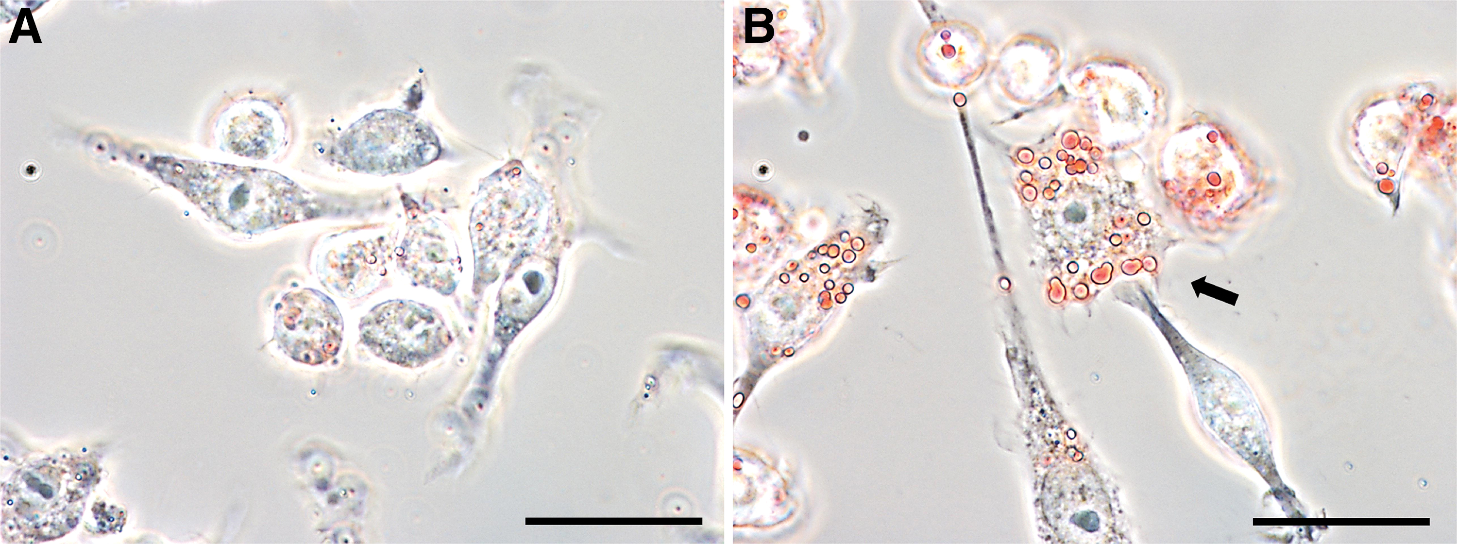
Accumulation of intracellular lipid droplets in CoCl2-treated RAW264.7 macrophages observed by using oil immersion microscopy. RAW264.7 macrophages were treated with 300 μM CoCl2 for 24 h and stained with oil red O.
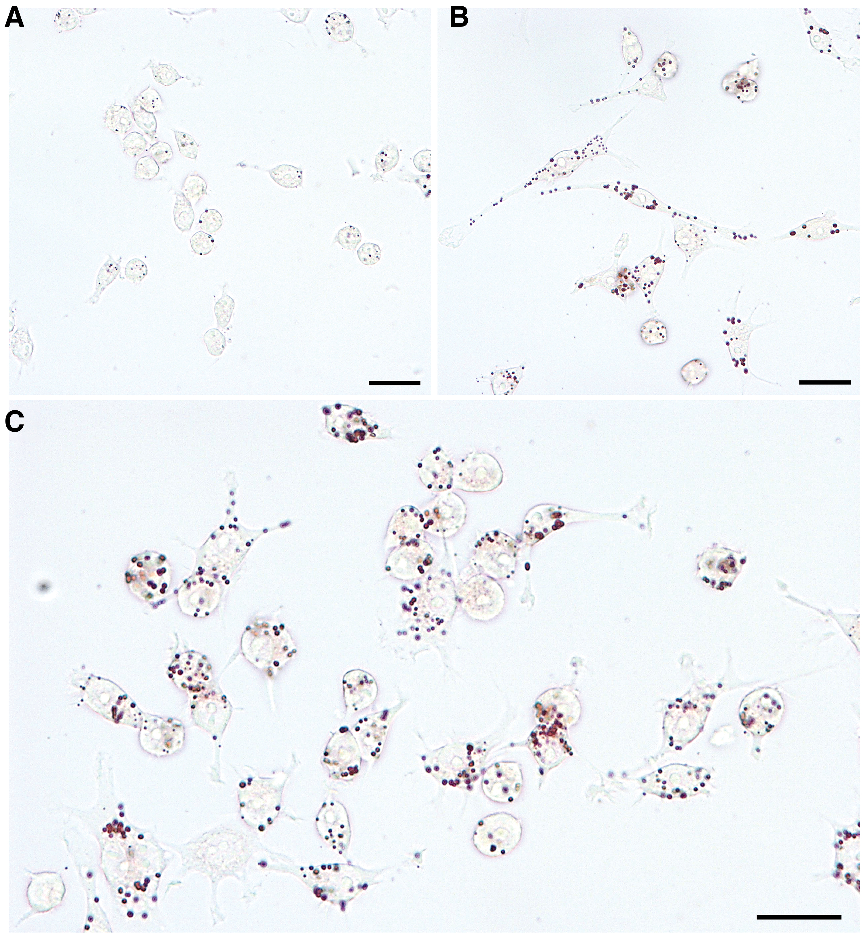
Dose-dependent treatment with CoCl2 triggered lipid droplet formation in RAW264.7 macrophages. RAW264.7 macrophages were treated with 150 or 300 μM CoCl2 for 24 h and stained with oil red O.
CoCl2 Increased the Number and Size of Intracellular Lipid Droplets in RAW264.7 Macrophages
To quantify the effect of CoCl2 on foam cell formation, the intracellular lipid droplets were selected and measured by digital image processing software ImageJ (Fig. 4). The results of quantification of lipid droplets in Figure 4 showed that treatment with 150 and 300 μM CoCl2 for 24 h increased almost 40- and 38-folds of total area of cytoplasmic lipid droplets in macrophages, respectively. Incubation with 150 and 300 μM CoCl2 both significantly elevated the averaged number and area of cytoplasmic lipid droplets compared with the control group, but there were no significant differences between 150 and 300 μM CoCl2-treated macrophages (Figs. 5 and 6). Moreover, the quantified results showed that treatment with 150 μM CoCl2 could significantly increase the lipid droplet area in RAW264.7 macrophages compared with the control group (Table 1). It supposes the proatherosclerotic effect of CoCl2 on lipid droplet formation in macrophages.
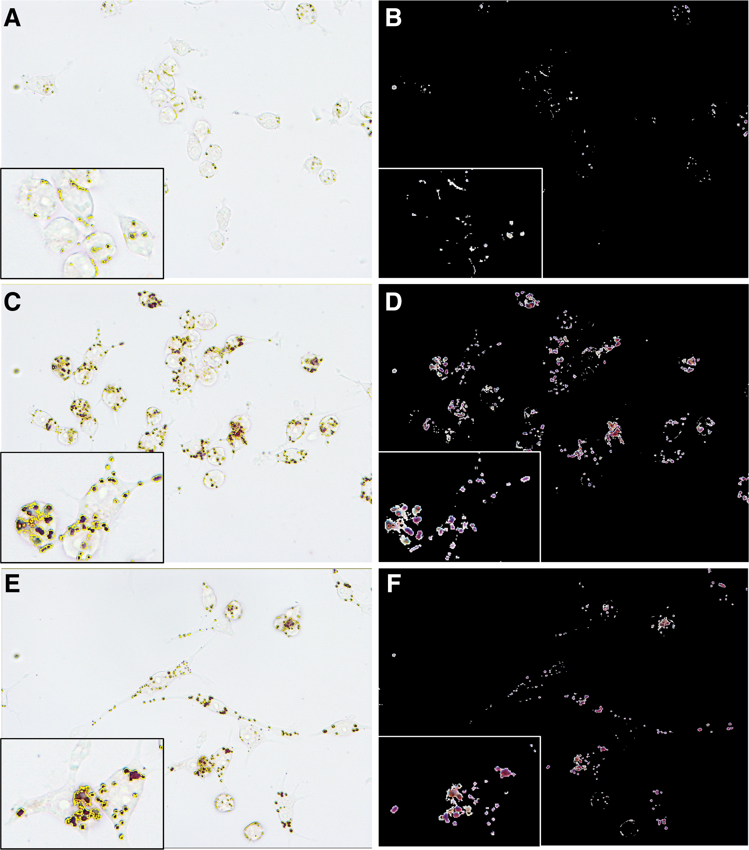
Selection of cytoplasmic lipid droplets in CoCl2-induced macrophage foam cells using the ImageJ software.

Quantification of lipid droplets in Figure 4 revealed that treatment with CoCl2 increased the averaged number of cytoplasmic lipid droplets in RAW264.7 macrophages. ***p < 0.001 compared with the control group. ### p < 0.001 compared with the control group. Color images are available online.
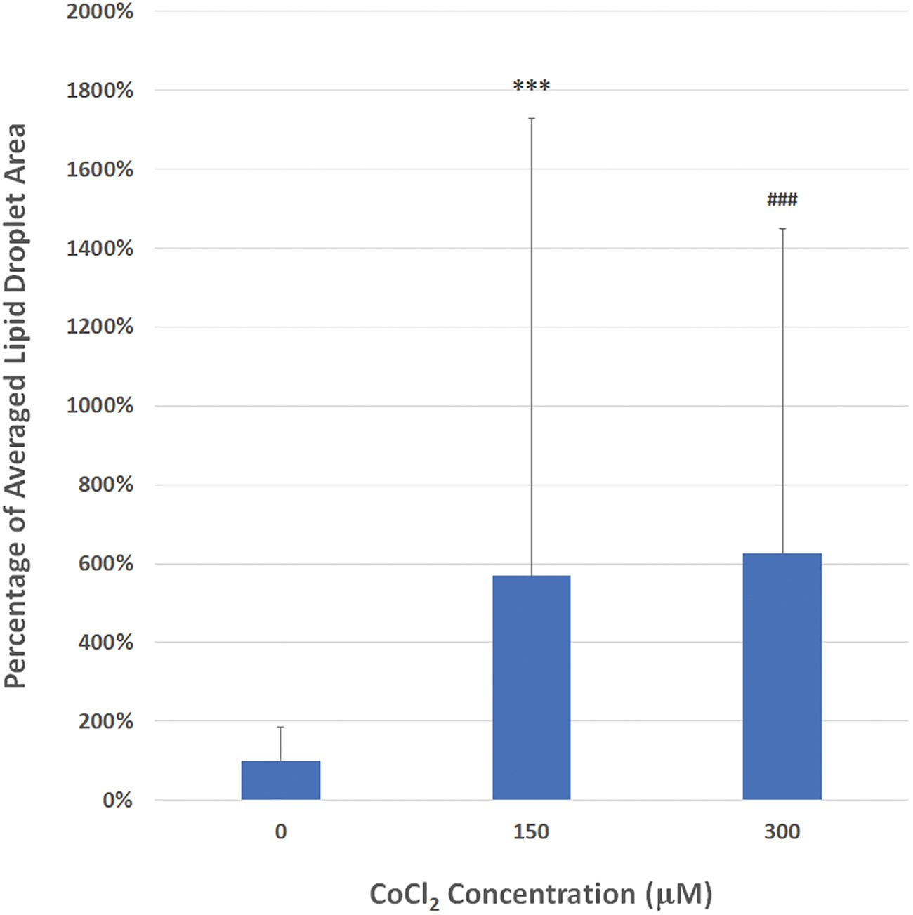
Quantification of lipid droplets in Figure 4 revealed that treatment with CoCl2 increased the averaged area of cytoplasmic lipid droplets in RAW264.7 macrophages. ***p < 0.001 compared with the control group. ### p < 0.001 compared with the control group. Color images are available online.
Quantification of Cobalt Chloride-Induced Lipid Droplet Accumulation in RAW264.7 Macrophages
RAW264.7 macrophages were treated with or without 150 μM CoCl2 for 24 h and stained with oil red O. After quantification, data represent the mean ± SD of three independent experiments. ** p < 0.01 compared with the control group. CoCl2, cobalt chloride; Conc., concentration; Exp., experiment; LD, lipid droplet; SD, standard deviation.
Discussion
This study is the first one that demonstrates and quantifies CoCl2-mediated lipid droplet formation in macrophages. In summary, we found that the chemical hypoxia agent CoCl2 triggered lipid droplet formation and cell transformation in RAW264.7 macrophages. Treatment with CoCl2 induced morphologic and physiologic changes in macrophages, and stimulated cell transformed to lipid-rich foam cells. The quantified results showed that incubation of CoCl2 led to a significant increase of the number and size of cytoplasmic lipid droplets in macrophage foam cells.
Treatment with CoCl2 triggered cell transformation and lipid droplet formation in M1/M2 RAW264.7 macrophages. It is known that alternative stimulation into M2 macrophages results in increased foam cell formation by inducing scavenger receptor expression. 15 CoCl2 exposure enhances alternative M2 activation in resting murine J774A.1 macrophages, and remodel the classical M1 phenotype of macrophage activation by changing the balance of inducible nitric oxide synthase, nicotinamide adenine dinucleotide phosphate oxidase 2, and interleukin-6. 16 Moreover, cobalt (II) ions modulate macrophage migration and cytoskeleton organization, and lead to induction of podosome formation and enhancement in macrophage spreading, adhesion, and inhibition of migration, which could induce prolonged immune cell retention and consequent chronic inflammation. 17 Collectively, incubation of CoCl2 would induce macrophage transformation into foam cells accompanied by accumulation of intracellular lipid droplets and inflammatory response.
CoCl2-stimulated macrophage foam cells should be a potential hypoxic cell model applied in anti-atherosclerotic drug screening. It is reported that treatment with 100 μM CoCl2 results in marked expression of HIF-1α protein, a subunit of a heterodimeric transcription factor HIF-1, in the nuclei of RAW264 macrophages. 18 HIF-1 exerts its effect on the pathogenesis of atherosclerosis, and should be a therapeutic target on the disease development. 19 Taken together, these studies imply that not only accumulation of intracellular lipid droplets but also stimulation of HIF-1 expression in CoCl2-mediated macrophage foam cells have therapeutic values for atherosclerosis drug discovery. In addition, it is investigated that foam cell formation in vivo converts macrophages to a profibrotic phenotype, which could contribute to atherosclerotic plaque stability, especially in early lesions of atherosclerosis that majorly consists of cholesterol-engorged macrophages but not vascular smooth muscle cells. 20 The in vivo results suggest that the candidates that could attenuate CoCl2-induced macrophage foam cells may prevent the progression of atherosclerosis.
CoCl2-mediated macrophage foam cell formation is similar to that induced by ox-LDL as shown in our previous study. 14 It is reported that hypoxia would contribute markedly to lipid metabolism in atherosclerotic lesions. 21 Hypoxia enhances lipid uptake into RAW 264.7 macrophages and regulates the expression of ox-LDL receptors, which may affect lipid droplet formation in macrophages. 22 In THP-1 cell line-derived and human monocyte-differentiated macrophages, hypoxia causes an accumulation of triglyceride-containing cytosolic lipid droplets and conversion of macrophages into foam cells. 23,24 Theses results suppose that macrophages may accumulate oil red O-stained lipid droplets containing triacylglycerol in response to CoCl2 exposure. However, it is demonstrated that incubation of a small volume of Watanabe heritable hyperlipidemic rabbit serum could induce foam cell formation with an increase of cellular content of cholesteryl ester in J774A.1 macrophages. 25 It suggests that CoCl2-mediated lipid accumulation may derive from supplement of serum in the cell culture medium. The components of lipid droplets in CoCl2-induced macrophage foam cells need to be further investigated.
There are several studies investigating the regulatory mechanism of foam cell formation in macrophages. It is reported that peroxisome proliferator-activated receptor (PPAR)-α and γ agonists, but not the PPAR-β agonist, inhibit macrophage foam cell formation in vivo through distinct ATP-binding cassette subfamily A member 1-independent pathways. 26 On the contrary, cluster of differentiation (CD) 146, expressed on the macrophages in human and mouse atheroma, could trigger macrophage activation by driving the internalization of scavenger receptor CD36 during lipid uptake, subsequently promote macrophage foam cell formation and retention during atherosclerosis. 27
Our findings suggest that CoCl2-induced macrophage foam cells should be as a surrogate target for rapid screening of anti-atherosclerotic effects in response to small molecule drugs. The in vitro hypoxic macrophage-derived foam cell model combined with high content screening technology may provide more probability on atherosclerosis drug discovery.
Footnotes
Acknowledgment
The authors thank Prof. Shwu-Fen Chang for offering RAW 264.7 macrophage cell lines.
Disclosure Statement
No competing financial interests exist.
Funding Information
This work was partly funded by a grant from Putian University (Grant No. 2019114).