




Abstract
Mesalamine is the first-line choice of drug for ulcerative colitis management. However, due to the nontargeted delivery of mesalamine, it shows side effects. The possible impact of mesalamine can be improved by coated microparticles in combination with S. boulardii for targeted delivery to the colon with the prevention of unwanted side effects. In this work, pectin-based mesalamine and S. boulardii loaded microparticles were prepared by dehydration technique and coated by an oil-in-oil solvent evaporation method and characterized by Scanning electron microscopy (SEM), X-ray diffraction, and zeta analysis. 2, 4, 6-Trinitrobenzenesulfonic acid was used for the induction of colitis. The anti-inflammatory effects of coated microparticles on Caco-2 cells were assessed by the determination of interleukin (IL)-8 concentration. In addition, the impact of coated microparticles on the concentration of colonic enzymes, including myeloperoxidase (MPO), lipid peroxides, and glutathione (GSH), were also evaluated. Moreover, hematological parameters, including white blood cell (WBC), erythrocyte sedimentation rate (ESR), and C-reactive protein (CRP), were assessed. SEM data revealed that all the prepared coated microparticles had an almost spherical shape. The X-ray powder diffraction analysis of uncoated and coated microparticles showed maximum stability without any interaction. The particle size of uncoated and coated microparticles was 9.14 and 15.61 μm, respectively. The zeta potential of uncoated and coated microparticles was observed to be −26.78 and −29.36 mV, respectively. The prepared coated microparticles decreased the levels of lipid peroxides, MPO, and GSH significantly in colitis. In the Caco-2 cell culture model, the concentration of IL-8 is decreased significantly. The hematological observations confirmed that the prepared formulation showed a promising decrease in the levels of WBC, CRP, and ESR in diseased animals. Animal experiments revealed that cellulose acetate phthalate coated microparticles of mesalamine and S. boulardii significantly improved the colitis disease conditions of Wistar rats. Hence, cellulose acetate phthalate-coated microparticles of mesalamine and S. boulardii could be recommended as adjuvant therapy to achieve a synergistic effect in the management of UC.
Lay summary
Mesalamine is the drug of choice for the management of ulcerative colitis (UC), which inhibits mediators responsible for inflammation. We investigated the in vivo effects of cellulose acetate phthalate-coated microparticles of mesalamine with Saccharomyces boulardii (probiotic) for their efficacy against UC. Our findings evidenced that the combination of mesalamine with S. boulardii showed a synergistic effect in the 2,4,6- trinitrobenzene sulfonic acid-induced colitis model by reducing the inflammation and maintains the macroscopic features. From the observed results, it can be concluded that S. boulardii can be used to enhance the individual drug's effect in the therapeutic management of UC.
Introduction
For the pharmaceutical industry, successful ulcerative colitis (UC) management strategy development remains a significant problem. 1 In a conventional treatment strategy, tablets and capsules may release the drug in the stomach region instead of the colon, which causes various side effects. 2 Many approaches were used in the novel drug delivery system for colon targeting, including pH, time-dependent, and bacterially degradable. However, it is challenging to provide the drug specifically to the colon's diseased site due to the body's complex physiological system, as there is a pH difference in each gastrointestinal tract (G.I.T) compartment. UC is a chronic recurrent condition of the colon marked by bloody diarrhea, weight loss, and frequent urge for feces. 3,4 Mesalamine is the primary drug for UC management. 5 However, due to the lack of targeted drug delivery, long-term colitis treatment leads to negative impacts on the patient's quality of life. 6 Diarrhea, nausea, and vomiting are frequently observed side effects of mesalamine that further induce the removal of drugs from the body. 7 In UC prevention, probiotics play an imperative role. Probiotics cover the intestinal mucosal layer against the invasion of microbes. It also helps to retain the concentration of mucus, which reduces colitis. 8 From the literature survey, it has been clear that in UC, the concentration of probiotics at the colonic site is decreased. So, probiotic formulations are given to patients to manage the disease condition. 9 Probiotics, including Saccharomyces, Lactobacillus, and Bifidobacterium, are widely used for UC management 10 –12 Saccharomyces boulardii belongs to the Saccharomycetaceae family. It has excellent anti-inflammatory properties. 10 Dong and his research group 13 concluded that S. boulardii was as efficient as mesalamine in decreasing serum inflammatory markers and maintaining the histological structures in dextran sodium sulfate-induced colitis in mice. Accordingly, by the research group of Guslandi et al., 14 the effect of S. boulardii has been studied in patients with UC, where preliminary findings indicated that S. boulardii can be effective in the management of UC. Thomas et al. 15 reported that S. boulardii inhibited (interferon-γ and tumor necrosis factor-α) and promoted interleukin (IL)-8 and transforming growth factor-β- dependent mucosal healing in patients with chronic colon inflammation. The combination of probiotics with mesalamine would be the best novel solution for UC management. 16 Various novel strategies have been used to optimize drug delivery, particularly to colon sites, including pro-drug, time-dependent release, microparticles, nanoparticles, and pellets. 4,14 –17 One of the most successful methods for colon-specific delivery is the microparticle approach. 17 –19 The particle size of microparticles was ranging from 4 to 15 μm, providing maximum mucoadhesion at the colon site. 20 –23 Pectin is a cell wall structural polysaccharide, which is dispersed richly in some fruits and vegetable sugars. Pectin is enriched in blocks of (1 → 4) linked galacturonic acid and galacturonic acid methyl ester units interrupted by single (1 → 2) linked rhamnose units. 24 –26 Pectin has a beneficial effect on the prevention of UC. 27 It is degraded by the colon-specific enzyme (bacteroids, bifidobacterium, eubacterium). 22
Cellulose acetate phthalate (CAP) is widely used as an enteric-coated polymer by the pharmaceutical industry for the coating of tablets and pellets. 27,28 In previous literature findings, coating of prepared microparticles with Eudragit-S100 was reported for specific colon drug delivery. However, the Eudragit S-100 pH-based single colon delivery system is not adequate to deliver the drug to the colon site as per recent studies.
Due to premature dissolution of the formulation in the small intestine leading to the insufficient release of drugs in the colon region, there are chances of drug accumulation at the duodenum instead of the colon due to the higher pH of the duodenum. Moreover, in UC, the colon's pH becomes acidic and only an enzyme-based strategy can work properly without any pH effect in such a situation. 29 The best strategy for specific colon targeting may be a combination of enteric-based polymer and enzyme-based approaches. 30 Therefore, polysaccharide carriers like pectin, which are microbially degraded by a particular colonic enzyme, have a higher potential for drug release at the colon site. 31,32 Bhatt et al. prepared Eudragit S100-coated calcium pectinate microbeads that can retard the release of p53 polyplex in upper G.I.T while releasing the polyplexes in the colon. 33
Accordingly, the purpose of this study was to prepare and evaluate the therapeutic effect of pectin-based CAP-coated microparticles loaded with mesalamine and S. boulardii for UC management.
Materials and Methods
Materials
Mesalamine was procured from Loba Chemie Pvt. Ltd (Mumbai, India). Probiotic S. boulardii was purchased from Hi-Tech BioSciences (Pune, India). CAP, pectin, alcohol, acetone, sodium chloride, disodium hydrogen phosphate, and potassium dihydrogen phosphate were purchased from CDH (Central Drug House (P) Ltd, New Delhi, India). Isooctane and Span 80 were procured from S D Fine Chemical Limited (Ambala, India). 2, 4, 6-Trinitrobenzenesulfonic acid (TNBS), dimethyl sulfoxide (DMSO), and light liquid paraffin were bought from Loba Chemie Pvt. Ltd (Mumbai, India). Caco-2 cell lines and Sandwich Enzyme-Linked Immunosorbent Assay (ELISA) Kit were obtained from Sigma-Aldrich (Mumbai). C-reactive protein (CRP) turbilatex test kit was procured from Anamol laboratories Pvt ltd (Maharastra, India). Analytical-grade chemicals were used during the whole study.
Methods
Preparation of pectin microparticles
As Jain et al. 34 reported, the pectin microparticles were prepared by dehydration technique with necessary modifications. Mesalamine was dissolved in DMSO, followed by probiotic S. boulardii (109 cfu/mL) in the phosphate-buffered saline (PBS) 7.4 solution, and then this suspended solution was added to the mesalamine solution. A solution of pectin was prepared in another beaker, and drug-probiotic dispersion was poured into it. Around 10 mL was dispersed from this dispersion into 50 mL of isooctane containing a span 80 (1.0% w/v) and continuously stirred to achieve stable water/oil emulsion at various speeds. The dispersion was rapidly cooled to 15°C, and about 50 mL of acetone was then poured into it to dehydrate the pectin droplets. Microparticles were then continuously stirred for 30 min at 100 g at a temperature of 30°C to allow the solvent to be fully evaporated, followed by drying, and then stored in a desiccator.
Coating of pectin microparticles
Pectin microparticle coating was done using the oil-in-oil solvent evaporation method reported by Lorenzo-Lamosa and others, 34 with few modifications. In this method, the dispersion of 50 g pectin microparticles was prepared in 10 mL of organic solvent mixture (acetone: ethanol, 9:1) containing CAP. Addition of the prepared organic phase into 70 mL of light liquid paraffin containing 1% w/v span 80 was done, followed by constant stirring for 3 h at 150 g at room temperature to evaporate the solvent. Finally, n-hexane has been used for the washing of coated microparticles. After this, microparticles are filtered, dried, and stored in a desiccator.
Characterization of Prepared Microparticles
Shape and surface morphology
Scanning electron microscopy (SEM) was used to evaluate the prepared uncoated and coated microparticles' shape and surface morphology. This procedure was completed by sprinkling dried powdered microparticle sample on double adhesive tape, fixed on an aluminum stub. Coating of stubs was done with gold to a thickness of ∼300Å, and SEM obtained the images of samples at 1,300 × magnification (JSM-840; Joel, Tokyo, Japan). 16
Particle size analysis, polydispersity index, and zeta potential analysis
Zetasizers (Nano NS, Malvern, PA) were used to estimate the particle size of microparticles at pH (1.2 and 7.4). The polydispersity index of the optimized microparticles was determined by dynamic light scattering. For estimation of zeta potential, the same instrument was used. 35
X-ray diffraction determination
X-ray diffraction (XRD) analysis was conducted for optimized formulations of uncoated and coated microparticles. Measurements of X-ray scattering angle were conducted with a copper anode fixed to the diffractometer (45 kV, 40 mA) in a wide-angle X-ray Diffractometer (D8 Advance; BRUKER, Germany) with 2θ angle. 36
Cell Culture
Culturing of Caco-2 cell lines of human colon carcinoma was done in a medium consisting of RPMI 1640 (50% v/v), fetal bovine serum (15% v/v), Dulbecco's modified Eagle's medium (DMEM) (35% v/v), and 1% (w/v) penicillin–streptomycin. NIH3T3 cells were cultured with DMEM supplemented with10% fetal bovine serum and 1% (w/v) penicillin–streptomycin in 5% of CO2 atmosphere conditions with 95% relative humidity and at 37°C.
Inflamed Intestinal Barrier Model
For the cultivation of Caco-2 cells, 96-well tissue plates were used. The cells were exposed to pro-inflammatory cytokines consisting of IL-1β and lipopolysaccharide at 50 and 1,000 ng/mL for 24 h at 37°C for inflammation induction. The untreated Caco-2 cells act as a control. For the confirmation of inflammation in the cell model, the centrifugation of extracellular media was done at 20000 g for 5 min, and IL-8 production was tested using Sandwich ELISA Kits (Sigma-Aldrich) as directed by the manufacturers. 37
In Vivo Studies
The study was carried out on 36 either sex Wistar rats weighing 180–260 g. Animals were obtained from ISF College of Pharmacy (ISFCP), Moga, and Punjab, India. Animals were kept at ambient temperature (21°C ± 10°C) and relative humidity (55% ± 5%) with a fixed 12-h light/12-h dark cycle. The experimental layout was approved as ISFCP/Institutional Animal Ethics Committee (IAEC)/control and supervision of experiments on animals (CPCSEA)/Meeting No 26/2020/Protocol No.434 by the IAEC as per the committee's guidelines for CPCSEA.
Induction of TNBS Colitis
As per the procedure mentioned by Morris et al., 38 colitis was induced with slight modifications. Fasting of rats was done for 48 h with free access to water and then anesthetized with Ketamine (50 mg/kg). A rubber cannula was rectally inserted into the colon with 8 cm proximal to the anus. TNBS (10 mg dissolved in 0.25 mL of 50% ethanol) was administered into the colon lumen using a rubber probe (total volume 0.5 mL solution). Disease control group received 0.5 mL 50% ethanol (v/v). The animal division was carried out in six groups. Group I served as normal control, and Group-II served as disease control. Group III was treated with colitis +placebo group (Inert material formulation) through the oral route. Group IV received colitis+mesalamine microparticles (23 mg/kg) through the oral route. Group V was treated with colitis+probiotic (109 cfu) microparticles through the oral route. Group VI was treated with colitis+coated microparticle formulation (mesalamine [23 mg/kg]+probiotic [109 cfu]) through oral route. Fifteen days of oral treatment were given using oral gavage, as shown in Figure 1.
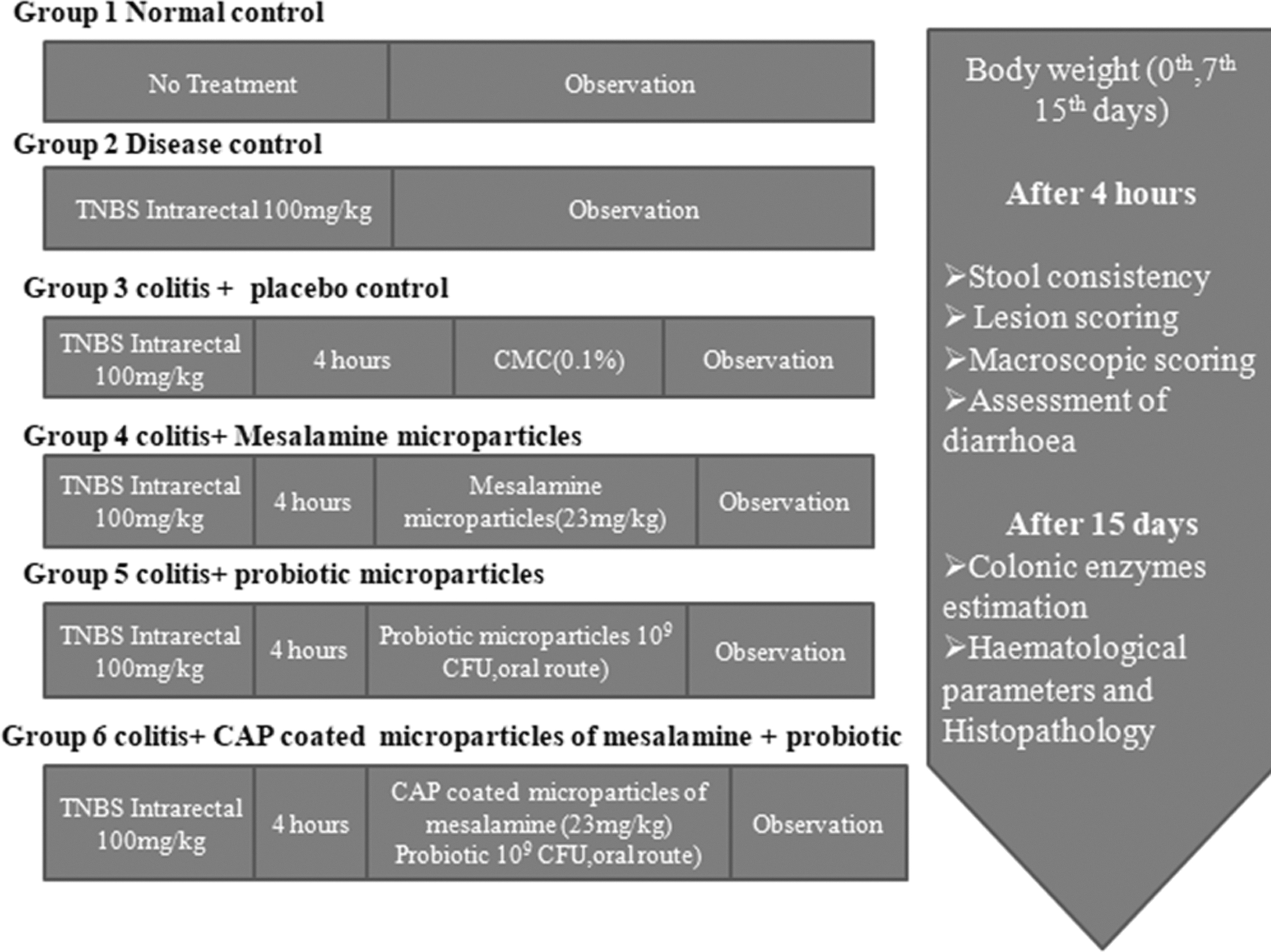
The disease was induced by TNBS, a gap of 4 h was given, and then various treatment formulations were given followed by various observations as mentioned in figure. TNBS, 2,4,6-Trinitrobenzene sulfonic acid.
Body Weight Evaluation
Body weights of rats were evaluated before sacrificing animals at the beginning of the 0th day before induction and after colitis induction after (0th day), 7th day, and day (15th day). 39,40
Macroscopic Character Assessment
The macroscopic observation of the colon is a significant part of UC severity monitoring. The severity of colitis was measured by an observer who is unfamiliar with the schedule of treatment. The 10 cm colon portion was removed and cut longitudinally for all animals and then washed to remove fecal residues. After the weight inflammation scoring was completed, inflammation scoring was permitted based on the colon's clinical characteristics. Zero points were counted for no visible change, 1 point was counted for hyperemia at the site, and 2 points were counted for lesions with a diameter of 1 mm or less. Scores are given in Table 1. For lesions with a diameter of 2 mm or less, 3 points were given, and for lesions with a diameter of more than 2 mm, 4 and 5 points were assigned. 41 The extent of colon inflammation has been visually assessed, and the treatment's effectiveness can be directly judged. 42
Scores Are Given for Weight Change, Consistency of Stool, and Lesions
Diarrhea Assessment
Wistar rats were housed in a cage with a clean white sheet after colitis induction by TNBS and were assessed for 4 h for the occurrence of diarrhea, fecal discharge, fecal consistency, and fecal bleeding. From each group, fecal material was analyzed regularly, and the recordings were ranked accordingly for diarrhea evaluation, as shown in Table 1. 43
Biochemical Marker Estimation
Estimation of myeloperoxidase, lipid peroxidase, and glutathione concentration
For the colitis severity estimation, colonic enzymes myeloperoxidase (MPO), lipid peroxidase (LPO) level, and glutathione (GSH) concentrations were measured. Approximately 600 mg of colon tissue was measured, added immediately to the test tube, and stored at 4°C. Saline (5.5 mL) that was added in the mixture was homogenized and centrifuged for a total of 15 min at 850 g. For MPO, LPO, and GSH measurement, the supernatant was collected. 16,43,44
Determination of CRP
For withdrawing blood from each animal, retro-orbital puncturing was used. A latex agglutination method was used to conduct the CRP assay. CRP Turbilatex Test Kit was used for the same. 45
Measurement of Hematology Parameters
The total leukocyte white blood cell (WBC) count was analyzed using diluted blood samples, and the solution of Turk (3% acetic acid) was used at a ratio of 1:200 for dilution. Counting was done using a hemocytometer. 46,47
A collection of 0.8 mL for the erythrocyte sedimentation rate (ESR) measurement in Westergren's method was used. From each group, around 0.8 mL blood sample collection was obtained in tubes containing ethylenediaminetetraacetic acid, and ESR level was assessed. 48
Histopathology study
The colon (2 cm) of each rat (n = 6) was taken and fixed with 10% formalin, followed by cutting into 5 μm thickness, staining with hematoxylin–eosin dye, and finally, the interpretation was evaluated. For any inflammatory changes such as tissue damage, cell infiltration, nucleus damage, and necrotic focal points, these colon sections were analyzed. 48
Statistical evaluation
Data were expressed as mean ± standard deviation (SD). For statistical evaluations, one-way analysis of variance (ANOVA) and Tukey–Kramer post-test were applied. A value of p-value <0.05 was considered significant.
Results and Discussion
Particle Shape and Morphology
As shown in Figure 2, SEM of prepared uncoated and coated microparticles was performed. Due to the presence of pectin, uncoated microparticles were slightly spherical with a slightly porous character. An almost complete spherical shape without porous visibility with a smooth surface has been evaluated in CAP coated microparticles. The uncoated and coated microparticles were between 8 and 15 μm in the size range. For efficient drug delivery in the colonic site, microparticles with sizes varying from 5 to 15 μm are quite suitable.

SEM images of uncoated microparticles
Determination of Particle Size, PDI, and Zeta Potential Analysis
In the Zeta Sizer, the mean particle size and size distribution were calculated. The particle size was 7.06 ± 0.8 and 10.31 ± 0.9 at pH 1.2, whereas at pH 7.4 the particle size was found to be 9.26 ± 1.3 and 11.64 ± 0.5 μm for uncoated and coated microparticles, respectively. The polydispersity index of microparticles coated and uncoated was found to be 0.245 and 0.267, respectively. It concluded a homogeneous state of the microparticles and an equal distribution of the same particle size. The zeta potential of uncoated and coated microparticles was −26.78 ± 4.66 Mv and −29.36 ± 3.36 Mv, respectively, which ensures that the uncoated and coated microparticle formulations were stable. 49
XRD Analysis
The XRD of uncoated and coated microparticles was performed to scrutinize any alters in the drug's physical state during the formulation process. XRD studies of uncoated and coated microparticles had shown a peak at 20°, as shown in Figure 3A. The polymer has amorphous nature. No massive difference was monitored in uncoated and coated microparticles. The uncoated microparticles in Figure 3B showed diffraction peaks at 2θ; 5.86°,10.56°, 22.26°,27.38°, and 32.89°. However, coated microparticles showed diffraction peaks at 2θ; 5.92°, 10.15°, 22.79°, 27.79°, and 32.97. The XRD peak showed that the intensity remains almost the same in CAP-coated microparticles, which confirmed no chemical reaction in drug and polymers.

XRD of uncoated
The Cellular Model of the Inflamed Intestinal Mucosa
The Caco-2 model of inflammatory cells was used to examine the beneficial effect of mesalamine and S. boulardii loaded CAP-coated microparticles. The secretion detection of IL-8 validated this model in both inflamed and untreated Caco-2 cells. The multifunctional chemokines IL-8 are secreted by enterocytes in response to several inflammatory mediators during the acute phase of inflammation and used as a standard inflammation biomarker in comparative studies. The inflamed Caco-2 cells treated with pro-inflammatory mediators consist of significant amounts of IL-8. The concentration of IL-8 was decreased from 2,491 ± 211 pg/mL to 368 ± 32 pg/mL after stimulation, verifying the inflammation in existence in vitro model.
In Vivo Studies
Assessment of bodyweight
Compared to the control group, a decrease in Wistar rats' bodyweight exposed to the treatment schedule for TNBS-induced UC was observed. Each treatment schedule's effects were observed in six animal groups on the 0th day, 7th day, and 15th day. Improvement in body weight during the treatment period is considered a sign of the diseased condition's recovery, as shown in Table 2. The weight of diseased animals was significantly decreased (p > 0.001) compared to the control group. While upon treatment with CAP coated microparticle formulation, the weight was significantly improved. Bodyweight assessment of different groups was performed. Each data represents mean ± SD (n = 6). Significance was tested using one way ANOVA and Tukey–Kramer post test. ***p < 0.001 (normal control vs. disease control group), ns p > 0.05, ## p < 0.01, and ### p < 0.001 (disease control vs. treatment groups).
Bodyweight Assessment of Different Groups
Each data represents mean ± SD (n = 6). Significance was tested using one way ANOVA and Tukey–Kramer post test. *** p < 0.001 (normal control vs. disease control group), ns p > 0.05, ## p < 0.01, and ### p < 0.001 (disease control vs. treatment groups).
ANOVA, analysis of variance; SD, standard deviation.
Macroscopic activity score
The TNBS model of colitis has been used to induce colon inflammation. The percentage change in weight, stool consistency, lesion score, and macroscopic scores for the colitis group was observed to be 4 ± 0.07, 4 ± 0.04, 4 ± 0.07, and 4 ± 0.09 correspondingly, while for CAP coated microparticles, the scores for the percentage change in weight, stool consistency, score of the lesion, and macroscopic scores for the colitis group were found to be 1 ± 0.3, 1 ± 0.08, 1 ± 0.13, and 1 ± 0.21 correspondingly. The Wistar rats' weight was measured at the beginning and end of the treatment schedule, as shown in Table 3 (15th day).
Macroscopic Parameter Assessment to Evaluate Disease
Values are statistically significant at p < 0.05 compared to normal and p < 0.05 compared to disease control animals (n = 3).
Assessment of macroscopic injury of rat colon
Macroscopic observation is an essential indicator for assessing the severity of colitis. Figure 4 shows the macroscopic picture of the colonic segment. Figure 4A demonstrated a normal colon with negligible macroscopic damage. Figure 4B shows severely damaged and mucosal congestion, hemorrhage, and deep ulcers that have been observed in the disease control group. Figure 4C Placebo group where almost same sign and symptoms have been observed as in group B. Figure 4D Slight reduction in hyperemia and the necrotic zone has been observed with improvement in inflammation after treatment with orally administered plain mesalamine microparticles. Figure 4E Slight reduction in inflammation, hyperemia, and the necrotic zone has been observed after treatments with orally given plain probiotic microparticles. Figure 4F Almost negligible inflammation and hyperemia have been observed in colons of rats of orally given coated microparticles of mesalamine and probiotic. All over, remarkable results have been observed. The colon of these rats was very similar to those of healthy rats.
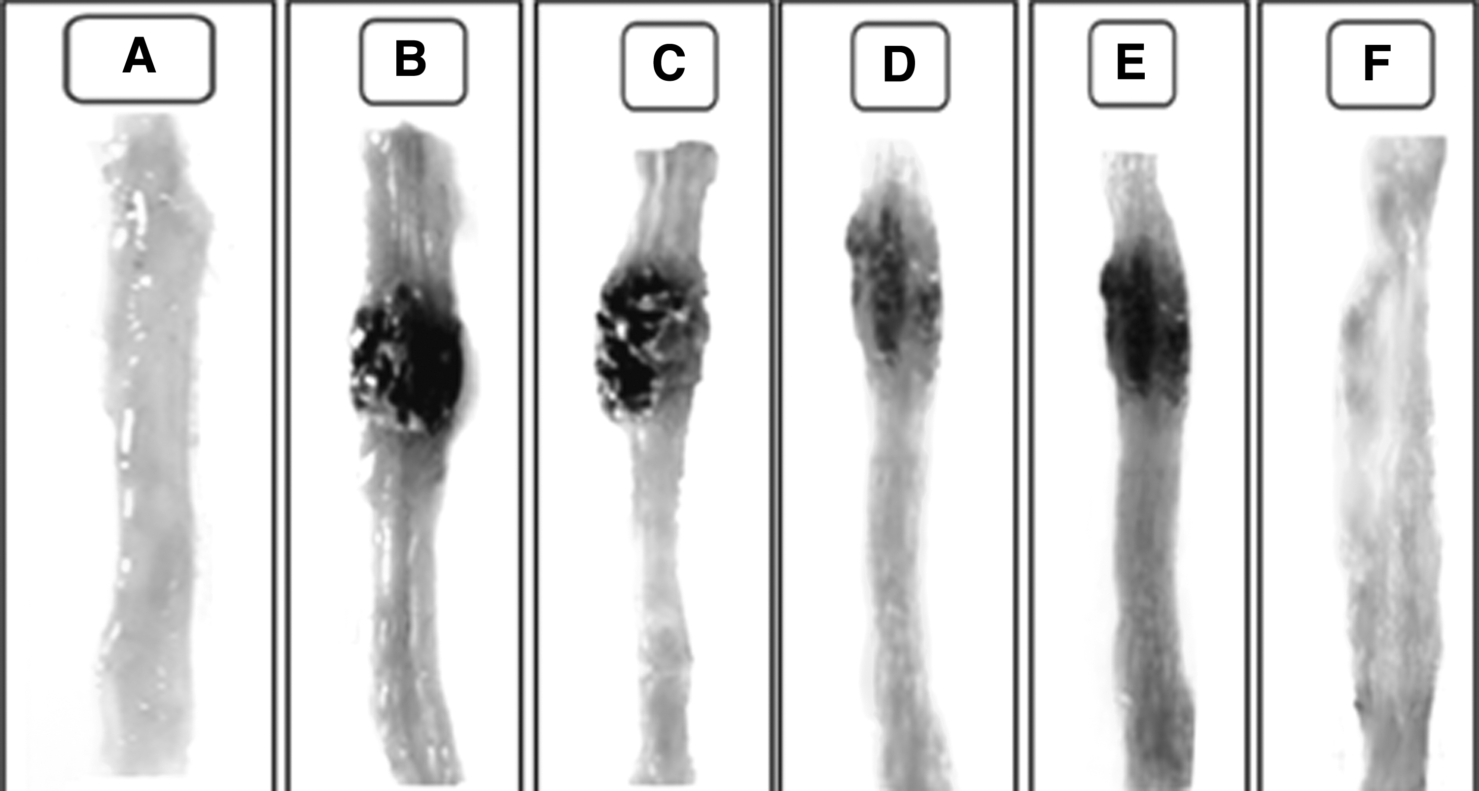
Treatment efficacies of various formulations after oral administration in TNBS induced colitis in Wistar rats.
Effect of Microparticle Formulation on MPO in TNBS Induced Colitis in Rats
Due to the colitis induction, the colonic enzyme concentration becomes changed. MPO is a colonic enzyme. The activity of this enzyme is concerned with the concentration of neutrophils in the inflamed tissue. Therefore, colonic enzyme determination can act as a parameter for the assessment of acute intestinal inflammation. TNBS using intrarectal administration showed a significant MPO concentration increase, that is, 19.06 μmol/min/mg tissue, while the MPO concentration is almost the same in the normal group and placebo group. Plain mesalamine microparticles reduce MPO concentration, that is, 11.6 μmol/min/mg, which is near the value obtained after giving plain probiotic microparticles, that is, 13.2 μmol/min/mg. However, coated microparticles give a remarkable reduction in MPO concentration, that is, 7.9 μmol/min/mg compared to disease control, as shown in Figure 5.

Effect of microparticle formulation on MPO level in TNBS induced colitis in Wistar rats. Values are given as mean ± SD; values are statistically significant at p < 0.005.# p < 0.001 versus normal, *p < 0.01 versus disease control, and @ p < 0.05 versus coated mes+pro microparticles. mes+pro, mesalamine+probiotic; MPO, myeloperoxidase; SD, standard deviation.
Effect of Microparticle Formulation on LPO in TNBS Induced Colitis in Rats
LPO is a colonic enzyme. An increase in LPO concentration can lead to reactive metabolites, which can further cause inflammation. TNBS through intrarectal administration demonstrated a considerable rise in LPO concentration in the disease control group, that is, 153.11 μmol of malondialdehyde (MDA)/mgpr, while the placebo group has the almost same concentration of LPO in comparison with normal control. Plain mesalamine microparticles give a reduction in LPO concentration, that is, 125.16 μmol of MDA/mgpr. The effect of plain probiotic microparticles is 129.7 μmol of MDA/mgpr. However, CAP coated microparticles of mesalamine with probiotics give a remarkable decrease in the LPO concentration up to 74.37 μmol of MDA/mgpr compared to disease control, as shown in Figure 6.

Effect of microparticle formulation on LPO level in TNBS induced colitis in Wistar rats. Values are given as mean ± SD. Values are statistically significant at p < 0.05. # p < 0.001 versus normal, *p < 0.01 versus disease control, and @ p < 0.05 versus coated mes+pro microparticles. LPO, lipid peroxidase.
Effect of Microparticle Formulation on GSH in TNBS Induced Colitis in Rats
GSH is a colonic enzyme that is involved in DNA repair and also responsible for antioxidant activity. Intrarectal administration of TNBS has shown an extensive decrease in GSH concentration in the disease control group, that is, 4.1 μmol of GSH/mgpr, which is nearly the same as the placebo group. Plain mesalamine microparticles increase the GSH concentration, that is, 5.4 μmol of GSH/mgpr, but probiotic microparticles raise the GSH concentration, that is, 4.7 μmol GSH/mgpr, which was lesser than the mesalamine effect. CAP coated mesalamine microparticles loaded with probiotic showed an impactful increase in GSH concentration, that is, 6.2 μmol of GSH/mgpr compared to disease control, as shown in Figure 7.

Effect of microparticle formulation on GSH level in TNBS induced colitis in Wistar rats. Values are given as mean ± SD. Values are statistically significant at p < 0.05 compared to normal and p < 0.05 compared to disease control rats. GSH, glutathione.
Determination of CRP
CRP is a sensitive marker of inflammation; Table 4 showed that CRP levels are increased in UC, but treatment with CAP coated mesalamine and probiotic microparticles showed a reduction in CRP levels very effectively of mesalamine microparticles, as well as plain probiotic microparticles. However, the level of CRP was almost similar for disease control, as well as placebo.
Determination of White Blood Cell, C-Reactive Protein, and Erythrocyte Sedimentation Rate Level After Treatment Schedule
CRP, C-reactive protein; ESR, erythrocyte sedimentation rate; WBC, white blood cell.
Determination of ESR
ESR is an easy test for the assessment of inflammatory response. The observed results have shown that CAP coated microparticles of mesalamine and probiotic have the property to considerably reduce the ESR compared with plain mesalamine microparticles and plain probiotic microparticles. However, the ESR level was comparatively similar to disease control and the placebo, as shown in Table 4.
Determination of WBC
The body releases WBCs when an infection or inflammatory disease arises to help fight the infection. As shown in Table 4, WBC levels are increased after UC due to the cells' proliferation. However, coated microparticles of mesalamine and probiotic have shown a marked reduction in the level of WBCs in comparison with plain microparticles of mesalamine and probiotic, respectively. However, the results are almost the same for the placebo and disease control groups.
Histopathology Examination
Histological examination of tissues can help diagnose disease conditions because each condition causes characteristic changes in the tissue structure. Histopathology studies (Fig. 8) have shown that the colon tissue of Group I (A) the mucosal lining appears normal, showing a normal ionic gland with minimal stroma with goblet cells and no infiltration, muscularis mucosae, and normal submucosa. Group II (B)'s colon tissue revealed histopathological changes, including severe forms of mucosal ulceration replacement by inflammatory cellular infiltrate and fibrinoids like necrosis in all colonic wall layers. There was a marked rise in the number of inflammatory cells, mainly in lamina propria, consisting of lymphocytes with lymphoid follicle formation–neutrophils, eosinophils–goblet cell, depletion, and distribution mucosal gland featuring. Group III (C)'s colon tissue showed an increase in inflammatory cell number in lamina propria, with the invasion of the base of crypts and progress in the direction of crypt lumina to form crypt abscess, eosinophils –goblet cell, depletion, and distribution mucosal gland featuring. Group IV (D) expressed better reemission, and a slight improvement in the infiltration of cells has been observed. In Group V (E), orally administered plain probiotic microparticles showed a slight change in histopathological conditions. In Group VI (F), orally administered coated microparticles of mesalamine and probiotic histopathological examination revealed intact mucosal lining with mucosal line gland and showed almost 100% recovery of colonic mucosa from TNBS induced colitis damage in comparison with other groups. Especially, coated microparticles of mesalamine and probiotic showed the advantages of synergistic effects for colitis rats.

Histopathological change in colon experimental of Wistar rat;
Conclusion
In the presented work, a combination of mesalamine with S. boulardii for specific colon targeted drug delivery in the form of CAP coated microparticles has been developed. Up to the best of our knowledge, it is the first report of the combination of mesalamine with probiotic S. boulardii, in which natural polysaccharide pectin was used to prepare microparticles. The prepared microparticles were coated with CAP to prevent drug release in gastric fluid. The SEM evaluation ensured that the coated microparticles were spherical. The evaluation of coated and uncoated microparticles with a zeta sizer confirmed that formulations have the desired size range, PDI, and zeta potential.
Furthermore, the XRD evaluation confirmed that the drug and polymer have no interaction with each other. The in vivo anti-inflammatory effect of orally administered CAP coated microparticles was evaluated utilizing Wistar rat groups. A significant rise in body weight has also been observed in the case of the colitis group. It was worth noting that CAP coated microparticles could reduce the amount of MPO and LPO in a diseased state and increase the GSH level. The histopathological study of colitis-induced rat colon was very similar to those of healthy rats. Macroscopic evaluation and hematology estimation confirmed that the CAP coated microparticle loaded with mesalamine with S. boulardii has a synergistic effect in colitis. Despite the number of advantages offered by this microparticle formulation, few limitations are also there, including the high cost of the material, as well as more processing charges. Moreover, it also required proper process parameter optimization. For the future perspective to confirm the therapeutic efficacy of coated microparticles in humans, clinical trials are mandatory.
Footnotes
Authors' Contributions
A.S., U.K.M., and R.K.N. planned the study and wrote the article. A.S. performed all of the experiments. All authors have read and approved the article.
Acknowledgment
The authors are thankful to the management of ISF College of Pharmacy, Moga (Punjab) for providing research facilities and support.
Disclosure Statement
No competing financial interests exist.
Funding Information
No funding was received for this article.