




Abstract
It is hypothesized that etoposide/VP-16 nanomicellar formulation (VP-16 NMF) utilizing D-α-Tocopherol polyethylene glycol 1000 succinate (TPGS) can improve etoposide solubility and anticancer activity. The following four different concentrations of TPGS: 3, 6, 8, and 10 wt% were used to solubilize the drug. Among these four formulations, 10 wt% of TPGS loaded with VP-16 NMF dramatically enhanced etoposide apparent solubility by 26-folds compared with the native drug. The physicochemical properties of the optimized formulation were further analyzed by dynamic light scattering, X-ray powder diffraction, scanning electron microscopy, proton nuclear magnetic resonance (1HNMR) and Fourier transform infrared spectroscopy. Liquid chromatography tandem-mass spectrometry (LC-MS/MS) was used to assess solubility and intracellular uptake of the drug from the NMF. Cell viability assay ([3-(4,5-dimethylthiazol-2-yl)-5-(3-carboxymethoxyphenyl)-2-(4-sulfophenyl)-2H tetrazolium solution [MTS]) was performed on MCF-7 and MCF-10A cell lines to assess intracellular uptake and anticancer activity of etoposide. The MTS assay results showed that the VP-16 NMF platform provides a higher anticancer activity than the native VP-16 on the MCF-7 cells line as it integrates a dual anticancer activity of VP-16 and TPGS. LC-MS/MS data showed a threefold increase in cellular uptake of VP-16 NMF in MCF-7 cell line compared with the native etoposide. These data suggest that an optimal TPGS concentration can improve VP-16 bioavailability and efficacy with potential benefits for chemotherapy.
Introduction
Derived from a perennial herbaceous plant, Podophyllum peltatum, the mayapple, 1 etoposide is described as an inhibitor of the enzyme topoisomerase II. Etoposide is known to be frequently utilized in the treatment of tumors, specifically in treating brain tumors. 2 Furthermore, other cancers treated by etoposide include small cell lung cancer and testicular cancer, where the drug can be administered either intravenously or orally. 3 Similar to many other drugs, etoposide has several side effects, including, and are not limited to, vomiting, nausea, persistent metallic taste, hypotension, fever, and burning sensation where it is injected. 4 Hypersensitivity reaction that could occur due to the solvent or etoposide itself has been well defined. Dyspnea, chest pain, hypotension, bronchospasm, and/or flushing are symptoms of this condition. 5 Moreover, high amount of ethanol is required to solubilize etoposide, which may cause substantial hypotension and alcohol intoxication. 6
Etoposide (VP-16) [with low water solubility (80 μg/mL) and low permeability/low absorption range (10.16%) is a BCS class IV drug. To solve the etoposide poor aqueous solubility issue, the U.S. Food and Drug Administration (FDA) approved an intravenous salt form of etoposide phosphate in 1996. 7 However, there have been a lot of arguments regarding etoposide phosphate treatment. As a water-soluble drug, it does not consist of solvents that can act as causative agents. Also, as given in such literature, researchers failed to present any evidence of hypersensitivity reactions as for polysorbate 80, which is found and administered in IV etoposide formulations. 8 The main downside of oral etoposide is its incomplete and uncertain bioavailability. Compared with the intravenous dosage forms, ∼50% (30%–97%) of the oral dose is bioavailable. 9 In addition, variability in bioavailability has been observed within patients. 10 Variability in bioavailability of etoposide, especially from the oral dosage form can be the results of various reasons. Intrinsic and acquired resistance are some of the shortcomings of the success of the treatment as used in chemotherapy. Previous studies reported that altered topoisomerase II expression, decreased topoisomerase II sensitivity to etoposide, decreased expression of DNA mismatch repair genes, increased expression of ABC transporters such as multidrug resistance (MDR)1 and MRP1 may be responsible for etoposide resistance. 11 –13 It was also reported that MRP1 that encodes efflux pump, are notably upregulated in sublines causing resistant to etoposide. 14 It has been reported that the concomitant use of etoposide with D-α-Tocopherol polyethylene glycol 1000 succinate (TPGS) resulted in decreased in vitro etoposide permeability across artificial membrane. 15
Nanotechnology provides some advantages to overcoming the poor bioavailability of the low water-soluble drugs. Among several technology platforms, polymeric nanomicelles has attained attention of scientists throughout the world as a multifunctional delivery system for poorly water-soluble drugs. 16 Polymeric nanomicelles are composed of amphiphilic polymers that self-assemble into hydrophobic core and hydrophilic shell. The hydrophobic core allows to solubilize poorly water-soluble drugs, whereas the hydrophilic shell protects the encapsulated drug from the in vivo environment. In addition, the small particle size of the nanomicelles increases the blood residence time, helps in bypassing liver and spleen filtration and glomerular filtration, increases intracellular uptake all of which results in increased bioavailability. 17 Vitamin-E-TPGS or TPGS is a derivative of alpha-tocopherol which is gaining interest in nanomicellar system development. 4 It offers a variety of benefits in drug delivery system formulations and expands drug's systemic half-life. 6 Considered as a waxy solid, which can fully dissolve in water and form its own micelles, the water-soluble parts of the molecule are all in interaction with the external solvent, sequestrating the micelle center of the hydrophobic tail parts. 18 Known for its potentials to have applications in pharmaceuticals, TPGS exhibits a melting point of around 37–41°C and also heat notably up to 199°C. 6 As such, the TPGS can be comfortably processed at high temperature and also known to be quite stable at pH 4.5–7.5. 19 In addition, TPGS itself is reported to enhance the solubility of poorly water-soluble drugs. 20 It may also serve as an inhibitor of P-glycoprotein (P-gp) that has been used as an excipient to resolve MDR and to improve many anticancer drugs' oral bioavailability. 21
This study aims to improve the water solubility of etoposide (VP-16) with TPGS, which, in turn, could potentially help overcome the drug's resistance issues, and minimize its side effects while maintaining its efficacy. In this study, etoposide/VP-16 nanomicellar formulation (VP-16 NMF) was prepared with solvent evaporation thin-film rehydration technique. The optimized formulation was characterized by Fourier transform infrared spectroscopy (FTIR), proton-nuclear magnetic resonance ( 1 H-NMR), X-ray powder diffraction (XRD), and scanning electron microscopy (SEM). An MTS assay of VP-16 NMF was carried out on breast normal cells (MCF-10A) as well as identified breast cancer (MCF-7) cells to assess the anticancer efficacy. Liquid chromatography tandem-mass spectrometry (LC-MS/MS) has been used to assess the solubility enhancement of etoposide and the intracellular uptake from the VP-16 NMF in MCF-7 cell line.
Materials and Methods
General
Etoposide (VP-16) was purchased from 001Chemical (Hangzhou, China). TPGS, phosphate buffered saline (PBS) pH 7.4, 0.1 N HCl, acetonitrile, D2O, CDCl3, formic acid, ethanol, Dulbecco's modified Eagle's medium (DMEM), DMEM/F12, high glucose, non-essential amino acid (NEAA),
Preparation of VP-16 NMF
VP-16 NMF were prepared following solvent evaporation technique in which a known amount of VP-16 (10 mg) with different TPGS wt% (3, 6, 8, and 10 wt%) were dissolved in ethanol. Then, vitamin TPGS was added slowly to obtain a homogenous solution. The ethanol was then evaporated with rotavap equipped with a VC3000D speed vacuum (GeneVac Technologies, Warminster, PA) for 9 h, resulting in a solid layer, which was then mixed and rehydrated with deionized water. After 10 minutes, the resulting solution was filtered through a nylon filter membrane (0.22 μm) (Fig. 1). 22
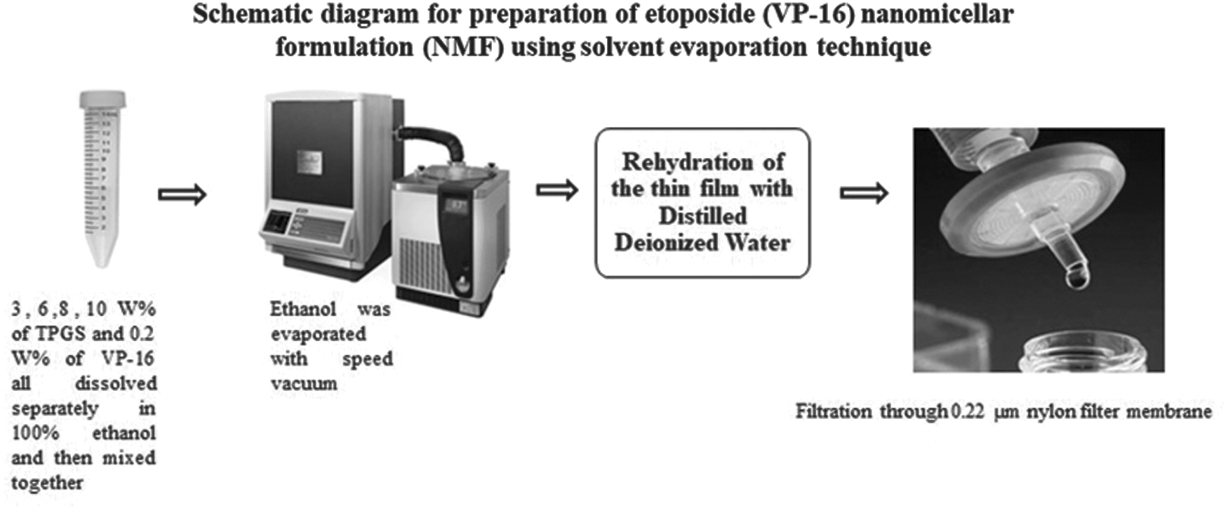
Preparation scheme for etoposide (VP-16) NMF NMF, nanomicellar formulation.
Finally, the filtrate solution was lyophilized with a laboratory-scale freeze dryer (Labconco Corporation, Kansas City, MO).
Characterizations of VP-16 NMF
Size, polydispersity index, and surface charge
The average hydrodynamic nanomicellar size, polydispersity index (PDI), and Zeta potential of surface charge density of VP-16 NMF were measured by employing Zetasizer Nano ZS (Malvern Instruments Ltd., Worcestershire, UK) using 1,000 μL of sample in triplicate measurements. 23
Entrapment and loading efficiency
LC-MS/MS was used to calculate VP-16 loading and entrapment efficiency. Entrapment efficiency was calculated indirectly by subtracting the overall amount of unentrapped drug from total amount, which we started with. Then, equations (1) and (2) were used to calculate the entrapment and loading efficiency.
24,25
Fourier transform infrared spectroscopy
The FTIR experiment was conducted following a published method. 26 The infrared spectroscopy (IR) spectra were acquired on the Nicolet IS10 FTIR instrument (Thermo Fisher Scientific, Madison, WI) equipped with a zinc selenide (ZnSe) crystal. For IR spectra acquisition, the instrument was used in attenuated total reflectance mode. Moreover, a resolution of 4 cm−1 and a sample scan of 228 were selected as basic acquisition parameters. Then, the IR transmittance was acquired to a spectrum between 4,000 and 650 cm−1. FTIR spectra were then analyzed with the OMNIC Spectra Software (Thermo Fisher Scientific, Waltham, MA, USA). Typically, the average IR spectrum acquisition time was 6 min.
1 H-NMR spectroscopy
To perform the 1 H-NMR experiment, the freeze-dried powder of VP-16 NMF and blank nanomicelles formulation were dissolved in D2O. Varian 400 MHz spectrometer (Palo Alto, CA) with a Varian 2-channel probe NMR instrument were employed to acquire all NMR spectra. VNMRJ software (version 4.2A) was used to process and compare the NMR spectra of VP-16 NMF with blank nanomicellar formulation (BNF) and native VP-16 spectra. 14
XRD crystallography
To elucidate the VP-16 NMF in its solid state, XRD was performed using a MiniFlex, which is an automated X-ray diffraction instrument (Rigaku, The Woodlands, and TX) at room temperature. Ni-filtered Cu-K alpha radiation was employed at 30 kV and 15 mA. The diffraction angle was covered from 2θ = 5° to 2θ = 60° with a step size of 0.05/step, and a count time of 2.5 s/step (effectively 1.1/min for ∼46 min/scan). The diffraction patterns were processed using Jade 8+ software (Materials Data, Inc., Livermore, CA). 23,27
Scanning electron microscopy
SEM was used to visualize the morphology of both native etoposide, blank nanomicelles, and VP-16 NMF. Several freeze-dried samples were sputtered with ∼20 nm thickness in a gold-palladium alloy and then later visualized using an FEI/Philips XL30 Field-Emission Environmental SEM. This was done at 5 kV and also, for further investigation, computerized pictures were acquired utilizing a relatively small amount of powder. This was in turn mounted on a 1.27 cm aluminum stub with two-ORIUS™ SC 100 huge arrangement (II Megapixel) CCD camera, by Gatan. The length and width in the SEM images were also evaluated utilizing Picture Pro Plus programming (Image Pro in addition to 6.0; Media Cybernetics, Silver Spring, MD). 18
Determination of VP-16 NMF Apparent Solubility
Ten milligrams of native etoposide and 10 mg of VP-16 NMF with different weight ratios (3, 6, 8, and 10 wt%) of vitamin E TPGS were dissolved in five separate beakers containing 5 mL deionized water. Then, a 0.22-μm filter membrane was used to remove any foreign undissolved particulates. The final concentration in both etoposide and VP-16 NMF were measured by LC-MS/MS using a Sciex 3200 QTrap Spectrometer (Foster City, CA) coupled to a Shimadzu HPLC system (Columbia, MD) using electrospray ionization in negative mode with Analyst v.1.4.2 software. Data were acquired in multiple reaction monitoring mode in 15 min run using an ammonium acetate-based neutral solvent system consisted of Solvent A (10 mM ammonium acetate, pH 6.5) and Solvent B (100% acetonitrile). The optimized gradient was 35% C (0–3 min), 35%–100% B (3–10 min), 100% B (10–10.5 min), 100%–35% B (10.5–11 min) with a 4 min post-equilibration at the end. The linearity, sensitivity and the matrix effect of the developed method were analyzed according to the protocol described in Ayon et al. 28
Cell Culture
MCF-7 (human breast adenocarcinoma), estrogen positive cells, were carefully cultured in DMEM with high glucose. The culture media was supplemented with 10 μg/mL insulin, 10% FBS, 1% v/v Pen-Strep solution, 1% v/v
Cell Cytotoxicity Study
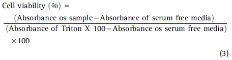
The cytotoxicity of VP-16 NMF was evaluated by [3-(4,5-dimethylthiazol-2-yl)-5-(3-carboxymethoxyphenyl)-2-(4-sulfophenyl)-2H tetrazolium solution (MTS) assay on human breast cancer cells (MCF-7) and human mammary gland cells (normal breast cells, MCF-10A). In brief, cells were added to 100 μL of DMEM culture media containing 10% FBS and seeded in a 96-well plate at a density of 5,000 cells/well. Cells were further incubated and allowed to attach for 24 h at 37°C in 5% CO2 environment. Then, VP-16 NMF and BNF were prepared in serum-free media and filtered with a sterile 0.22-μm nylon filter membrane under laminar flow inside a biosafety cabinet hood. DMEM media was subsequently removed and cells were rinsed thrice with Dulbecco's phosphate-buffered saline (DPBS). The cells were then treated with different concentrations of VP-16, VP-16 NMF, and BNF alone for 24 and 48 h. Triton-X 100 and serum-free media served as a positive and negative control, respectively. The BNF and VP-16 NMF were dissolved in serum-free media, whereas etoposide was dissolved in DMSO since it has poor solubility in aqueous media. After 24 and 48 h of exposure, 10 μL of a 5 mg/mL of MTS (cell titer 96® Aqueous), (Promega, Madison, WI) stock solution was added to each well and according to the manufacturer's protocol. 19,24,30 Cell viability percentage was further determined by measuring the absorbance of each well at 450 nm using a DTX 880 multimode microplate reader (Beckman Coulter, Brea, CA). The number of viable cells is directly proportional to the amount of formazan formed. Cell viability was expressed according to equation (3):
Drug Uptake Study
To evaluate the utility of the NMF assisting in cellular uptake, a drug uptake study was conducted on the breast cancer cell line (MCF7). In brief, MCF7 cells were grown in a 12-well plate until it reached confluence. Each well approximately contained 20,000 cells. The cells were then treated with native etoposide and VP-16 NMF at a concentration of 500 μM and incubated overnight. The next day, the cells were washed with cold DPBS solution twice and the plates were kept at −80°C overnight. Then, 3 mL of 100% cold acetonitrile were added to each well and kept on ice for 5 min, mixed well with pipetting the liquid up and down to detach the cells. The cellular suspension in acetonitrile was then transferred to prechilled 15-mL falcon tubes, vortexed well, and centrifuged at 5,000 rpm for 30 min. The supernatant was collected into a fresh prechilled 15-mL falcon tube. To the remaining cellular pellets was then added 2 mL of cold 100% acetonitrile, and were processed as described before. The supernatant was collected and combined with the first supernatant. The combined supernatant was then freeze-dried using Genevac Quattro centrifugal concentrator (SP Industries, Warminster, PA) and reconstituted in 100 μL water/acetonitrile (65:35, v/v) for LC-MS/MS analysis.
In Vitro Drug Release from Etoposide NMF
In vitro drug release study was conducted to assess the release of etoposide (VP-16) from the nanomicelles in PBS (8 mM Na2HPO4, 150 Mm NaCl, 2 mM KH2PO4, 3 mM KCl, pH 7.4). At predetermined time points, 1,000 μL of the samples was collected and filtered using a dialysis bag with a molecular weight cut-off of 1,000 Da. It is important to note that the next step was to tie both ends of the dialysis bag before immersing them into 5 mL of PBS at 37°C. With notably predetermined time points, the entire buffer was switched with fresh PBS. A calibration curve, presented in equation 1, was performed at the maximum absorption λmax = 250 nm, using UV spectrometry (Thermo Electron Corporation, Waltham, MA) to determine the amount of etoposide released.
where A and C represent the absorbance and drug concentration, respectively. The coefficient of determination describing the correlation between the absorbance value and the drug concentration was R 2 = 0.9996. The release of VP-16 was measured in triplicate, and the data were plotted as mean ± standard deviation. 31
Statistical Analysis
All values are expressed as mean ± SDs from three independent replicates (n = 3). Unpaired t-test and one-way analysis of variance in combination with Tukey test were used to compare samples with unequal variances and identify means of the data that are significantly different from each other. All the statistical analyses were carried out using GraphPad Prism 8 (GraphPad Software, San Diego, CA) where p-value <0.05 is considered statistically significant.
RESULT AND DISCUSSION
Loading and Entrapment Efficiency of Etoposide NMF
It has been reported that the critical micelle concentration (CMC) value of TPGS is 0.02 wt/wt%. 32 As it is shown in Table 1, the %wt concentrations of TPGS (3, 6, 8, and 10%wt) were higher than the CMC value. As the TPGS concentration was increased from 3 to 10% wt, more micelles were formed, which eventually incorporated more VP-16 molecules. This can be confirmed from Table 2, by observing that the encapsulation efficiency of VP-NMF was increased as the TPGS' concentration (micellar formation) was increased. The drug loading and the entrapment efficiency were measured by LC-MS/MS. The VP-16 NMF in 10% wt of TPGS was found to have the highest drug loading and entrapment efficiency. Various polymers such as pluronic F127 and PEG phosphatidylethanolamine have been used in conjunction with TPGS and showed changes in CMC of TPGS containing nanomicelles. 33,34 In this study, it is possible that loading etoposide could change the CMC value of TPGS. In future studies, this could be monitored by techniques such as potentiometry or pyrene fluorescence analysis.
Size Distribution, Polydispersity Index, and Surface Charge Values of VP-16 Nanomicellar Formulation in Different Percentage of D-α-Tocopherol Polyethylene Glycol 1000 Succinate
PDI, polydispersity index; TPGS, D-α-Tocopherol polyethylene glycol 1000 succinate.
Drug Loading and Entrapment Efficiency Values of VP-16 Nanomicellar Formulation in Different Percentage of D-α-Tocopherol Polyethylene Glycol 1000 Succinate
Size, PDI, and Surface Charge
It is well documented that as the concentration of surfactant increases, the micelle aggregation number increases significantly. 35 The higher concentration of surfactant may cause morphological changes and decrease the micellar size as well as the micelle aggregation number. Also, as the surfactant concentration increased, the hydrophobic–hydrophobic interaction between the poorly water-soluble drug and the lipophilic block of the amphiphilic polymer increased, which might significantly reduce the size of the nanomicelles. In this study, D-α-tocopheryl polyethylene glycol succinate (Vitamin E TPGS, or simply TPGS) acts as surfactant. In fact, TPGS has an amphiphilic structure of lipophilic alkyl tail and hydrophilic polar head with a hydrophile/lipophile balance value of 13.2 and a relatively low CMC of 0.02% w/w. 32 The VP-16 NMF was determined to be a self-assembling nanosized (sized in the range of 9–11 nm) colloidal dispersions with a hydrophobic core and hydrophilic shell as shown in Table 1. 36 Furthermore, we observed that increasing the TPGS amount decreased the size of the nanomicelles. The resulting aqueous solution of the etoposide nanomicelles (VP-16 NMF) was homogeneous and clear in appearance. Such smaller size of the VP-16 NMF is noteworthy and can aid in achieving relatively high concentrations of the drug. Besides, the uniform small size of VP-16 NMF potentially can be very suitable for the IV formulation. 37 The uniform smaller size of VP-16 NMF might be attributed to the strong interaction of the lipophilic drug with the hydrophobic portion of the amphiphilic polymer. As the amount of hydrophobic moities of the TPGS increased with an increase in the concentration, the size, PDI, and surface charge were decreased. As it is shown in Figure 2, VP-16 NMF at 10 wt% of TPGS has an approximate size of 9.2 nm.
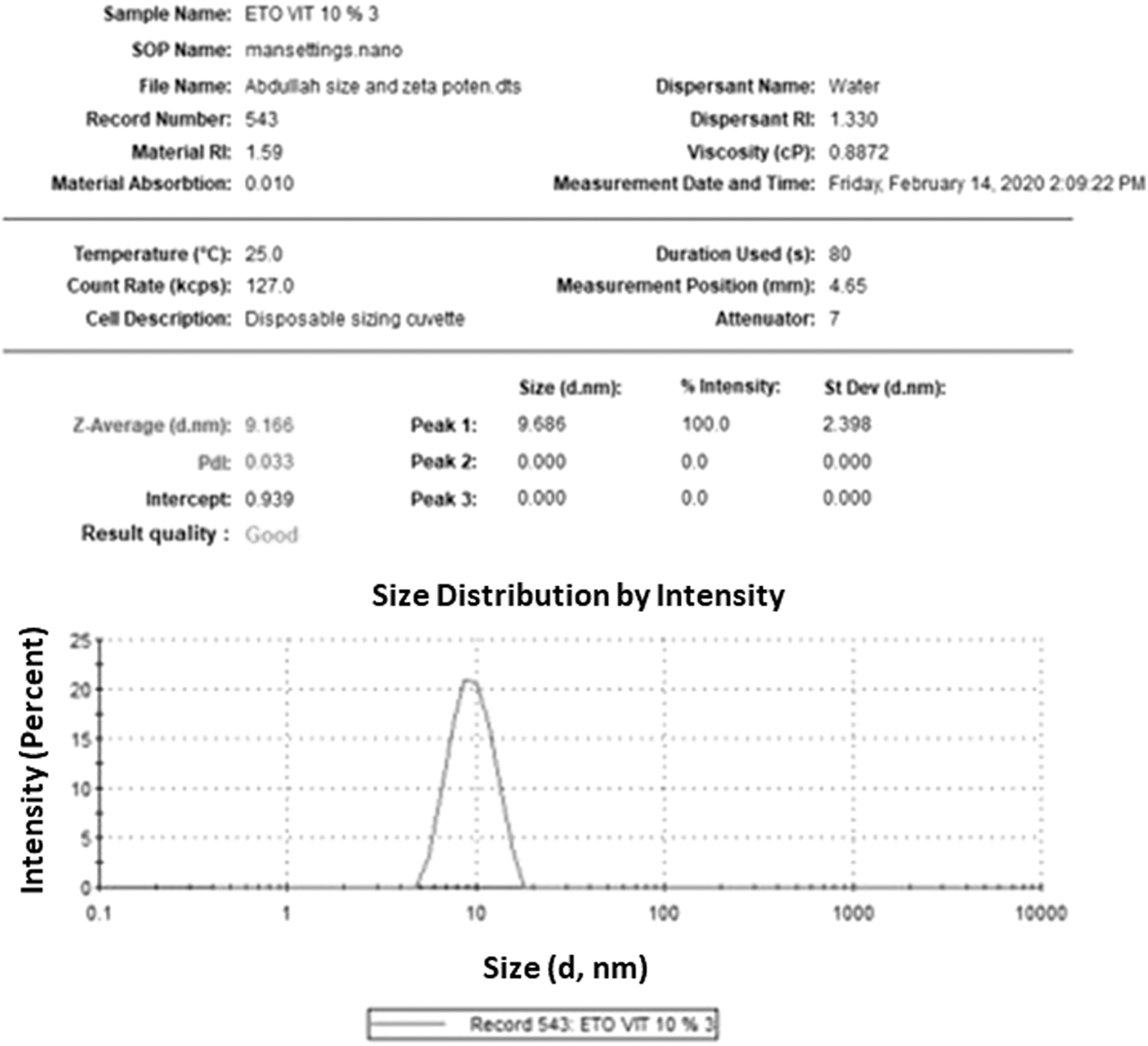
Size distribution for etoposide (VP-16) NMF in 10 wt% of TPGS. TPGS, D-α-Tocopherol polyethylene glycol 1000 succinate.
Average monomer size of TPGS is reasonably estimated to 1,530.78 Da (530.78 Da for D-α-Tocopherol +1,000 for PEG part, according to manufacturer specification). The etoposide's molecular weight is 588.56 Da. The built in calculator in the Zetasizer software produced by Malvern Instrument can be used to estimate particle size from their MW. Particle size for a number of proteins such as lysozyme (∼14–15 kDa), cytochromes (∼40–60 kDa), and bovine serum albumin (∼66.5 kDa) were experimentally measured and validated for other globular proteins. Based on these data, the molecular weight of 10, 20, 40, and 100 kDa corresponds to particle radius of 1.59, 2.14, 2.88, and 4.26 nm, respectively. Therefore, for our small TPGS molecules (of 1,530.78 Da) the micelles mean diameter of about 10 nm (∼5 nm radius) would correspond micelle with MW of 100 kDa, which give us an estimate of the average aggregation number of 100 kDa/1,530.78 or 65 molecules per blank micelle. Considering the drug size of about a 1/4 the size of a blank micelle, this aggregation number would be 1/4 lower per drug loaded micelle. In future studies, this number could be determined by luminescent probes. 38 Based on our spectral analysis of the NMF (Fig. 3), the drug was indeed encapsulated within the micelle and not merely surface bound as it showed no characteristic peak in the formulation.
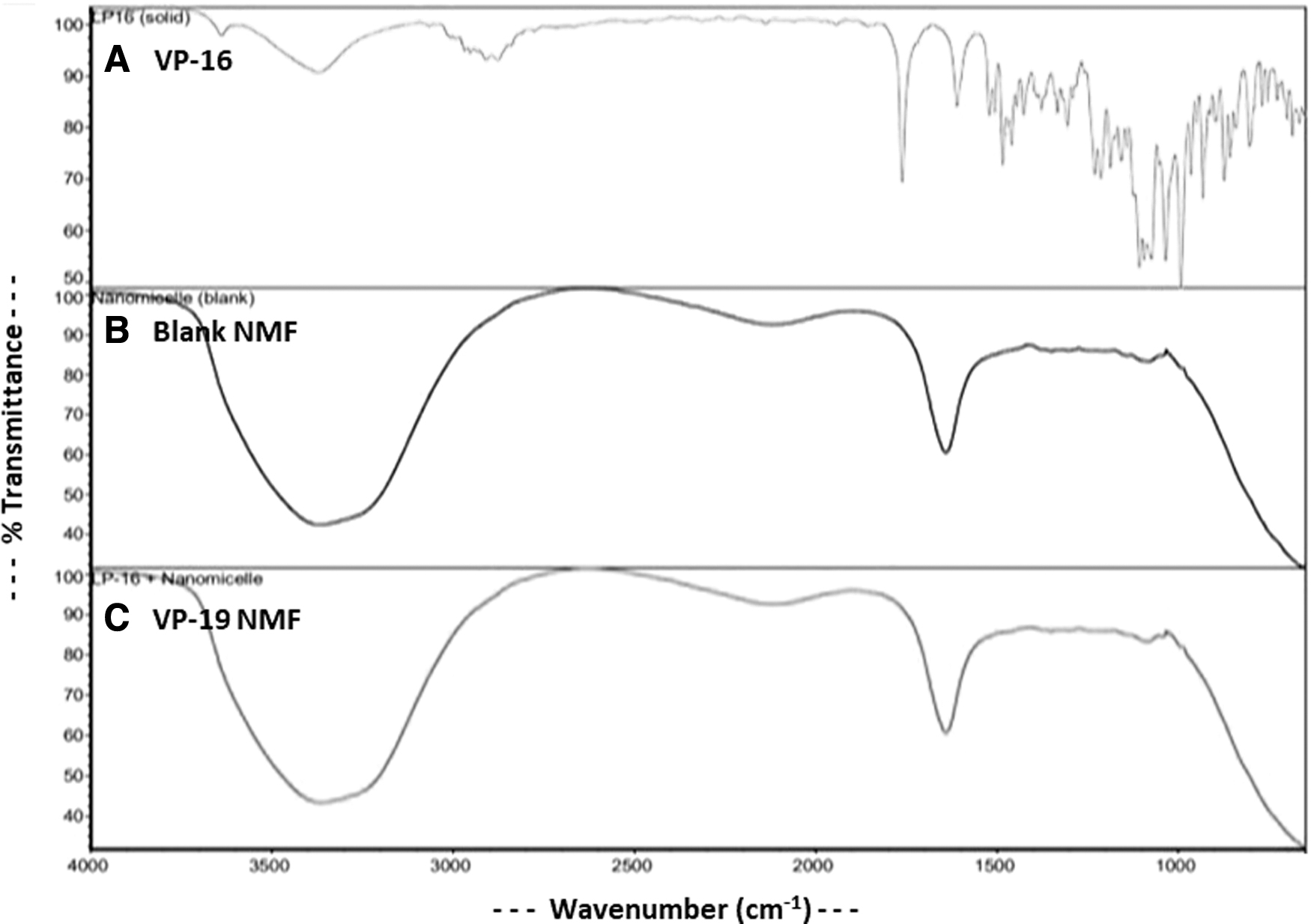
FTIR spectrum of
FTIR Analysis
Fourier Transformed Infrared (FTIR) Spectroscopy analysis was conducted to compare native etoposide (VP-16), BNF, and VP-16 NMF as shown in Figure 3.
In the FTIR spectrum of native VP-16, a distinct peak at 3,446 cm−1 referred to the phenolic–OH group stretching; bands at 1,614, 1,504, and 1,459 cm−1 were attributed to the aromatic C = C stretching; and the band at 1,760 cm−1 indicated the ester group. 39 However, these characteristic peaks of etoposide were not observed in the FTIR spectrum of VP16-NMF. Both BNM and VP-16 NMF have a broader OH peak between 3,500 and 3,200 cm−1, which was attributed to the OH group stretching of PEG. 40 There were no differences in the peaks of BNF and VP-NMF, which means that the VP-16 molecules were effectively incorporated inside the hydrophobic core of the NMF.
1 H-NMR Analysis
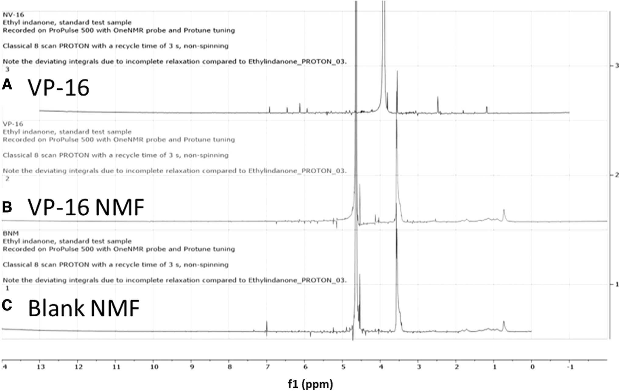
1 HNMR spectra were recorded on a Varian 400 MHz spectrometer (Varian) in deuterated water (D2O) for BNF and VP-16 NMF, and deuterated DMSO (d6-DMSO) for the native VP-16 as shown in Figure 4.
1
H-NMR spectrum of
The NMR data identified the major peaks of etoposide in the native VP-16 sample in d6-DMSO, including CH3 peak was present at 2.240 ppm, CH2 peak is shown at 2.883 ppm, CH peaks at 6, 6.183, 6.524, and 7.004 ppm. These characteristic peaks of VP-16 were absent in VP-16 NMF (Fig. 4B), which proved that all VP-16 molecules were effectively entrapped inside the hydrophobic core of the micellar formulation. 41
XRD and SEM Analysis
XRD analysis was performed to examine the solid state of VP-16 in the NMF. As it is shown in Figure 5, the NMF modified VP-16 solid state from crystalline structure to amorphous one, which can be observed by the presence of the peaks in the native etoposide XRD spectrum and absence of these peaks in the VP-16 NMF XRD spectrum.

XRD graph of
Also, no difference was observed between the peaks of BNF and VP-16 NMF, which further proved that all VP-16 molecules are effectively entrapped in the nanomicellar formulation. 42
SEM was performed to visualize the morphology of VP-16, BNF, and VP-16 NMF, as shown in Figure 6.
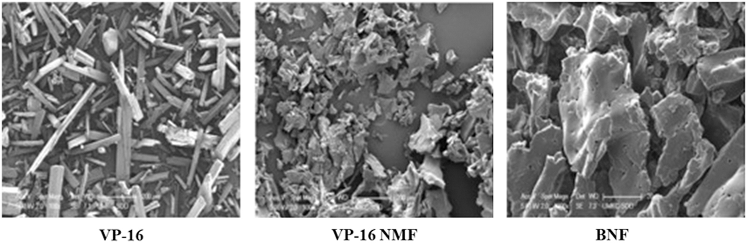
SEM of native VP-16, VP-16 NMF and BNF. Scale bars represent 20 and 500 μm, respectively. SEM, scanning electron microscopy.
The SEM image of native etoposide (VP-16) exhibited a needle-shaped crystalline structure, whereas BNF, as well as VP-16 NMF, revealed irregular shape amorphous structure, which was consistent with XRD finding. 43 Figure 6 also strongly suggested that both blank and drug-loaded nanomicelles exhibited lamellar structures. Lamellar structure of self-assembling systems have been also observed with lipid-peptide nanostructures. 44
Apparent Solubility Study
VP-16 NMF has been prepared in 3, 6, 8, and 10 wt% of TPGS to enhance VP-16 water solubility, which was analyzed by LC-MS/MS. As it is shown in Figure 7, the NMF enhanced VP-16 solubility to 767.8, 843.43, 1,533.61, and 2,148.28 μg/mL in 3, 6, 8 and 10 wt% of TPGS.

Amount of etoposide (VP-16) in different nanomicelles formulations with varying concentrations percentage of TPGS determined by LC-MS/MS. LC-MS/MS, liquid chromatography tandem-mass spectrometry.
The solubility folds enhancement was calculated by dividing the VP-16 solubility in the NMF by the native VP-16 solubility in the DI water at room temperature at the same condition. The NMF enhanced VP-16 solubility by 9.25-, 10.16-, 18.48-, and 25.88-folds in 3, 6, 8, and 10 wt% ratio, respectively.
Cell Cytotoxicity Assay of VP-16 NMF
As shown in Figure 8, the MTS data revealed that at 50 μM native etoposide, the cell viability was 50%–60% on the MCF-7 cells line after 24 h of exposure.
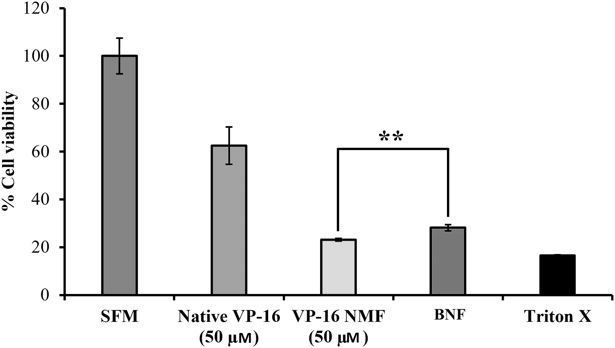
Cell viability assay (MTS) carried out on breast cancer MCF-7 cells after 24-h exposure to native etoposide (VP-16), VP-16 NMF, and BNF. [**p < 0.01 of native VP-16 vs. VP-NMF.] MTS, [3-(4,5-dimethylthiazol-2-yl)-5-(3-carboxymethoxyphenyl)-2-(4-sulfophenyl)-2H tetrazolium solution.
However, when cells were exposed to 50 μM of VP-16 NMF and BNF, the cytotoxicity was higher than the native etoposide. The cytotoxicity observed with BNF was due to the presence of TPGS, which was reported to selectively induce cell cycle arrest or apoptosis in breast cancer cell lines, which was associated with the upregulation of P-21 protein and downregulation of antiapoptotic proteins. 45 The VP-16 NMF has higher cytotoxicity than the BNF due to the additive anticancerous activity effect of both VP-16 and TPGS. After 48 h of cell exposure to VP-16 NMF, the cell cytotoxicity has significantly increased due to the cumulative release of VP-16 from nanomicellar formulation gradually increased with the time as it is shown in Figure 9.
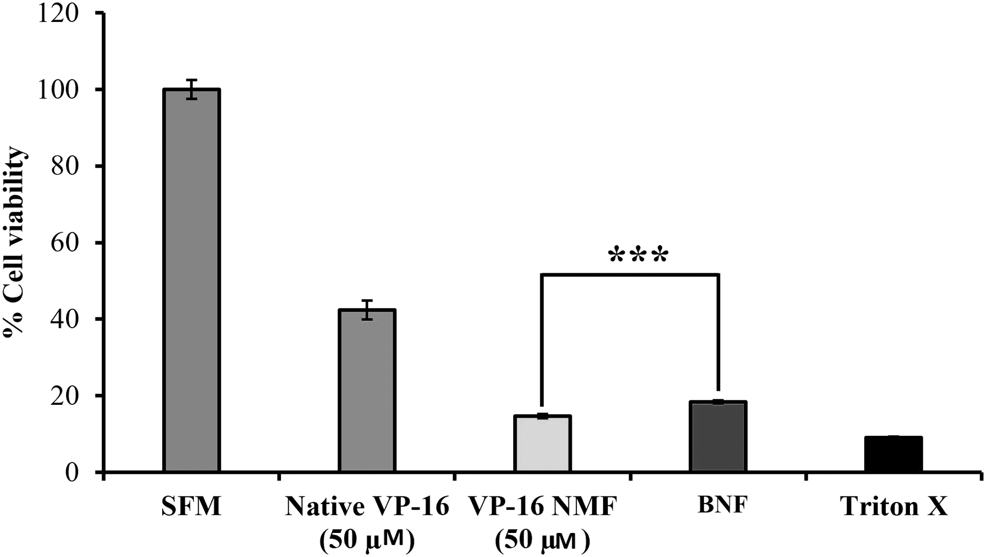
Cell viability assay (MTS) carried out on breast cancer MCF-7 cells after 48-h exposure to native VP-16, VP-16 NMF, and BNF. [***p < 0.001 of native VP-16 vs. VP-NMF.]
VP-16 cytotoxicity on normal breast cell lines was consistent with the previous finding. As it is shown in Figure 10, BNF has relatively lower cell cytotoxicity, where ∼90% of the cells were viable, which indicated that TPGS did not reduce the cell viability of the normal breast cell lines. 45

Cell viability assay (MTS) carried out on normal breast MCF-10A cells after 24 h exposure to native VP-16, VP-16 NMF, and BNF. [****p < 0.0001 of native VP-16 vs. VP-NMF.]
Also, VP-16 NMF exhibited relatively less cytotoxicity or the normal breast cell line compared with native VP-16 because the polymer used to prepare the nanomicelles is cancer cells specific. 46
Intracellular Uptake of VP-16 from VP-16 NMF into MCF7 Cell
It was observed that NMF as a delivery system provided a significant increase in etoposide uptake in MCF-7 cell line. Uptake of etoposide was 1.02 μM (±0.02 μM, SE) and 2.8 μM (±0.01 μM, SE), respectively, from the native drug and VP-16 NMF, respectively, which was about threefold higher as it is shown in Figure 11. It has been reported that the concomitant use of etoposide with TPGS resulted in decreased in vitro etoposide permeability across artificial membrane. 15 However, the VP-16 NMF provides a significant increase in etoposide uptake in MCF-7 cell line, perhaps due to the NMF unique composition and properties.
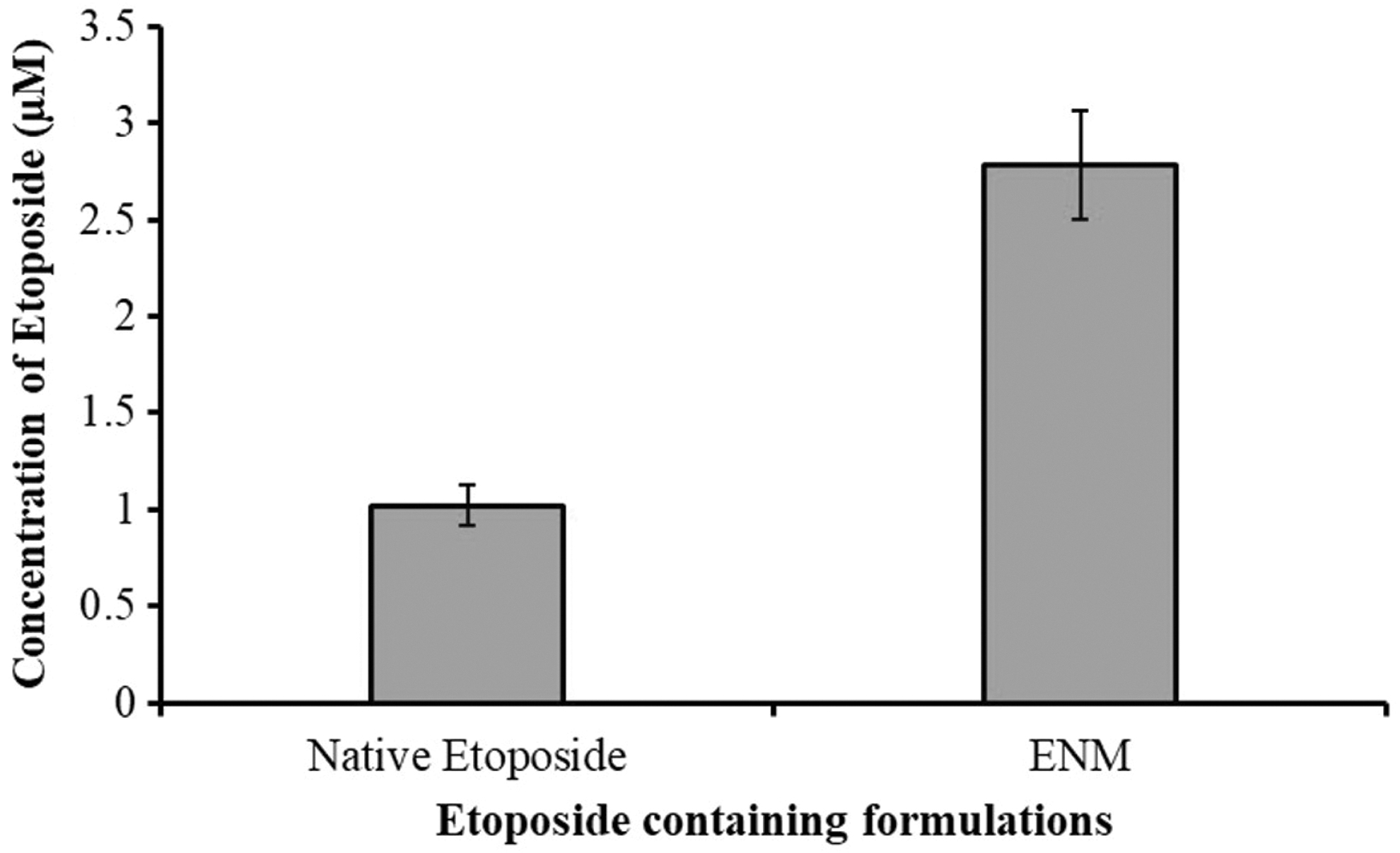
Uptake of VP-16 from native drug solution and VP-16 NMF into MCF-7 cells. Data are expressed in mean ± SD where n = 3. SD, standard deviation.
The results indicated that the uniform smaller size (9 nm) of the NMF facilities the permeation of VP-16 uptakes through multiple routes such as passive diffusion. Also, the major hydrophobic components of the lipid bilayer are cholesterol, diacylglycerol, and ceramide. The hydrophobicity of cell membranes assists in the cellular uptake of many chemical entities, especially small molecules, which can easily diffuse across the plasma membrane into the cells since they are soluble in the hydrophobic region of the phospholipid bilayer. Lipophilicity is one of the most important factors in determining how small molecules are taken up by cells. When small molecules cross a lipid bilayer by simple diffusion, they usually accumulate at high concentration in the hydrophobic regions of the bilayer due to hydrophobic interaction. 47 Collectively, the FTIR, 1 H-NMR, and XRD analysis confirmed that etoposide (VP-16) is incorporated in the hydrophobic core of TPGS resulting in a very small and uniform sized nanomicelles, which could facilitate VP-16 transport and uptake by the hydrophobic interaction between the cell membrane lipid bilayer and the hydrophobic core of the polymer. Lastly, TPGS as a delivery system suppresses P-gp and MDR proteins that are highly overexpressed in breast cancer cell lines, 21 which can result in less cellular efflux and more etoposide to remain inside the cells and explain the higher cellular uptake from the NMF and better anticancer activity observed with the same against MCF-7 cell lines compared with native etoposide.
In Vitro Drug Release Study from Etoposide NMF
In vitro drug release study was conducted in PBS at a physiological pH 7.4, and 37°C under sink conditions. As it is shown in Figure 12, the cumulative % release of VP-16 from NMF was very slow, and it increased with time that is, 53% of the drug was released over 1 week without any burst effect.
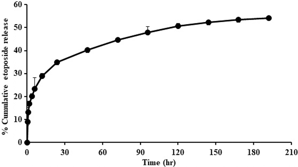
In vitro release for etoposide (VP-16) from NMF in 10 wt% of TPGS/PBS (pH 7.4), at 37°C under sink conditions. Data are expressed in mean ± SD where n = 3.
The observed release profile is characteristic of a sustained release drug delivery system. This, in turn, notably offered evidence that it can indeed help to achieve the therapeutic concentration at the physiological condition in breast cancer cells with less frequent dosing. The incremental release might be attributed to the noted lipophilic nature of the drug. Furthermore, the hydrophobic–hydrophobic interaction between the hydrophobic drug and the hydrophobic portion of the amphiphilic polymer might have contributed to the slow release of etoposide.
Conclusion
In this study, TPGS was selected to prepare VP-16 NMF through solvent evaporation method to develop a polymeric NMF to improve the solubility and anticancer activity of etoposide (VP-16). The following concentrations of TPGS: 3%, 6%, 8%, and 10% were used to solubilize the drug. The 10 wt% TPGS can solubilize 100% of VP-16 with the highest drug loading and entrapment efficiency. The nanomicelles were found to be very small in size (9–11 nm), and the resulting aqueous solution is completely homogeneous and clear in appearance. Collectively, the physicochemical analysis using FTIR, NMR, and XRD shows that the VP-16 molecules were effectively entrapped in the NMF. XRD along with SEM analysis confirms the amorphous status of the NMF. The MTS data show that the VP-16 NMF platform provided higher anticancerous activity than the native VP-16 on MCF-7 cells line as it integrated a dual anticancer activity of VP-16 and TPGS. VP-16 NMF has less cytotoxicity on MCF-10A cell line than native etoposide. LC-MS/MS data revealed that VP-16 NMF has a threefold increase in cell uptake in MCF-7 cells line compared with the native etoposide which is consistent with the higher anticancer activity in MCF-7 cells compared with native etoposide. This study shows the utility of TPGS NMF to increase the solubility and anticancer activity of etoposide. These findings can help in developing drug delivery system to achieve sustained release of etoposide and provide a safe and effective means of delivering etoposide as a chemotherapeutic agent. Furthermore, future in vivo study using the developed system can provide more data of direct clinical relevance.
Footnotes
Acknowledgments
Abdullah Alsalhi is a recipient of The Saudi Arabia Government Scholarship for Graduate Studies in United States of America. The authors would like to thank Dr. Kun Cheng's lab (University of Missouri-Kansas City, School of Pharmacy, Kansas City, MO), Dr. Fariba Behbod's lab (University of Kansas Medical Center and Cancer Center, Kansas City, KS) and Dr. James Murowchick (University of Missouri Kansas City, Department of Geosciences) for supporting with MCF-7, MCF-10 cell lines and XRD analysis, respectively.
Disclosure Statement
No competing financial interests exist.
Funding Information
This study was supported by the University of Missouri-Kansas City Technology Jump and University of Missouri System Fast Track grants.