




Abstract
This study aimed to develop a nanoparticle drug delivery system using poly (lactic-co-glycolic acid) (PLGA) for enhancing the therapeutic efficacy of lurasidone hydrochloride (LH) in treatment of schizophrenia through intramuscular injection. LH-loaded PLGA nanoparticles (LH-PNPs) were prepared using the nanoprecipitation technique and their physicochemical characteristics were assessed. Particle size (PS), zeta potential, morphology, % encapsulation efficiency, % drug loading, drug content, and solid-state properties were analyzed. Stability, in vitro release, and in vivo pharmacokinetic studies were conducted to evaluate the therapeutic efficacy of the developed LH-PNPs. The optimized batch of LH-PNPs exhibited a narrow and uniform PS distribution before and after lyophilization, with sizes of 112.7 ± 1.8 nm and 115.0 ± 1.3 nm, respectively, and a low polydispersity index. The PNPs showed high drug entrapment efficiency, drug loading, and drug content uniformity. Solid-state characterization indicated good stability and compatibility, with a nonamorphous state. The drug release profile demonstrated sustained release behavior. Intramuscular administration of LH-PNPs in rats resulted in a significantly prolonged mean residence time compared with the drug suspension. These findings highlight that intramuscular delivery of the LH-PNP formulation is a promising approach for enhancing the therapeutic efficacy of LH in treatment of schizophrenia.
INTRODUCTION
Schizophrenia is a severe mental illness characterized by disruptions in thinking, emotions, and behavior. 1 Individuals with schizophrenia often experience a disconnection from reality. According to the World Health Organization, ∼21 million people worldwide are affected by this condition. 2 Antipsychotic drugs are commonly prescribed to alleviate symptoms associated with psychiatric disorders such as schizophrenia and bipolar disorder. However, many of these drugs are associated with a wide range of adverse effects. Typical antipsychotic agents, in particular, tend to produce fewer extrapyramidal side effects compared with conventional antipsychotics. 3 –5
Lurasidone hydrochloride (LH) is a novel atypical antipsychotic that has demonstrated efficacy in treating various symptoms of schizophrenia, including cognitive deficits. 6 Currently, LH is available commercially as an oral immediate-release tablet marketed under the trade name Latuda. However, LH is classified as a Bio-pharmaceutical classification system class II drug, which means it has poor solubility and low bioavailability (only 9%–19% absorption after oral administration). 7 To address these limitations, nanotechnology has emerged as a promising approach to enhancing the pharmacokinetic properties of drugs. 8
Various carrier systems, such as nanocrystals, nanosuspensions, polymeric nanoparticles, amorphous nanoparticles, nanosponges, and liposomes, have been utilized to improve the bioavailability of poorly bioavailable drugs. 9 Among these, biodegradable polymeric nanoparticles, particularly those based on polylactic acid (PLA), polyglycolic acid (PGA), and poly (lactic-co-glycolic acid) (PLGA), have gained significant attention as long-acting injectable (LAI) formulations. 10
PLGA, a biocompatible and biodegradable polymer, is commonly used for formulating nanoparticulate carriers in drug delivery. 11,12 It comprises monomers derived from PLA and PGA. PLA has higher lipophilicity than PGA, enabling control over drug release rates at the target site. The biodegradability of PLGA into metabolite monomers, glycolic acid, and lactic acid, along with its tissue compatibility, has made it a favorable choice for nanoparticulate drug delivery systems.
These monomers can be easily metabolized in the body through the tricarboxylic acid cycle. 13 –15 LAI depot systems using PLGA offer advantages such as consistent plasma drug concentrations, improved medication compliance, and the ability to accurately adjust the lowest effective dose, addressing issues related to absorption variability and first-pass metabolism. 16 –19
The objective of this study was to develop and characterize PLGA nanoparticles loaded with LH (LH-PNPs) to improve the bioavailability, reduce the dosing frequency, enhance pharmacokinetics, and increase the therapeutic efficacy of LH. Response surface methodology in conjunction with central composite design (CCD) was employed to optimize the formulation of LH-PNPs, allowing for a systematic approach to design and deliver the active pharmaceutical substance.
Physicochemical characterization of the optimized LH-PNPs involved evaluating % drug entrapment, drug loading, particle size (PS), zeta potential, and shape, using techniques such as ultraviolet–visible (UV-Vis) spectrophotometry, dynamic light scattering (DLS), and transmission electron microscopy (TEM). The biocompatibility analysis was performed using Fourier transform infrared (FTIR) and differential scanning calorimetry (DSC) measurements. Additionally, in vitro release studies and in vivo oral pharmacokinetics in rats were conducted.
The study aimed to develop and characterize LH-PNPs as a means to enhance the therapeutic properties of LH, including its bioavailability, dosing frequency, pharmacokinetics, and overall therapeutic efficacy.
MATERIALS AND METHODS
Materials
A sample of LH was generously provided by Alembic Pharmaceuticals (Vadodara). PLGA 75:25 was received as a gift sample from Corbionpurac (the Netherlands). Tween 80 and methanol were purchased from S.D. Fine Chemicals. All other necessary chemicals were procured through commercial means.
Preparation of LH-PNPs
LH-PNPs were prepared using the nanoprecipitation method, optimizing process parameters (stirring time, speed, and rate of addition) and formulation variables (drug-to-polymer ratio, organic-to-aqueous phase ratio, surfactant type, and concentration). LH and PLGA were dissolved in acetonitrile, while an aqueous phase with 0.2% Tween 80 was used. The organic phase was rapidly injected into the aqueous phase, with stirring at 1,500 rpm for 4 h.
Lyophilization was employed to convert a liquid formulation into a free-flowing powder, enhancing its stability.
Experimental Design
The optimization process was conducted using Design-Expert 7.0.0 software and employed the CCD methodology to obtain nanoparticles with minimal size and higher entrapment efficiency. The concentration of PLGA (A) and concentration of Tween 80 (B) were selected as independent variables and they varied at three discrete levels. The goal was to understand the individual and interactive effects of these variables on the dependent variables, namely entrapment efficiency (Y1) and PS (Y2).
The central point in the design was replicated five times to assess reproducibility and identify potential errors (S3 section in Supplementary Data).
% Encapsulation Efficiency, % Drug Loading, and % Drug Content
The % encapsulation efficiency (%EE) and % drug loading (%DL) of LH-PNPs were determined by separating the unentrapped drug using low-speed centrifugation and quantifying the absorbance of the dissolved pellet and the nanoparticle suspension at 315 nm using a UV spectrophotometer; %EE and %DL were calculated as follows:

For estimating the total drug content, the formulation was appropriately diluted with acetonitrile and its absorbance was measured at 315 nm using a UV spectrophotometer.
Characterization of Optimized LH-PNP Formulations
Dynamic light scattering
The PS, zeta potential, and polydispersity index (PDI) of optimized LH-PNPs were determined at 25°C ± 0.5°C using the Malvern Zetasizer Nano ZS instrument. To minimize multiple scattering effects, the samples were diluted with ultrapure water before measurement. The measurements were conducted in triplicate, employing a scattering angle of 90° relative to the incident beam. 20,21
Transmission electron microscopy
The morphological aspects of optimized LH-PNPs were observed using a transmission electron microscope (JEM-1400, Jeol, Japan) at 200 kV. The optimized LH-PNPs were deposited on a copper grid, any excess was removed by filtration, and negative staining with a 2% w/v phosphotungstic acid solution was performed before drying at room temperature for improved contrast before TEM examination. 22
Fourier transform infrared
FTIR spectra of free LH, PLGA, and optimized LH-PNPs were obtained using an IR spectrophotometer (Shimadzu, Japan) with a resolution of 4 cm−1. The samples were scanned in the wavenumber range of 4,000–500 cm−1 at room temperature, allowing the identification of functional groups and evaluation of drug-PLGA interactions. 23
Differential scanning calorimetry
In this study, DSC analysis was utilized to investigate the physical state of LH in nanoparticles. Pure LH, PLGA, and optimized LH-PNPs were examined, and the analysis was conducted under nitrogen-purged conditions with a temperature range from 20°C to 300°C and heating rate of 10°C per minute. 24
Stability study
Stability studies were conducted on the nanoparticle (NP) suspension and lyophilized NPs under two temperature conditions: room temperature (25°C ± 2°C) and refrigerated conditions (2°C–8°C). The NP suspension was stored at refrigerated condition and room temperature, and the PS distribution and %EE were assessed immediately and at various time points within a 1-month storage period. 25,26
In vitro drug release studies
The in vitro release of LH from PNPs was assessed using the dialysis bag diffusion technique, as previously described by Yang et al. 27 Before the study, the dialysis membrane was activated. Two dialysis sacs were prepared, one filled with a drug suspension and the other with optimized LH-PNPs, both at a concentration of 2 mg/mL, serving as donor compartments.
These sacs were immersed in 30 mL of pH 7.4 phosphate buffer solutions containing 1% sodium lauryl sulfate, acting as the receptor compartments. Stirring was achieved using a magnetic bead, and the beakers were covered to prevent evaporation. To prevent fungal growth, 0.2% sodium azide was added as a preservative. Samples were withdrawn from the receptor compartments at predetermined intervals, and the amount of drug released was assessed by UV-Vis spectrophotometry at a λmax of 319 nm.
In vivo pharmacokinetic study
The study employed male Sprague–Dawley rats weighing between 200 and 300 g. The animal study performed was authorized by the Institutional Animal Ethics Committee (IAEC) with approval number MSU/IAEC/1651.
The study involved 12 female Wistar rats, divided into two groups of six rats each. The first group received an intramuscular dosage of 2.066 mg/kg LH suspension. The second group received the LH-PNP formulation through intramuscular injection. Blood samples were collected from the retro-orbital plexus at specific time intervals after drug administration. The first group was sampled at 0.25, 0.5, 1, 3, 5, and 8 h, while the second group was sampled at 0.5, 1, 3, 7, 10, 14, and 18 days.
The collected blood samples were centrifuged, and plasma was extracted and examined using high-performance liquid chromatography after spiking with a 2-ppm standard solution to address the limit of detection and limit of quantification. 28 Kinetica 5.0 software was utilized to calculate the pharmacokinetic parameters for the LH suspension and LH-PNP formulations.
RESULTS AND DISCUSSION
Experimental Design
Influence on dependent factors
Effect on PS
The following second-order quadratic regression equation of the fitted model was used to minimize PS:
The analysis of the equation indicates that all factors have varying effects on PS. Factor A, representing concentrations of the PLGA, has a positive impact on PS. Increasing the concentration of PLGA relative to the concentration of Tween 80 (Factor B) leads to a larger PS, while decreasing the PLGA concentration results in a smaller PS.
On the other hand, Factor B has a negligible influence on PS compared with Factor A. In this study, increasing the PLGA concentration may lead to a larger PS due to higher viscosity and lower solubility, while Tween 80 may have a negligible effect due to its surfactant properties. The significance of the factors was determined through analysis of variance (ANOVA). The obtained model F-value of 5.00 indicates the significance of the model with a probability of 2.61%, suggesting that the model is not likely a result of random noise.
The Prob > F values <0.0500 suggest the significance of Factor A as a model term. However, values >0.1000 indicate the potential insignificance of certain model terms, which could be improved through model reduction. The lack of fit F-value of 1.74 suggests that the lack of fit is not statistically significant when compared with the pure error, indicating a satisfactory fit.
However, the difference between Pred R-Squared (0.0627) and Adj R-Squared (0.4999) raises concerns about a possible block effect or issues with the model and data. To address these concerns, it is advisable to explore options such as model reduction, response transformation, and outlier analysis.
Effect on entrapment efficiency
The following second-order quadratic regression equation of the fitted model was used to maximize %EE:
The analysis of the equation indicates that all factors have an influence on the %EE to some extent. Factor A has a positive effect on %EE, while Factor B has a negligible impact compared with Factor A. The ANOVA results provide further insights into the significance of the factors. The significant model F-value of 17.40 indicates that the model is statistically significant, with only a 0.08% chance of such a large F-value occurring due to noise. Prob > F values <0.0500 confirm the significance of model terms.
The lack of fit F-value of 4.09 suggests that the lack of fit is not significant relative to pure error, indicating a good model fit. However, the difference between Pred R-Squared (0.5719) and Adj R-Squared (0.8723) raises concerns about possible block effects or issues with the model and data. The Adeq Precision measure indicates that the model has an adequate signal-to-noise ratio, with a value of 14.661, suggesting effective navigation of the design space.
Figure 1 presents three-dimensional response graphs illustrating the effects of factors on responses, while the S4 section in Supplementary Data shows an overlay plot indicating the combined effect of all independent factors on the responses. The ANOVA, observed and predicted values, and regression variances for all responses are summarized in Table 1.
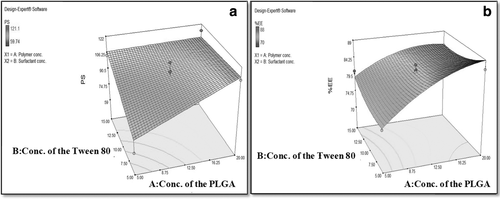
3D response surface graph for the effect of the factor on
Factors and Responses Selected for the Experimental Design and Model Summary Statistics for the LH-Loaded PLGA Nanoparticle Formulation and Their Predicted and Observed Values
%EE, % encapsulation efficiency; PLGA, poly (lactic-co-glycolic acid); PS, particle size.
%EE, %DL, and % Drug Content
The %EE of optimized LH-PNPs was 84.3 ± 1.2%. This indicates that a significant proportion of LH was successfully encapsulated within the NPs. LH loading in the optimized batch of NPs was determined to be 15.45%, indicating the amount of LH present in relation to the total weight of the NPs. Moreover, the LH content in the optimized batch of NPs was measured at 99.6 ± 1.5%, confirming the high concentration of LH within the NPs.
Characterization of Optimized LH-PNP Formulation
Size distribution, PDI, and zeta potential
PS analysis of the optimized LH-PNP formulations was performed using DLS, which provided information on the apparent particle hydrodynamic diameter (Dh) and PDI. As depicted in Figure 2a and b, the PS distribution and zeta potential graph of the optimized LH-PNP formulations were obtained. The Dh of the optimized LH-PNP formulation was determined to be 102.7 ± 4.5 nm, indicating a relatively uniform PS.
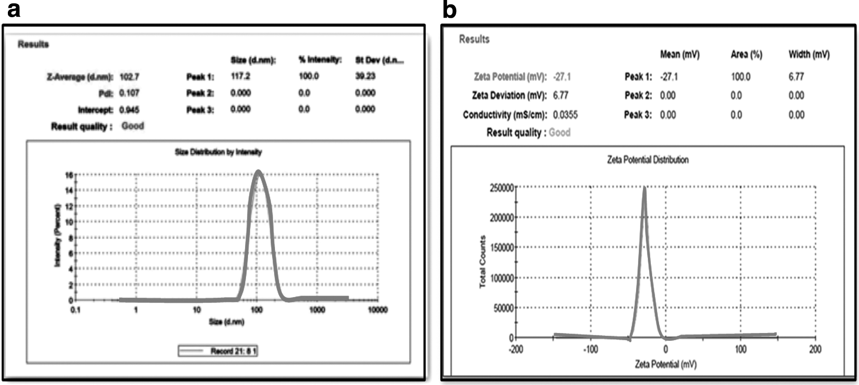
Moreover, the PDI value of 0.107 ± 0.07 indicated low polydispersity and high uniformity within the particle population. 29 Evaluation of the zeta potential, a crucial parameter for assessing nanoparticle suspension stability, revealed that the LH-PNP formulations exhibited a zeta potential value of −24.2 ± 3.0 mV. The negative charge on the nanoparticle surface was attributed to the presence of carboxylate end groups of PLGA. 30 This negative zeta potential value suggested good stability of the nanoparticles in suspension as it reduced the likelihood of particle aggregation. 31
Transmission electron microscopy
Figure 3 exhibits the submicron size and morphology of optimized LH-PNPs. TEM images provided insights into the NP characteristics, revealing a smooth surface and size distribution below 200 nm. 32 The NPs displayed a spherical shape, indicating a well-defined and uniform structure. 33

Transmission electron microscopy image of optimized LH-PNP formulation at 30°C ± 0.5°C.
Fourier transform infrared
FTIR spectra of LH, PLGA, and optimized LH-PNPs are displayed in Figure 4. The FTIR spectrum analysis of optimized LH-PNPs revealed the absence of peaks corresponding to LH, suggesting the successful entrapment of the drug within the polymer matrix. However, distinct peaks attributed to PLGA were observed, specifically at wavenumbers of 2,949.64 cm−1, representing the C-H stretching; 1,764.56 cm−1, indicating the characteristic stretching of the carbonyl group; and 1,096.58 cm−1, corresponding to the C-O stretching.

These findings indicate that the outer layer of NPs predominantly consists of the PLGA polymer. 34
Differential scanning calorimetry
DSC analysis provided valuable insights into the thermal behavior and drug state within the NPs. The DSC thermogram of pure (LH) displayed a distinct melting peak (Tm) at 272.25°C, indicating its crystalline nature. The DSC thermogram of the physical mixture revealed a glass transition temperature (Tg) of ∼45°C, consistent with the reported Tg of the empty PLGA 75:25 in the literature (between 40°C and 60°C). The endothermic melting peak of the drug was preserved at 276.82°C.
In contrast, the DSC curve of optimized LH-PNPs showed the absence of the native LH melting peak, suggesting conversion of the drug into an amorphous state within NPs. 35 Notably, a sharp peak at 164.42°C was observed, corresponding to the Tm of mannitol, which served as a cryoprotectant during the lyophilization process of NPs (Fig. 4).
Stability study
The short-term stability studies presented in Table 2 show that the PS of LH-PNP suspension increased under both room temperature and refrigerated conditions. However, the increase in PS was relatively lower for lyophilized LH-PNPs compared with the suspension. Additionally, the %EE stability assessment indicated a faster and higher reduction at room temperature compared with refrigerated conditions.
Storage Stability Study of Suspension and Lyophilized LH-Loaded PLGA Nanoparticles
LH, lurasidone hydrochloride; LH-PNPs, LH-loaded PLGA nanoparticles.
Lyophilized LH-PNPs demonstrated greater stability when stored under refrigerated conditions. Therefore, lyophilization of the optimized LH-PNP suspension and refrigerated storage are recommended to minimize PS increase and %EE reduction. 35,36
In vitro drug release
The in vitro release study of LH and LH-PNPs was conducted at pH 7.4, representing physiological conditions. The release profiles revealed interesting findings for the LH-PNP formulation. Figure 5a shows that LH was rapidly released within 8 h, indicating easy diffusion through the dialysis membrane. In contrast, Figure 5b displays a biphasic release pattern for LH-PNPs, with an initial burst release, followed by sustained release over 27 days.
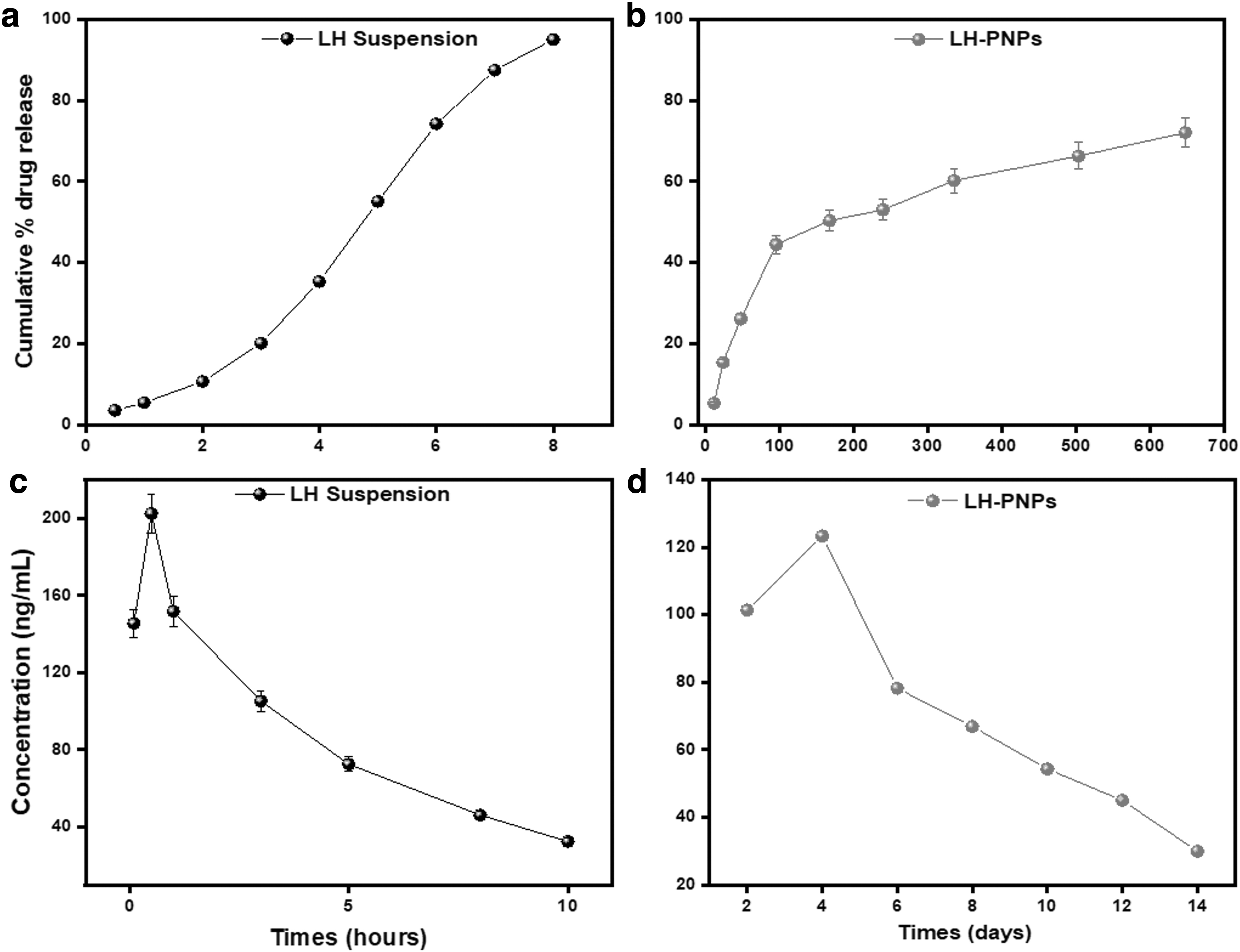
In vitro release profile of
The initial burst release is attributed to surface-adsorbed drug molecules, while sustained release indicates gradual release from the polymer matrix. 37,38 Notably, LH-PNPs exhibited prolonged release compared with the LH suspension, indicating controlled release through drug diffusion in the polymer matrix. Various release models were applied, with zero-order release for LH suspension and the Korsmeyer–Peppas model best fitting the LH-PNP release data (Table 3).
Regression Coefficients of Different Drug Release Models (pH 7.4) at 37°C ± 0.5°C and Mean Pharmacokinetic Parameters of LH Suspension Formulation and LH-Loaded PLGA Nanoparticle Formulation
AUC, area under the curve; Cmax, maximum concentration attained; MRT, mean residence time; Tmax, time at which maximum concentration is attained.
In vivo pharmacokinetic study
Figure 5a and b shows an in vivo plasma study of the LH and LH-PNPs. Based on the pharmacokinetic study of a single-dose administration of LH-PNPs through the intramuscular (I.M.) route, the plasma concentration of LH exhibited a peak concentration (Cmax) of 123.2 ng/mL at 1 day, followed by a gradual decline. Compared with administration of the drug suspension through I.M. injection, LH-PNPs demonstrated a prolonged mean residence time (MRT) of 13.85 days, indicating sustained-release properties.
The time to reach peak concentration (Tmax) also increased significantly from 0.5 h to 1 day for the formulated NPs compared with the drug suspension. Furthermore, parameters such as area under the curve (AUC0→∞), half-life (t1/2), and MRT showed an increase from a few hours to several days (Table 3), confirming the sustained-release characteristics of LH-PNPs.
CONCLUSIONS
The LH-PNP formulation was prepared and thoroughly characterized using a multitechnique approach. To optimize the formulation, a CCD was employed, and the nanoprecipitation method was utilized for synthesis. Based on the highest desirability value, the optimal formula was selected and evaluated. The optimized LH-PNP formulation exhibited a PS of 102.7 ± 4.5 nm, PDI of 0.107 ± 0.07, and zeta potential of −24.2 ± 3.0 mV, displaying a spherical shape.
The results demonstrated that the LH-PNP formulation achieved enhanced %EE, %DL, and % drug content, along with improved compatibility and stability over a period of 1 month. These characteristics make it suitable for pharmacological formulations and result in improved in vitro/ex vivo release profiles in simulated fluids. The in vitro release studies exhibited a biphasic profile with an initial burst release, followed by sustained release over 27 days.
The in vivo pharmacokinetic study confirmed the prolonged drug release and increased Tmax, highlighting the potential of LH-PNPs as an effective drug delivery system for psychiatric disorders.
Footnotes
AUTHORs' CONTRIBUTIONS
N.M. was involved in investigation, methodology, data analysis, and writing—original draft. H.S.P. supported the experimental work, software, and artwork. R.K.S. was involved in review and editing. N.J. was involved in artwork and review and editing. H.T. was involved in conceptualization, supervision, project administration, and writing—original draft, review, and editing. All authors read and approved the final manuscript.
ETHICS APPROVAL
The animal study protocol was approved by the Institutional Animal Ethics Committee (The Maharaja Sayajirao University of Baroda, Gujarat, India), No. MSU/IAEC/1651).
CONSENT FOR PUBLICATION
The authors declare that they provided consent for publication.
AVAILABILITY OF DATA AND MATERIALS
The authors confirm that all data underlying the findings are fully available without restriction. All relevant data are within the article.
DISCLOSURE STATEMENT
The authors declare that they have no known competing financial interests.
FUNDING INFORMATION
This research did not receive any specific grant from funding agencies in the public, commercial, or not-for-profit sectors.
SUPPLEMENTARY MATERIAL
Supplementary Data