




Abstract
Androgen therapy has been shown to alleviate type 2 diabetes mellitus (T2DM) but is also associated with severe side effects such as prostate cancer. The present study aims to identify the best hit selective androgen receptor (AR) modulator by in silico studies and then investigates its antidiabetic effects in high-fat diet– and streptozotocin (STZ)-induced T2DM male rat model. Molecular docking and molecular dynamics (MD) studies were carried out using Maestro 13.1 and Desmond (2023–2024). Cytotoxicity and insulin secretion were measured in MIN6 cell lines. T2DM was induced using high-fat diet (HFD) for 4 weeks, followed by single STZ (40 mg/kg, intraperitoneally). OneTouch Ultra glucometer was used to measure fasting blood glucose. Gene expression was determined using reverse transcription polymerase chain reaction. Histopathology was carried out using hematoxylin and eosin stain. Through molecular docking, we identify ligandrol as a potential hit. Ligandrol showed a good binding affinity (−10.74 kcal/mol). MD showed that ligandrol is stable during the 100 ns simulation. Ligandrol increases insulin secretion in a dose-dependent manner in vitro in 2 h. Ligandrol (0.3 and 1 mg/kg, orally) significantly decreased the body weight and fasting blood glucose levels compared with the HFD and STZ group. Gene expression showed that ligandrol significantly increased the AR-targeted gene, neurogenic differentiation 1, compared with the HFD and STZ group. Histopathological staining studies showed that ligandrol prevents pancreatic islet degeneration compared with the HFD and STZ group. Our findings suggest that ligandrol’s protective effect on pancreatic islets leading to its antidiabetic effect occurs through the activation of AR.
INTRODUCTION
Diabetes mellitus, also commonly referred to as diabetes, is a long-term metabolic disease marked by increased blood glucose levels (hyperglycemia) driven by impairments in the action or secretion of insulin, or both. 1 Among different types of diabetes, type 2 diabetes mellitus (T2DM), often considered an epidemic, has been a major threat to world health. 2 The International Diabetes Federation (IDF) estimates that 463 million persons (20–79 years old) worldwide have diabetes in 2019. Remarkably, 90%–95% of the 463 million cases of diabetes are estimated to be T2DM. 3 T2DM has a substantial economic impact because of associated healthcare expenses and diminished productivity. According to the IDF, diabetes-related healthcare costs worldwide are expected to reach over USD 760 billion in 2019. T2DM is a result of a complex interaction between genetic (insulin resistance, β-cell dysfunction), environmental, and lifestyle factors (visceral obesity). 4 The treatment’s main aim is to control blood sugar levels, minimize complications, and enhance overall health. To achieve optimal blood sugar control, a combination of medicines may be prescribed in a lot of cases. Lifestyle changes, such as a healthy diet and regular physical activity, are also important components of controlling T2DM. 5 However, despite the successful rate of antidiabetic drugs, side effects such as gastrointestinal issues, hypoglycemia, weight gain, genital yeast infections, and urinary tract infections were also reported. 6
According to specific research, men who have low testosterone levels may be prone to develop T2DM. Reduced testosterone levels can result in insulin resistance as testosterone influences insulin sensitivity. According to research, testosterone levels and the risk of developing T2DM in men have a complicated relationship. Testosterone is a male sex hormone that contributes to numerous biological processes, including metabolism and glucose regulation. 7 In men with low testosterone levels, testosterone replacement therapy (TRT) has been investigated as a potential intervention to improve insulin sensitivity and glycemic control. However, TRT is still being studied for its long-term safety and efficacy in the management of T2DM. 8 TRT can be beneficial for men suffering from hypogonadism, or low testosterone levels. TRT, like any medical intervention, has a likelihood for undesirable effects. It is crucial to take into account that the severity and likelihood of adverse effects might differ between individuals. Cardiovascular events, erythrocytosis, gynecomastia, testicular atrophy, and prostate cancer are some of the adverse effects associated with TRT. 9 Selective androgen receptor modulators (SARMs) are a class of compounds that selectively interact with the androgen receptor (AR). Androgens are hormones that activate AR and play an important role in the development and maintenance of male characteristics. SARMs, in contrast to traditional anabolic steroids, are designed to selectively target ARs in specific tissues, such as muscle and bone, while minimizing the risk of negative effects on other organs. 10,11 SARMs are generally designed to treat muscle atrophy and osteoporosis. SARMs have minimal to neutral effects on prostate organs, thereby avoiding severe side effects such as benign prostate hyperplasia and prostate cancer. Furthermore, SARMs have minimal effects on cardiovascular risk. 10,12
In this study, we hypothesize that SARMs, which selectively target ARs, may prevent pancreatic damage and improve fasting blood glucose (FBG) levels in T2DM rat model. Virtual screening using extra precision mode docking was performed to identify the best SARM, which was then evaluated for efficacy in an in vivo T2DM rat model.
MATERIALS AND METHODS
Molecular Docking
Molecular docking is a computerized structure–based method used in drug discovery for predicting the binding affinity and poses, and ligand–receptor interactions. The docking studies were performed using the Glide module of Maestro 13.1 (Schrodinger LLC). SARMs such as Ligandrol, LGD-3033, testolone, stanolone, ostarine, and stenabolic were downloaded from PubChem database. These ligands were prepared using LigPrep (Schrodinger) using default settings. A target pH of 7.2 ± 0.2 was set and EpiK was used to generate the ionization states of the ligands. The co-crystal ligands within the proteins were used as standard for the comparison of binding score. The X-ray crystallographic structures of AR-bound partial agonist (PDB ID: 3v49) were imported from the protein data bank. The protein crystal structure was prepared using Schrodinger’s protein preparation wizard using default settings. Missing side chains and loops were added using a feature in protein preparation wizard called Prime. Hydrogen bonds were assigned and optimized using ProtAssign. Finally, restrained minimization of the proteins was done using optimized potentials for liquid simulation 4 (OPLS4). The grid was generated around the co-crystallized ligand using the receptor grid generation module of Maestro 13.1. The grid is used to confine the ligand within the workspace of the co-crystallized ligand. The size of the grid was kept at 20 Å. 13
Molecular Dynamics
The receptor–ligand complex was subjected to molecular dynamics (MD) simulation to check the stability of the binding. Desmond was used to carry out the MD simulation using default settings with minor modifications. The ligand–receptor complex was immersed in a 10 Å orthorhombic TIP4P water box. The system was neutralized and 0.15M sodium chloride was added to mimic the physiological condition. The simulation was run in a constant temperature and pressure environment. The default temperature and pressure, i.e., 300 K and 1.01325 Pa (atmospheric pressure), were used for the 100 ns simulation. The output is evaluated for trajectory, root mean square deviation (RMSD) of ligand with respect to the receptor, root mean square fluctuation (RMSF) of the protein and stability of the ligand–receptor interactions during the simulation period. 14
Cell Culture and Cytotoxicity Assay
The pancreatic β-cell line, mouse insulinoma 6 (MIN6), was purchased from the National Center for Cell Sciences, Pune. The cells were maintained in Dulbecco’s Modified Eagles Medium (DMEM), Sigma Aldrich, United States. The cell line was cultured in a T25 tissue culture flask with DMEM supplemented with 10% fetal bovine serum (FBS), L-glutamine, sodium bicarbonate (Merck, Germany), and an antibiotic solution containing penicillin (100 U/mL), streptomycin (100 µg/mL), and amphotericin B (2.5 µg/mL). Cultured cell lines were kept at 37°C in a humidified 5% CO2 incubator (NBS Eppendorf, Germany). The viability of cells was evaluated by direct observation of cells by an inverted phase-contrast microscope and followed by the cytotoxicity assay. For the cytotoxicity assay, six different concentrations (100, 50, 25, 12.5, 6.25, and 3.125 µM) were used. Approximately 5 × 10³ cells/well were seeded in the 96-well plate. After 48 h of the incubation period, the sample content in the wells was removed and 10 µL of (3-[4,5-dimethylthiazol-2-yl]−2,5 diphenyl tetrazolium bromide) solution (5 mg/mL) was added along with 90 µL of phenol red negative culture medium. The plate was gently shaken and then incubated for 4 h. After the incubation period, the supernatant was removed and 100 µL of dimethyl sulfoxide (DMSO) was added to solubilize the formazan crystals. The absorbance was measured by using a microplate reader at a wavelength of 570 nm.
Insulin Secretion
MIN6 cells were seeded at a density of 20,000 cells/well. The cells were first treated with various concentrations (6.25, 3.125, 1.56, 0.78, 0.39, and 0.19 µM) ligandrol for 24 h. This was followed by addition of streptozotocin (STZ; 5 mM) for 30 min and then glucose (20 mM) addition for 1 hour. Finally, insulin secretion was measured using an enzyme-linked immunosorbent assay as described previously. 15
Animal Acquisition and Care
Male albino Wistar rats weighing 200–230 g were acquired from the animal house facility of Jagadguru Sri Shivarathreeshwara College of Pharmacy (JSSCP), Ooty, Tamil Nadu. Animals were housed in polypropylene cages in groups of three rats per cage and were kept in a room maintained at 25°C ± 2°C with a 12 h light/dark cycle. Animals were acclimatized to laboratory conditions for 1 week before the commencement of experimental studies. The standard housing conditions were maintained for the entire period of research work. The experimental procedure for this research work was approved by the Institutional Animal Ethics Committee (IAEC) (approval number — JSSCP/OT/IAEC/17/2021–22). The research work was carried out according to regulations set by the Committee for Control and Supervision of Experiments on Animals.
Selection of Dose, Duration, and Route of Administration
For ligandrol, two doses of 0.3 and 1 mg were selected based on previous findings. 16 These doses showed no adverse or unwanted side effects. These doses were administered orally once daily for a period of 3 weeks. STZ of 40 mg/kg was administered intraperitoneally (i.p.). 17 This dose was selected based on a pilot study that showed that 40 mg/kg increases the glucose levels (>200 mg/dL) when compared with 35 and 45 mg/kg. 18
Induction of T2DM and Treatment Period
A total of 24 animals were used in the study. The animals were divided into four groups of six animals each. The grouping of animals was done as follows: group 1, normal control (received 1% DMSO); group 2, high-fat diet (HFD) + STZ (received 1% DMSO); group 3, HFD + STZ + ligandrol (0.3 mg/kg/day, oral); group 4, HFD + STZ + ligandrol (1 mg/kg/day, oral). Initially, except normal control, the animals were fed with HFD for 4 weeks. HFD was prepared by crushing the normal pellet diet and mixing it with fat/lard (20%), butter ghee (10%), coconut oil (10%) and casein (10%). 18 The mixture was made into small round balls and kept in a tray under shade to solidify. The dried round balls of HFD was stored in a refrigerator and supplied to rats for 4 weeks prior to STZ injection. 18 The animals were then fasted for 12 h before a single dose of STZ (40 mg/kg, i.p.) is administered. STZ was dissolved in 0.1 M citrate buffer (pH 4.5). 17 STZ was used within 5 min of dissolution because it undergoes degradation after 15–20 min of preparation. In addition, STZ was covered with aluminum foil because it is sensitive to light. 19 This was followed by 20% glucose solution administration to avoid hypoglycemic-induced mortality. FBG level was measured after 3 days of STZ injection and animals were fasted for 12 h before this procedure is carried out. The rats with FBG level above 200 mg/dL were classified as diabetic and used in the study. 17,18 Subsequently, 3 days after the confirmation of induction of diabetes, ligandrol (0.3 and 1 mg/kg/day) treatment was initiated and carried out for a period of 3 weeks (Fig. 1). 16 Ligandrol was dissolved in 1% DMSO and administered orally. 20

Study design.
Body Weight and Fasting Blood Glucose Assessment
The animals’ weight was measured on day 1, post-HFD or preligandrol treatment (day 29), and post ligandrol treatment (day 55). The FBG assessment was carried out following STZ induction at the end of treatment period. The animals were fasted for 12 h before FBG level is measured by OneTouch Ultra glucometer. 21
Pancreatic Islets Isolation and Gene Expression Studies by RT-PCR
Following the treatment period, the animals were euthanized by overdosing with ketamine (300 mg/kg, i.p.) and xylazine (30 mg/kg, i.p.). 22 The pancreas was extracted aseptically and rinsed with phosphate buffer saline (PBS) (Invitrogen) supplemented with 2% v/v antimycotic (Invitrogen). The pancreas was next enzymatically digested using 5 mL of 0.3% w/v cold collagenase V (Sigma-Aldrich), 2% v/v antimycotic and 10% v/v FBS (Invitrogen). This step was performed in a 50 mL conical plastic tube for 15 min at 37°C in a water bath shaker. The digestion was completed by adding 20 mL of PBS and centrifuged at 1,000 rpm for 10 min. After digestion, the islets were cultured in RPMI-1640 (Invitrogen) supplemented with 10% v/v FBS and 1% v/v antimycotic on a six-well plate at pH 7.2. To allow for recovery following the isolation method, the cells were incubated at 37°C in a 5% CO2 incubator overnight. 23 Total RNA was extracted using TRIzol according to the manufacturer’s instructions. The purity and concentration of total RNA were measured with NanoDrop. The cDNA production kit (G-Biosciences) was used to generate template complementary DNA. The cycling settings used were 20 min at 42°C for cDNA synthesis and 5 min at 85°C for inactivation using an Eppendorf Master Cycler. Reverse transcription polymerase chain reaction (RT-PCR) analysis was performed using PCR Master Mix (Merck, India) and an Eppendorf Master Cycler. The primer were designed as described previously 24 (Table 1).
The Gene Primers
Histopathological Analysis
At the end of the studies, the animals (n = 3) were sacrificed by overdosing with ketamine (300 mg/kg, i.p.) and xylazine (30 mg/kg, i.p.). 22 The pancreatic tissues were immediately stored in 10% buffered formalin. Paraffin sections of the pancreas were prepared and stained with hematoxylin and eosin (H&E) for the assessment of histopathological changes. 21
Statistical Analysis
The data analysis was carried out using GraphPad Prism (8.0 trial version). Body weight and FBG levels were measured using two-way analysis of variance, followed by Tukey’s test for multiple comparison. Data were expressed as mean ± standard error of the mean. p-Value < 0.05 was considered significant.
RESULTS
Molecular Docking
The SARMs were obtained from PubChem database and docked within the binding pocket of AR (PDB ID: 3V49). Molecular docking studies reveal that among SARMs, the co-crystal ligand showed the best binding score (−11.13 kcal/mol), which was followed by ligandrol (−10.75 kcal/mol) (Table 2). Co-crystal forms hydrogen bond with HIS874, while ligandrol forms hydrogen bond with THR877. Other SARMs did not form hydrogen bond with AR during our docking analysis (Fig. 2). Based on docking and receptor–ligand 2D interaction, we selected ligandrol for MD simulation.
Molecular Docking Score of SARMs

2D interactions of SARMs with the AR.
Molecular Dynamics
A 100 ns MD simulation was carried out in the explicit solvent for apoprotein and protein–ligand complex. The structures were evaluated for RMSD, RMSF, protein–ligand interactions fraction, and ligand–protein contacts. Our result indicates that the apoprotein converged to an acceptable deviation (1–3 Å) at the end of 100 ns (Fig. 3A). Similarly, the RMSF of apoprotein showed no significant fluctuation during the 100 ns simulation (Fig. 3C). The RMSD of protein–ligand complex is also stable during the 100 ns simulation and showed no significant deviation (Fig. 2B). The binding of ligandrol to AR maintains the RMSF of the protein as that observed in the apoprotein alone (Fig. 2D). Remarkably, instead of maintaining contact with THR877, ligandrol made a significant contact with ASN705 of AR at the end of 100 ns simulation (Fig. 3E and 3F).

RMSD, RMSF, and protein-ligand contact at the end of 100 ns simulation.
Cytotoxic and Insulin Secretion Effects of Ligandrol on MIN6 Cells
Ligandrol at 3.12 and 6.25 µM did not exhibit significant (p > 0.999) cytotoxicity compared with control (Fig. 4A). Based on cytotoxicity results, 6.25 µM, which is the highest nontoxic concentration, was chosen as the highest concentration for the insulin secretion assay. Cells treated with STZ and high glucose (HG) significantly decrease (p < 0.001) the insulin secretion compared with control. Treatment with ligandrol increases the insulin secretion in a concentration-dependent manner starting from 1.56 to 6.25 μM. Treatment with ligandrol significantly increases (1.56 μM: p = 0.003; 3.12 μM: p < 0.001; 6.25 μM: p < 0.001) the insulin secretion compared with HG- and STZ-treated group alone (Fig. 4B).
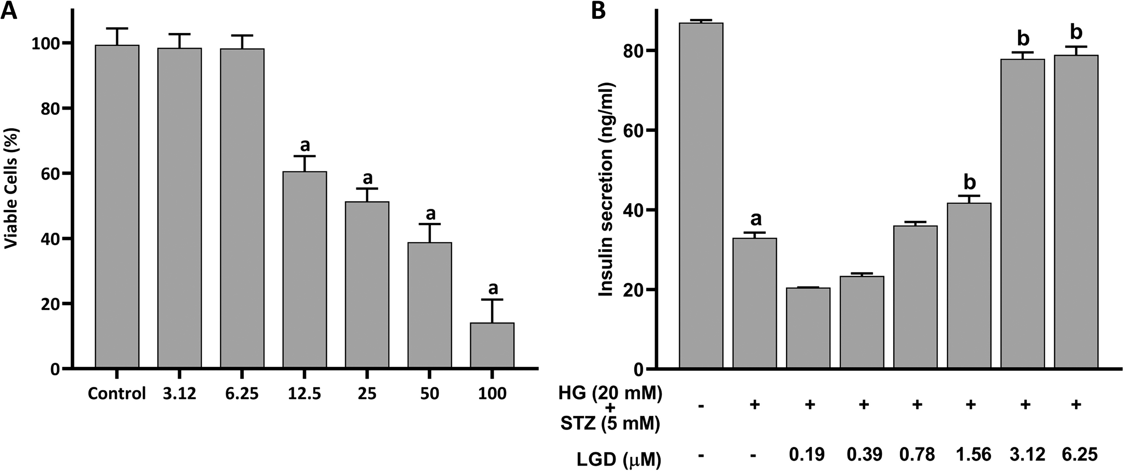
Cytotoxicity and insulin secretion effects of ligandrol on MIN6 cells.
Effect of Ligandrol on Body Weight and Fasting Blood Glucose Levels
The body weight of the animals was measured on day 1, day 29 (1 day post-HFD), and day 55 (1 day posttreatment). On day 29, following the HFD, the animals showed a significant increase (p < 0.001) in body weight compared with the control group. On day 55, the STZ-treated group showed a significant increase (p < 0.001) in body weight compared with the control group. However, the animals treated with ligandrol (0.3 and 1 mg/kg) showed a significant decrease (p < 0.001) in FBG level compared with STZ-treated group (Fig. 5A). The FBG levels of the animals were measured on day 31 (3 days post-STZ) and on day 55 (1 day posttreatment). On day 31, following the HFD, the animals showed a significant increase (p < 0.001) in FBG levels compared with the control group. On day 55, the STZ-treated group showed a significant increase (p < 0.001) in the FBG level compared with the control group. However, the animals treated with ligandrol (0.3 and 1 mg/kg) showed a significant decrease (p < 0.001) in FBG level compared with STZ-treated group (Fig. 5B).

Effect of ligandrol on body weight and FBG levels.
Effect of Ligandrol on the NEUROD1 mRNA Expression
The mechanism of action of the ligandrol was elucidated through quantifying the mRNA expression of genes coding for AR-targeted gene, neurogenic differentiation 1 (NEUROD1). The HFD + STZ significantly decreased (p < 0.05) the expression of NEUROD1 compared with control. Ligandrol at 0.3 and 1 mg/kg significantly increased (p = 0.003, p < 0.001) the expression of NEUROD1 (Fig. 6).
Gene expression of NEUROD1. 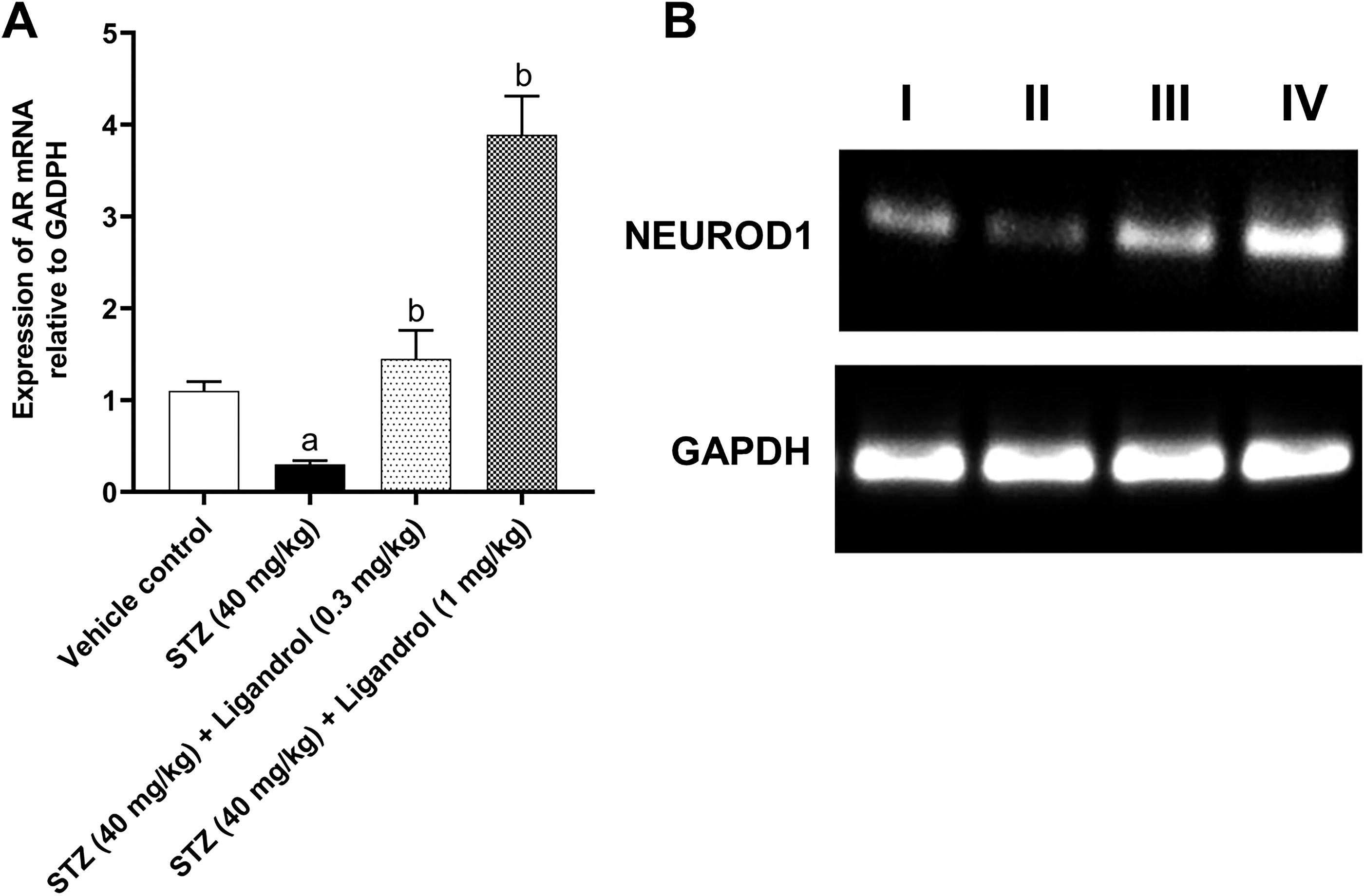
Effect of Ligandrol on Pancreatic Islets
The protective effect of ligandrol on pancreatic islet of diabetic male rats was assessed by H&E staining (Fig. 7). The pancreatic islets portion is lightly stained compared with acinar cells, which are darkly stained. The control group showed normal, bigger, and more prevalent pancreatic islets, with clear boundary of separation from acinar cells compared with HFD and STZ groups (Fig. 7A). The pancreatic islets morphology of HFD- and STZ-treated group is totally distorted and the islets cells are totally degenerated compared with the control group (Fig. 7B). The ligandrol (0.3 and 1 mg/kg)-treated group showed a normal pancreatic islets morphology and a more prominent boundary of separation compared with HFD- and STZ-treated group. These groups are also presented with round islets cells. This effect is more prominent in animals treated with ligandrol 1 mg/kg (Fig. 7C and 7D). The results obtained from these groups suggest the ability of ligandrol to regenerate the pancreatic islets, leading to antidiabetic effects against HFD- and STZ-induced T2DM.
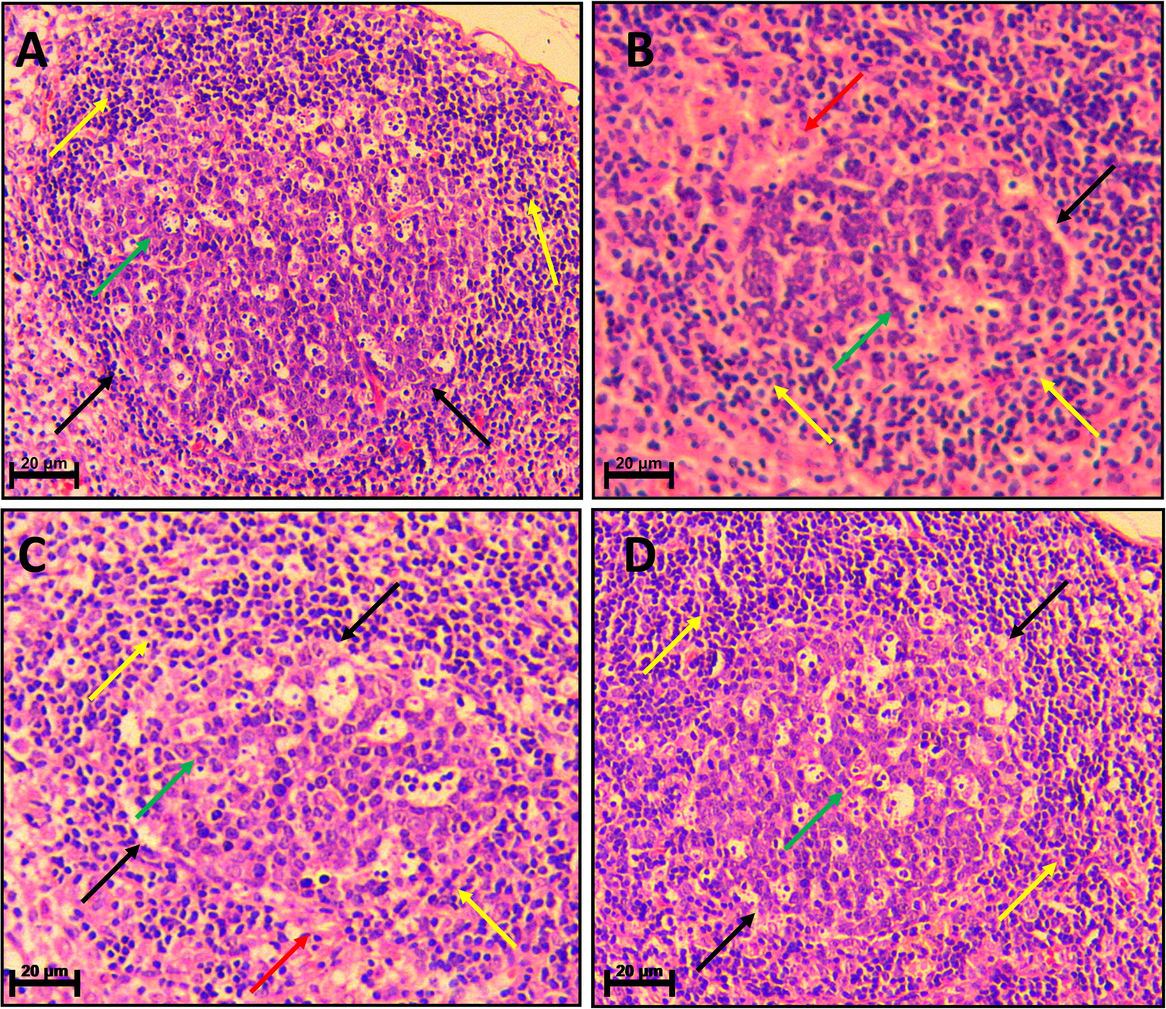
Micrograph showing H&E-stained sections of the pancreas (40× magnification).
DISCUSSION
According to IDF 2018, T2DM is more common in men compared with women. 25 This is reinforced by various studies from different countries such as China, Ghana, India, and Spain. 26,27 This higher prevalence of T2DM is men is primarily a result of reduced testosterone levels. 28 Low testosterone levels hampered the pancreatic islets resulting in apoptosis. 29,30 In line with this finding, compound activating the AR, i.e., testosterone, have shown to protect pancreatic islets apoptosis in males. 31 This effect of testosterone on pancreatic islets results in improvement of visceral obesity, insulin resistance, and lipid profile in males with T2DM. 32,33 However, full agonists of AR such as testosterone and dihydrotestosterone (DHT) are often associated with severe side effects such as benign prostatic hyperplasia and/or prostate cancer. 34 On the other hand, SARMs are considered to be much safer because they are devoid of these effects. 10,11 This prompted us to investigate the possible potential antidiabetic effects of SARMs in male rat model.
The selection of ligandrol for antidiabetic study was carried out through virtual screening. Among the widely known SARMs, we identified ligandrol as a potential hit molecule because of its low binding affinity (−10.75 kcal/mol). This indicate that ligandrol requires low energy to bind to AR when compared with other SARMs. Ligandrol also forms hydrogen bond with THR877, which is one of the key amino acids that interact with AR agonists. ASN705, ARG752, and THR877 are the key amino acids that form hydrogen bonds with AR agonists. 35 However, ligandrol poorly retains its interaction with THR877 as indicated by our 100 ns MD simulation. Remarkably, ligandrol showed a more prominent interaction (30%) with ASN705, which was not seen during molecular docking. This interaction of ligandrol with ASN705 is in agreement with previous findings, which showed that SARMs form hydrogen bond with ASN705 instead of ARG752 and THR877. 36
Following in silico studies, ligandrol was investigated for its effect on insulin secretion in MIN6 cells in vitro. These cells are commonly used for diabetes studies with reference to pancreatic β-cells. Although derived from transgenic mice insulinoma, MIN6 maintained their differentiation by regulating the expression of certain genes. For instance, MIN6 expresses liver-type glucose transporter (GT) but restrict the brain-type GT. The liver-type GT is required for glucose sensing and insulin secretion, while the brain-type GT is implicated in the cancer development. 37 Importantly, MIN6 expresses AR and acts as a suitable model to evaluate the effect of AR agonists and AR modulators in T2DM. 38 Using MIN6 cells, ligandrol was initially assessed for its cytotoxicity at various concentrations, prior to insulin secretion assay. Among different concentrations, 3.12 and 6.25 μM showed no significant toxicity compared with control. Hence, 6.25 μM was chosen as the highest concentration for insulin secretion assay. Among different concentrations of ligandrol, three concentrations, i.e., 1.56, 3.12, and 6.25 μM, displayed a significant effect on insulin secretion. With this result, we suggest that ligandrol could have a negative impact on hyperglycemia, which is a common condition associated with T2DM. 8
Rats are rodents that are commonly used in diabetes studies. They are more sensitive to chemically induced diabetes, STZ (LD50 ∼ 130 mg/kg), 39 compared with mice. 40 HFD and STZ (40 mg/kg) combination is more effective in mimicking the T2DM-like symptoms where obesity precedes the diagnosis of T2DM. 18,19 Hence, HFD was used to induce obesity, whereas STZ was used to induced pancreatic islets apoptosis. 19 Our study of 4 weeks of HFD-induced animals showed a significant effect, by increased in animals body weight, which is consistent with previous findings. 18 The treatment with ligandrol (0.3 and 1 mg/kg, orally) significantly decreased the animals body weight at the end of 3 weeks of treatment. Remarkably, our result is correlated with H&E staining where we observed the protective effect of ligandrol (0.3 and 1 mg/kg, orally) on pancreatic islet degeneration.
Several lines of studies have shown that AR agonists possess antidiabetic effect. 32,33 This is consistent with our current findings. Therefore, to determine the molecular mechanism behind the antidiabetic effects of ligandrol, gene expression studies using pancreatic islets tissue were carried out. In this study, we specifically choose to assess the effect of ligandrol on AR-targeted gene, NEUROD1. NEUROD1 is a transcription factor that aids in the maintenance of a mature phenotype of pancreatic β cells. In addition, it is necessary for the development and growth of β cells, and expansion of pancreatic islet cells mass. 41 In the HFD + STZ group, we observed a significant decreased of NEUROD1 mRNA compared with control. The inhibitory effect of STZ on NEUROD1 expression occurs possibly through the STZ-induced hyperglycemia, which further activates nuclear factor kappa B. 42 Studies have shown that the activation of AR increases the gene expression of NEUROD1. 41 Similarly in our studies we observed a significant increase of NEUROD1 mRNA expression in animals treated with ligandrol of 0.3 and 1 mg/kg compared with the HFD + STZ group. We speculate that ligandrol protects and increases pancreatic islet mass through the activation of AR, which is widely expressed in pancreatic islets 43 and consequently induces the NEUROD1 expression. Our study is supported by previous findings that showed that AR agonist testosterone prevents pancreatic islets from degeneration. 31 Furthermore, animals treated with ligandrol (0.3 and 1 mg/kg, orally) have significantly decreased FBG levels compared with those treated with HFD and STZ. We speculate that this effect of ligandrol could be a result of the protective effect of ligandrol on pancreatic islets. Previous findings have also shown the ability of AR agonist testosterone to improve the FBG levels in T2DM male rats model. 32
CONCLUSION
Through virtual screening, we identified ligandrol as potential hit molecule among other SARMs. We then investigated the possible antidiabetic effects of ligandrol in HFD- and STZ-induced T2DM male rat model. We found that ligandrol could increase insulin secretion in MIN6 cells. In addition, we showed that ligandrol (0.3 and 1 mg/kg, orally) could decrease the body weight and FBG levels induced by HFD and STZ respectively. Through H&E staining, we observed that ligandrol (0.3 and 1 mg/kg, orally) could protect pancreatic islets from degeneration, although this effect is more pronounced with ligandrol of 1 mg/kg. We speculate that ligandrol activated the AR in pancreatic islet and induced the expression of NEUROD1 that is involved in proliferation or differentiation mechanisms. Therefore, to ascertain the mechanism of action underlying ligandrol’s antidiabetic effects, further studies are warranted to validate our current findings.
Footnotes
ACKNOWLEDGMENTS
The authors would also like to thank the Department of Science and Technology—Fund for Improvement of Science and Technology Infrastructure in Universities and Higher Educational Institutions (DST-FIST), New Delhi, for the infrastructure support to our department.
AUTHORS’ CONTRIBUTIONS
D.S.: conceptualization; methodology; software, resources, visualization; writing original draft; writing-review, and editing. R.G.: conceptualization; writing-review and editing. E.R.: methodology; data curation; methodology, writing-review and editing. J.C. and P.T.K.: methodology; formal analysis. R.S.P. and S.S.: investigation. D.S.: visualization; supervision; funding acquisition; writing-review, and editing.
DISCLOSURE STATEMENT
The authors declare no conflict of interest.
FUNDING INFORMATION
The authors would like to thank the Indian Council of Medical Research (ICMR), Department of Health Research-Ministry of Health and Family Welfare for providing financial assistance for this work under the division “Senior Research Fellow” Award No. 2021–11508-F1.