




Abstract
Vulvovaginitis is a growing concern worldwide because of increasing drug resistance, drug toxicity, low bioavailability, and limitations of topical administration. The purpose of the study was to develop a novel mucoadhesive in situ gel of quercetin–cyclodextrin (CD)-loaded chitosan nanoparticles (NPs) for the treatment of vulvovaginitis. In this work, an innovative association between mucoadhesive (chitosan) and thermosensitive polymers (poloxamer 188 and 407) was used to treat vulvovaginitis. The formulation showed compatible pH (4.08) and a desired rheological behavior (viscosity—158.9 cps at 25.9°C, gelation temperature—37°C, and gelation time—65 s) suitable for vaginal application. The quercetin/sulfobutyl-ether-β-cyclodextrin-loaded chitosan NPs (QCD-NPs), prepared by the ionic-gelation method, showed higher in vitro antibacterial activity against Staphylococcus aureus and Pseudomonas aeruginosa compared with the quercetin solution because of the ionic interaction between chitosan NPs and bacteria. Moreover, upon incorporating the QCD-NP in in situ gel, the clearance from the vagina might further reduce and longer residence can be achieved at the site of vaginal infection because of sol–gel transition at body temperature. The developed formulation was confirmed to be biocompatible, nonirritant (hen’s egg chorioallantoic membrane assay), and bioadhesive (mucoadhesion test) with good antimicrobial efficacy and sustained drug release profile, paving the path for improved therapeutic interventions in the treatment of vulvovaginitis.

INTRODUCTION
Vulvovaginitis is a mucosal infection that causes inflammation of the vulva and vaginal epithelium in the lower reproductive tract of females. 1 Vaginitis often develops in women of reproductive age group, particularly in young and middle-aged (15–45 years). 2 Seventy percent of the vaginitis cases seen in premenopausal women are mainly due to bacterial vaginosis, vulvovaginal candidiasis, and trichomoniasis. 3 Bacterial vaginosis is a symptomatic infection that often involves the vulva swelling and causes itching, burning sensation, erythema, and abnormal white vaginal discharge with sloughed epithelium and immune cells. 4 –6 It can be primarily caused by aerobic bacteria such as Staphylococcus aureus (S. aureus), Pseudomonas aeruginosa (P. aeruginosa), Escherichia coli, Enterobacter species, and Klebsiella species. 7 Traditionally, the treatment of bacterial vaginosis is made with antibacterial drugs, including clindamycin, metronidazole, and tinidazole. The most commonly used is metronidazole, and many studies report the development of metronidazole resistance and increased chances of recurrence (58%), 12 months after oral metronidazole therapy. 8 Furthermore, with prolonged treatment, these drugs show toxic effects. Thus, the emerging situation has led to the development of many new drug moieties.
Quercetin is a polyhydroxy bioflavonoid with extensive antimicrobial properties and, therefore, can be used effectively for the treatment of vulvovaginitis. It is known to have antibacterial, antifungal, anti-inflammatory, antioxidant, anticancer, and vasodilator effects as well. 9 Despite the potential use of quercetin in clinical trials, its application in pharmaceutical and food industries is limited owing to poor absorption and low chemical stability. 10 These drawbacks can be overcome by encapsulating the drug in an inclusion complex (IC) of cyclodextrin (CD) or in biopolymer-based nanocarriers, which guarantee modified release at the site of administration, assuring an enhanced biological effect. 11
CDs are well-known cyclic oligosaccharides that possess a hydrophilic surface that makes them water soluble and a hydrophobic interior that facilitates the formation of ICs with hydrophobic substances. 12 Among the various CDs available, the modified β-cyclodextrin, sulfobutyl-ether-β-cyclodextrin (SBE-β-CD), known also under the commercial brand name Captisol®, is preferred because it enhances water solubility significantly, because of the larger distance between the charged groups (sulfobutyl anion and sulfobutyl-ether chain). 13,14 Furthermore, its higher water solubility and better biocompatibility have made it appropriate for the formulation of poorly soluble drugs. 15,16
The introduction of polymeric nanocarriers has gained substantial attention among the different drug delivery techniques designed to improve the bioavailability and solubility of poorly soluble medicines. Incorporating drugs into nanocarriers can be a suitable approach to provide prolonged release and chemical stability, avoid degradation of the active moieties, and promote their site-specific activity assuring enhanced biological effect. 17 Chitosan, a versatile mucoadhesive polymer, is biocompatible, biodegradable, and nonirritant, making it suitable for targeted local vaginal delivery. 18 In addition to these properties, chitosan has antibacterial properties and provides synergistic activity along with quercetin in the treatment of vulvovaginitis. Furthermore, the positively charged primary amino groups in chitosan interact with the negatively charged bacterial cell wall, which enables the drug to penetrate the cell wall improving the treatment efficacy. 19
However, effective vaginal delivery is challenged by high variation in anatomy and the presence of secretions and vaginal fluids that reduce the residence time of the formulation. Recent evidence suggests that in situ gel formulations are more convenient for vaginal applications than conventional dosage forms, as they provide several benefits, including ease of administration into desired body cavities, high spreadability at room temperature, reduced administration frequency, improved patient compliance, and comfort. 20 In situ gels are prepared using thermosensitive polymers such as poloxamer composition, which promotes in situ gelation of the formulation in the body cavity at 37°C. 21 Current strategies for vaginal drug delivery are focused on nanoparticle (NP)-based and mucoadhesive technology. 22,23
The present study was undertaken to develop an intravaginal formulation for quercetin. The IC of quercetin with SBE-β-CD was prepared (QCD) followed by ionotropic gelation with chitosan resulting in the formation of mucoadhesive NPs (QCD-NPs). Furthermore, the QCD-NPs were incorporated into an in situ gel of poloxamer 407/188 to obtain QCD-NP-ISG to provide longer residence at the infection site (vagina) and sustained release profile for quercetin.
MATERIALS AND METHODS
Materials
Quercetin (Mw: 302.24, percentage purity ≥95%), high-molecular-weight chitosan (HMW chitosan, Mw >75% deacetylated), and low-molecular-weight chitosan (LMW chitosan, Mw >75%–85% deacetylated) were purchased from Sigma-Aldrich (USA). Poloxamer 188 (Pluronic F68, F68) was purchased from Signet Enterprises and poloxamer 407 (Pluronic F127) was purchased from BASF (Germany). Captisol (SBE-β-CD) with an average molecular weight of 2,163 g/mol and average degree of substitution of 6.5 was obtained from Ligand Pharmaceuticals (USA). Analytical reagent-grade methanol and hydrochloric acid (35%) were purchased from Finar Ltd. (India). Acetic acid glacial 99%–100% was purchased from Merck Life Science Pvt. Ltd. (India), and sodium hydroxide (AR) was purchased from Spectrochem (India). The purified water used in this study was collected from the Millipore Milli-Q® Plus system (USA). The chemicals sodium chloride (NaCl), potassium hydroxide (KOH), calcium hydroxide (CaOH), lactic acid, acetic acid, glycerol, urea, and orthophosphoric acid of pharmaceutical grade were obtained from Merck. The regenerated cellulose dialysis membrane was obtained from Himedia with a molecular weight cutoff (MWCO) of 12,000–14,000.
Bacterial cultures of S. aureus (ATCC 25923) (Gram-positive) and P. aeruginosa (ATCC 27853) (Gram-negative) were used as test microorganisms in the study.
Methods
Preparation of QCD IC
The quercetin IC was prepared in a 1:1 molar ratio with SBE-β-CD. Briefly, an accurate amount of quercetin was dispersed in 1 mL of Milli-Q water containing an equimolar concentration of SBE-β-CD. The suspension was placed on a Rotospin (TBS India, Telimatic and Biomedical Services Pvt. Ltd., India) and rotated at 50 rpm for 72 h at room temperature (25 ± 3°C). Beyond 72 h or the equilibrium point, no further solubilization of quercetin in CD was observed. After saturation, the mixture was centrifuged at 8,000 rpm for 10 min to remove the undissolved quercetin sedimented. 24 To evaluate the quercetin content in the QCD complex, the supernatant was appropriately diluted and analyzed by an ultraviolet (UV) spectrophotometer at 364 nm. Furthermore, for structural characterization studies, the supernatant was lyophilized to obtain a powdered QCD complex.
Characterization of QCD
Phase solubility studies
Experiments on phase solubility were carried out according to Higuchi and Connors’ approach.
25
In this study, different concentrations of SBE-β-CD from 0 to 50 mM were weighed and added separately to 1 mL of Milli-Q water in 2 mL amber-colored Eppendorf tubes. To each, an excess amount of quercetin was added, and the suspension was sonicated using a water-bath sonicator for 5–10 min to break the lumps. This was then placed in a Rotospin at 50 rpm for 72 h. After equilibrating, the samples were centrifuged at 8,000 rpm for 10 min to remove the undissolved quercetin, and the resulting filtrates were measured for quercetin content using a UV spectrophotometer at 364nm. The callibration curve obtained for quercetin is shown in Supplementary Fig.S1. The phase solubility curve was plotted by taking the molar concentration of the solubilized drug (quercetin) versus the molar concentration of SBE-β-CD. To calculate apparent solubility constant (ks) and complexation efficiency (CE), the following equations were used:
Saturation solubility studies of QCD
The solubility of the quercetin and QCD in water was determined according to the method reported earlier. Briefly, excess quercetin (15 mg) and its complex (QCD) equivalent to 15 mg of quercetin were added separately in 1 mL of Milli-Q water in a 1.5 mL Eppendorf tube. The Eppendorf tubes were rotated for 72 h at room temperature (25 ± 3°C), on a Rotospin. 27 Furthermore, the suspensions were centrifuged, and the drug content was estimated by analyzing the supernatant using a UV spectrophotometer at lambda max 364 nm.
Structural characterization of QCD IC
X-ray diffractometry analysis
Pure quercetin, SBE-β-CD, and QCD were analyzed for their crystallinity using the X-ray diffraction beam (Cu-kαX radiation) over the 2⊖ range of 5°−80° on the Rigaku Miniflex 600 X-ray diffractometer (Rigaku Co., Japan). The instrument functioned with a graphite monochromator and standard scintillation counter as a detector. The scan rate was 3°/min, and the X-ray tube voltage was 40 kV. 28
1H-nuclear magnetic resonance measurements
1H-nuclear magnetic resonance (1H-NMR) experiments were performed using the Bruker DRX 400-Avance spectrometer (USA), operating at 400 MHz using dimethyl sulfoxide (DMSO) as a solvent. Tetramethysilane was used as an internal standard, and the chemical shifts were reported in parts per million (ppm). The 1H-NMR spectra of quercetin, SBE-β-CD, and lyophilized QCD were recorded to investigate the interacting groups involved in the process of inclusion complexation. 29
Preparation of QCD IC-loaded chitosan NPs
The QCD IC-loaded chitosan NPs (QCD-NPs) were prepared based on the ionic gelation method as described in the previously reported method, with minor modifications. 30 Briefly, 2 mL of HMW chitosan (6 mg/mL) was prepared in glacial acetic acid (2%v/v) and pH was adjusted to 4 using 4 M NaOH. Furthermore, 100 µL of the prepared QCD complex (equivalent to 230.2 µg quercetin) was added drop by drop to the beaker containing 2 mL of HMW chitosan solution. The HMW chitosan concentration (5–7 mg/mL), glacial acetic acid concentration (1–3% v/v), pH (4–6), and volume of QCD complex (50–150 μL) were varied, to optimize the NPs (Table 2). Upon varying the amount of the QCD complex added to the pH-adjusted chitosan solution (HMW chitosan), the QCD-NP that was stable and turbid without precipitation was selected. In the process of optimization, the influence of each parameter, the pH, the concentration of glacial acetic acid, and the quantities and types of chitosan (HMW chitosan and LMW chitosan) were also studied. The finally optimized QCD-NP suspension, which contains the encapsulated drug, was used for further loading into the in situ gel.
Characterization of QCD-loaded chitosan NPs
Particle size and surface properties
The particle size distribution and zeta potential were measured with Malvern Zetasizer (Nano ZS, Malvern Instruments, UK) using the dynamic light scattering technique. The experiments were performed using clear zeta cell in water (refractive index—1.33, viscosity—0.73 cP), recorded at a temperature of 25°C.
Field emission scanning electron microscopy
The surface morphology of prepared QCD-NP was examined using a field emission scanning electron microscope (Zeiss, JEOL-JSM5800LV, Japan), operating at an acceleration voltage of 15 kV and an appropriate magnification at room temperature (25 ± 3°C). In brief, a drop of QCD-NP was diluted with water (1:200), mounted on 5 mm silicon wafers, and sputter-coated with gold under an argon atmosphere to observe the surface topography of samples. 31
Structural characterization of QCD-NP
Differential scanning calorimetry
The differential scanning calorimetry (DSC) thermograms of the pure quercetin, freeze-dried samples of QCD, unloaded NPs, and QCD-NP were recorded on a differential scanning calorimeter (DSC-50, Shimadzu, Japan). The thermal behavior of the samples was studied by placing them on a flat-bottomed aluminum pan, sealed, and heated from 10°C to 350°C at a constant rate of 10°C/min with a nitrogen flow of 20 mL/min. The baseline calibration was done using empty aluminum pans as a reference. 30
Attenuated total reflectance–Fourier transform infrared spectroscopy
The attenuated total reflectance–Fourier transform infrared spectroscopy (ATR-FTIR) analysis for quercetin, SBE-β-CD, HMW chitosan, QCD, unloaded NP, and QCD-NP was done using ATR-FTIR (Bruker, USA). The measurements were made in absorption mode at a wavelength region of 4,000–500 cm−1 and a resolution of 4 cm−1 upon 64 scans. 24
Preparation of QCD-NP-ISG
The mucoadhesive in situ gel was prepared by the cold method with thermoresponsive polymers (poloxamer 407 and poloxamer 188). Briefly, 60 mg poloxamer 188 (3% w/v) and 460 mg poloxamer 407 (23% w/v) were slowly added to the 2 mL chitosan formulation containing the QCD-NPs and stirred continuously at 4 ± 0.5°C in an ice bath, until a clear solution was obtained. The solution was then slowly brought to room temperature (25 ± 2°C) and gradually increased up to 37 ± 0.5°C, using a thermostatic water bath, which resulted in the formation of a gel. The prepared gels were stored at 4 ± 0.5°C in tightly sealed glass vials until further use. 20
Preparation of simulated vaginal fluid and Ringer buffer
The simulated vaginal fluid pH 4.5 (SVF) and Ringer buffer pH 7.4 were prepared as reported in literature. 32
Characterization of thermosensitive QCD-NP-ISG
Drug content
The drug content was determined by dissolving 1 mL of in situ gel in 9 mL of methanol–water (30:70) mixture. After preparing appropriate dilutions, the solution was vortexed and centrifuged at 12,000 rpm for 10 min. The supernatant obtained was then analyzed for quercetin content by a UV spectrophotometer at 364 nm. 33,34
Gelation temperature and gelation time
Gelation temperature of in situ gel was measured by gradually increasing the temperature (by 0.2°C) of the formulation in a thin-walled 10 mL glass tube using a thermostatic water bath until it forms a gel. The gelation temperature is the temperature at which no flow occurred on the inversion of glass tubes (when tilted >90°), as shown in Figure 5a. Gelation time was estimated by a flow or no-flow criterion using the tube inversion technique. The formulation in a glass tube at room temperature (25 ± 2°C) was in a flow state. This tube, which was immersed in a thermostatic water bath maintained at 37 ± 0.5°C, was taken out at regular intervals and inverted to see the physical state. The time taken by a system to convert from flow to no-flow state was recorded as gelation time. 35
Determination of pH
The pH of the in situ gel was checked using a pH meter at room temperature (25 ± 2°C). 20
Viscosity measurement
The viscosity of in situ gel was measured using the Brookfield Viscometer (Brookfield Engineering Laboratories, Inc., USA) using a T-bar spindle (spindle-CPE 40) rotating at a speed of 1.9 rpm. The spindle was placed perpendicularly into the gel in such a way that the end of the spindle did not touch the lower surface of the beaker. The viscosity of the in situ gelling solutions at storage/room temperature was measured once the level of gel was stabilized (after 30 s) at room temperature (25 ± 2°C). The tests were performed in triplicate. 35
In vitro release study
The in vitro drug release for the formulations was carried out using modified Franz diffusion cells, maintained in sink condition with a water-jacketed receptor medium (receptor volume 5 mL, diameter 25 mm, surface area 4 cm2) placed on an eight-station platform (Orchid Scientifics, India). Pure quercetin 500 µg (500 µL of 1 mg/mL quercetin-water suspension), QCD-NP suspension equivalent to 219.2 µg quercetin (2 mL of 109.6 µg/mL QCD-NP), and QCD-NP-ISG equivalent to 217.2 µg quercetin (2 mL of 108.6 µg/mL QCD-NP-ISG) were placed on the regenerated cellulose dialysis membrane (12,000–14,000 MWCO), which was soaked for 24 h in Milli-Q water on the donor compartment before the start of the experiment. The receptor medium chosen was SVF, and the assembly was placed on magnetic stirring at 500 rpm. 36 At definite time intervals (0.5, 1, 2, 4, 6, 8, 10, 12, 24, 36, and 48 h), 1 mL sample was withdrawn from the receptor compartment for up to 48 h and replenished with an equal volume of SVF. The samples were filtered and analyzed for drug content using a UV spectrophotometer at 364 nm.
In vitro mucoadhesion test
A goat’s reproductive system was donated from a local slaughterhouse, and the vaginal canal was separated and cut open longitudinally. To preserve the vaginal mucus on the tissue surface, rinsing was not done. Furthermore, the tissue was divided into small pieces and placed on an empty Transwell platform with 1.5 mL of Ringer buffer (pH 7.4) to support tissue viability and consistency during the study. Two hundred microliters of formulations, quercetin (Q, equivalent to 500 µg/mL), QCD-NP (equivalent to 109.6 µg/mL), and QCD-NP-ISG (equivalent to 108.6 µg/mL), was carefully placed on the vaginal tissue and incubated at 37°C in a shaking incubator at 250 rpm. Following 3 h, the tissues were washed by pouring 1 mL of SVF containing 4% human serum albumin drop by drop over the vaginal tissue, and the quercetin concentration in the washed samples was collected and measured. The percentage of quercetin adhered to the vaginal mucosa was calculated by subtracting the amount of quercetin in washed samples from the initial quercetin concentration applied on the vaginal mucosa. 32
Hen’s Egg Chorioallantoic Membrane Test assay
To evaluate the vaginal irritancy potential of formulations, the Hen’s Egg Chorioallantoic Membrane Test (HET-CAM) was performed. Fertilized hen’s eggs were incubated in an automatic incubator for 9 days at 37°C and 50%–60% humidity. On day 9, the eggs were taken out from the incubator and candled to ensure fertility, which is confirmed by observing the blood vessels through the shell. Thereafter, the eggshell was carefully removed and 200 µL of the test formulations, 0.9% w/v NaCl as negative control and 0.1 M NaOH as positive control, was applied directly on the chorioallantoic membrane, and the reactions were observed for 300 s. The irritation score was calculated based on the endpoints of hemorrhage, lysis, and coagulation for each formulation.
The formulations with irritant scores of 0.0–0.9 are classified as nonirritant, 1.0–4.9 as slightly irritant, 5.0–8.9 as moderately irritant, and 9–21 as highly irritant. 37
Antimicrobial assay
Agar well diffusion method
In vitro antibacterial activity for free quercetin, QCD IC, QCD-NPs, blank cyclodextrin (BCD), and BCD-loaded NPs (BCD-NPs) was assessed against two clinical strains, a Gram-positive bacterium (S. aureus) and a Gram-negative bacterium (P. aeruginosa) using the modified agar well diffusion method. Mueller–Hinton (MH) agar plates were prepared and individually spread with standardized inoculum (100 µL) of each test microorganism (overnight grown broth culture was inoculated with 1 mL peptone water, incubated at 37°C for 2–3 h to obtain growth with an optical density of 0.08–0.1 at 600 nm). Wells were prepared using a well-making plunger on the agar plate with a diameter of 4 mm aseptically with a sterile tip, and a volume (100 µL) of the formulation equivalent to 240 µg of quercetin was introduced into the well. The formulations QCD and QCD-NP equivalent to 240 µg quercetin were prepared by lyophilizing the respective suspensions and further diluting them to get a required concentration (240 µg in 100 µL). These MH agar plates were incubated for 24 h at 37°C, and the antimicrobial activity was determined by measuring the zone of inhibition around the wells. 38
Determination of minimum inhibitory concentration
Minimum inhibitory concentration (MIC) was determined using a microdilution assay recommended by the Clinical Laboratory Standards Institute. 39 The MIC value is the lowest concentration of the assayed antimicrobial agent that inhibits visible growth of the microorganism tested as observed by the unaided eye, and it is commonly reported in microgram per milliliter or milligram per liter.
In this method, quercetin (10 mg) and lyophilized QCD-NP (equivalent to 240 µg quercetin) were added to MH broth (MHB) and made up to 2 mL so that the concentration in each of the stock solutions is 5 mg/mL and 120 µg/mL, respectively. From each stock solution, 1 mL was taken and was serially diluted to 10 tubes to prepare dilutions for quercetin in the range 5,000–2.4 µg/mL and for QCD-NP in the range 120–0.19 µg/mL. Positive and negative controls containing only MHB were also kept. Furthermore, 10 µL of bacterial inoculum of S. aureus (density was set to read OD 0.1 at 600 nm) was added to each tube except for the negative control. All the tubes were maintained in an incubator at 37°C for 24 h and observed for visible growth of the organism in terms of turbidity. The MIC value was recorded for the tube with the lowest concentration of antimicrobial agent in which no turbidity/growth was observed.
RESULTS AND DISCUSSION
Preparation and Characterization of QCD IC
Phase solubility and saturation solubility study
There was an increase in the solubility of quercetin with increasing concentration of SBE-β-CD, which is categorized as AL-type profile (Supplementary Table S1). The phase solubility curve for QCD is shown in Figure 1a. The intrinsic solubility (S0), which is the solubility of quercetin in the absence of SBE-β-CD, was found to be 0.0184 mM, whereas the apparent stability constant (Ks) of QCD was found to be 1,182 M−1, which was lesser than the Ks reported for QCD in previous literature with 4,032 M−1. 40 This difference could be due to the different setup used for the solubilization in the previous literature, in which a shaking water bath was used at 30°C. However, we carried out solubilization of quercetin at room temperature (25 ± 2°C) on a Rotospin. 40 The Ks value (1,182 M−1) of the QCD complex suggested that the complex formed between quercetin and SBE-β-CD is stable and crucial for improving the bioavailability of quercetin. 41 The slope value of 0.0213, which is <1, on the phase solubility diagram, suggests the formation of a complex having a 1:1 molar ratio. The saturation solubility of quercetin was found to be 39.9 µg/mL, whereas the solubility of its complex was found to be 2,302 µg/mL. Owing to inclusion complexation, there was an increase in solubility of quercetin by 58-fold compared with the solution without CD. Furthermore, the CE of QCD was calculated using Eq (2) and found to be 0.0217. 41

Structural characterization of QCD complexes
XRD analysis
Powder XRD is performed to detect the crystallinity of the pure drug and solubilized drug complex. 42 The XRD patterns for quercetin, SBE-β-CD, and QCD complex are shown in Figure 1b. The diffraction pattern corresponding to quercetin exhibited several intense and sharp peaks at 10, 12, 25, and 26° of 2⊖ values, which confirm its crystalline nature. In contrast, the SBE-β-CD showed no characteristic peaks and exhibited a hallow pattern indicating its amorphous nature, similar to the findings in the literature. 43 Furthermore, the XRD spectrum of the QCD complex showed a broadened diffraction peak and an amorphous hallow nature implying the formation of an amorphous IC. The drug amorphization could be due to lyophilization process, further favored by the presence of intrinsically amorphous substance SBE-β-CD. The observations are in accordance with that reported in the literature for the QCD complex with parent β-CD. 28,42
1H-NMR measurements
The 1H-NMR spectra of quercetin, SBE-β-CD, and QCD IC have been recorded in DMSO-d6 and shown in Figure 2. The SBE-β-CD has a wide toroid-like structure, and the protons H1, H2, and H4 located on the periphery of SBE-β-CD molecule are not much involved in the cavity interaction with the guest molecule, whereas H3 and H5 reside in the interior cavity of SBE-β-CD and might interact with the guest molecule. 29 The SBE-β-CD protons appeared in the range of δ 1.5–5.8 ppm, as reported previously in the literature. 29,44 A relatively significant chemical shift was observed in the δ values of H3 protons (from δ 4.58 to 4.56; Δδ, −0.02 ppm) after the complex formation (as shown in Supplementary Fig. S2). The dense electron cloud around the H3 proton created by the adjacent C=O group could be the cause of this upfield shift. In addition, the H1, H3′, and H4′ protons showed a downfield shift from δ 1.61 to 1.62 ppm (Δδ, 0.01 ppm), indicating the formation of an IC between quercetin and SBE-β-CD.
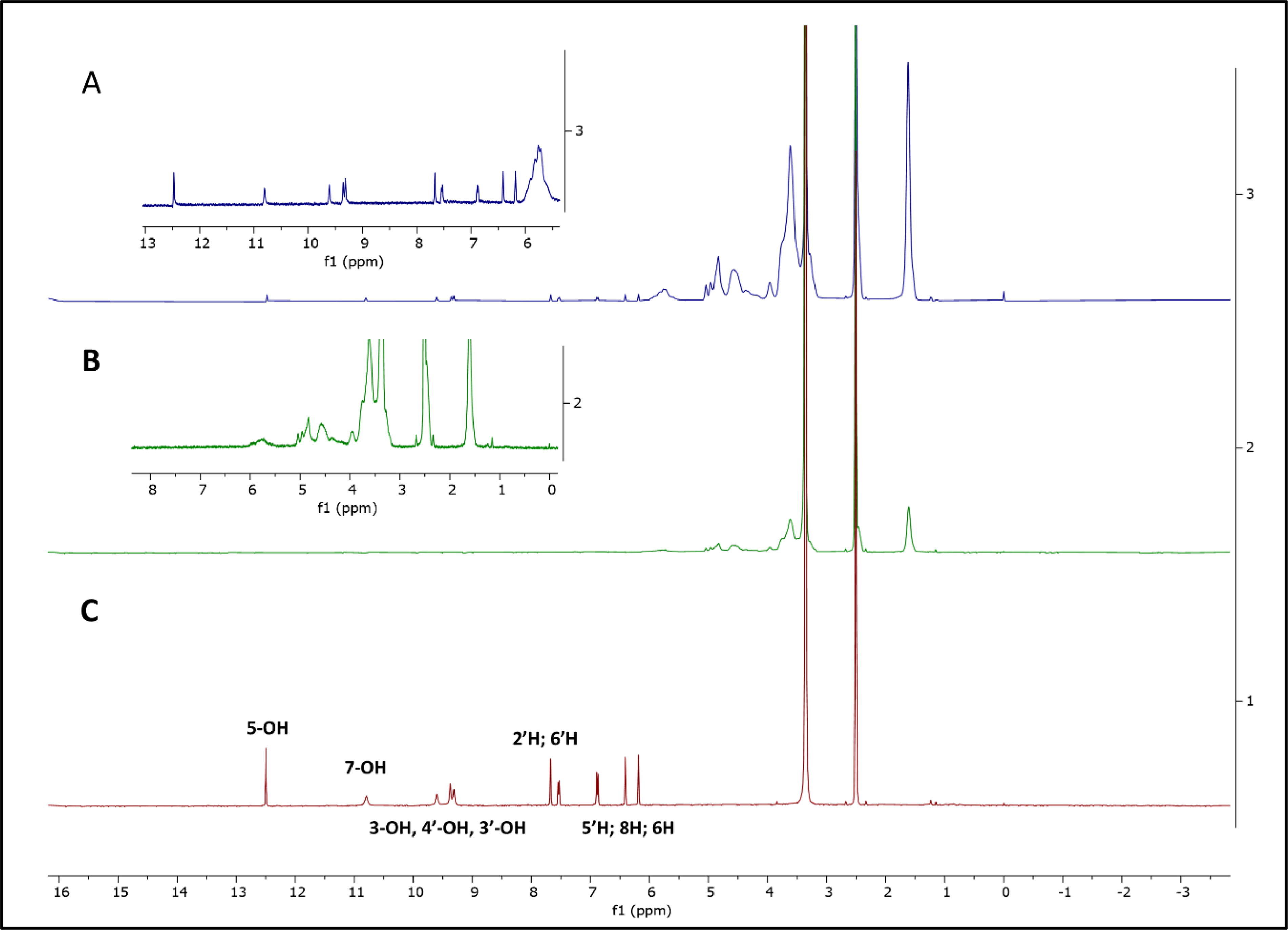
On the contrary, the spectra obtained for QCD in contrast to quercetin reveal a downfield shift for C3-OH, C4′-OH, and C3′-OH from δ 9.60 to 9.61 ppm (Δδ, 0.01 ppm); for C7-OH from δ 10.80 to 10.81 ppm (Δδ, 0.01 ppm); and for C5-OH from δ 12.48 to 12.49 ppm (Δδ, 0.01 ppm), suggesting that the A-ring, B-ring, and C-ring of quercetin interact with the hydrophobic cavity of SBE-β-CD. A sharp and extremely down shielded resonance was attributed to the C5-OH proton because of its intramolecular hydrogen bonding with carbonyl oxygen at the C4 position. 45 –47 Furthermore, as indicated in Table 1, an upfield shift for H2′ proton was also found from δ 7.68 to 7.67 ppm (Δδ, −0.01 ppm), indicating that the entire quercetin molecule was contained inside the SBE-β-CD cavity and, as a result, a stable QCD IC was produced.
Chemical Shifts of Protons of Quercetin and Sulfobutyl-Ether-β-Cyclodextrin in QCD Inclusion Complex
SBE-β-CD, sulfobutyl-ether-β-cyclodextrin.
Composition of Quercetin-Cyclodextrin Inclusion Complex-Loaded Chitosan Nanoparticles
HMW, high molecular weight; LMW, low molecular weight; PDI, polydispersity index; ZP, zeta potential.
Preparation and Characterization of QCD-NP IC
The NPs were prepared by the ionic gelation method, through electrostatic interactions between positively charged chitosan and negatively charged CD. 48 The NPs obtained in the preliminary trials were of size >500 nm. Therefore, the factors influencing particle size, which include the type and concentration of chitosan, the concentration of glacial acetic acid, and pH, were optimized as this greatly influenced the stability of the NPs. 49 Initially, the optimization was carried out by varying the molecular weight of chitosan (HMW chitosan and LMW chitosan were used). The HMW chitosan was selected because the NP formed was comparatively more stable and of less particle size than that prepared from LMW chitosan. It has to be noted that the preparation of QCD-NP was crucial as any slight fluctuation in the parameters affected the stability and size of NPs drastically. Therefore, the HMW chitosan concentration (5–7 mg/mL), glacial acetic acid concentration (1–3% v/v), pH (4–6), and volume of QCD complex (50–150 μL) were varied, to optimize the NPs (Table 2). As shown in F2, F7, and F8 of Table 2, pH is critical in the formation of NPs, as a rise in pH resulted in a significant increase in particle size, affecting the stability of NPs. This could be possible because of low-level chitosan protonation that reduced the electrostatic repulsion between the NPs. Furthermore, no NPs were formed at a pH <4, indicating that the surface charge density of the chitosan solution at this pH (<4) was not suitable for the preparation of QCD-NP. 50 It was also noted that the addition of a higher volume of QCD complex to chitosan solution resulted in the precipitation of NPs. From the above trials, it was concluded that 2% v/v of glacial acetic acid, 6 mg/mL of HMW chitosan, pH (4), and 100 μL of QCD complex gave formulation F2 a smaller particle size and better stability (as shown in Supplementary Fig. S3). Hence, this was selected as the final NP formulation for further studies. Similarly, unloaded NPs were also prepared by adding plain SBE-β-CD solution devoid of quercetin to the chitosan solution for the characterization studies.
Physicochemical characterization of QCD-NP
The optimized NPs showed a mean particle size of 478.9 ± 50 nm and a polydispersity index (PDI) 0.172. as summarized in Table 2.17 A slight increase or decrease in each of these factors—pH, volume of QCD complex, and the concentration of glacial acetic acid and chitosan—drastically affected the particle size and stability of the nanoformulation because of change in surface charge/residual amino groups of chitosan solution. The PDI reflects particle size distribution close to 0.2, which indicates that the optimized NP suspension was homogeneous. The zeta potential value was positive (29.6 mV), corresponding to a stable formulation (QCD-NP). The drug concentration in the optimized formulation (QCD-NP) was 109.6 μg/mL, based on the volume of QCD complex added. The particle morphology of the QCD-NP suspension was investigated using field emission scanning electron microscopy (Figure 3). The mean average particle size of the NPs obtained from SEM was 300 nm, which was slightly lesser than the hydrodynamic size obtained by the Malvern Zetasizer. This was possibly due to the swelling effect of the NPs upon dilution in water. 50
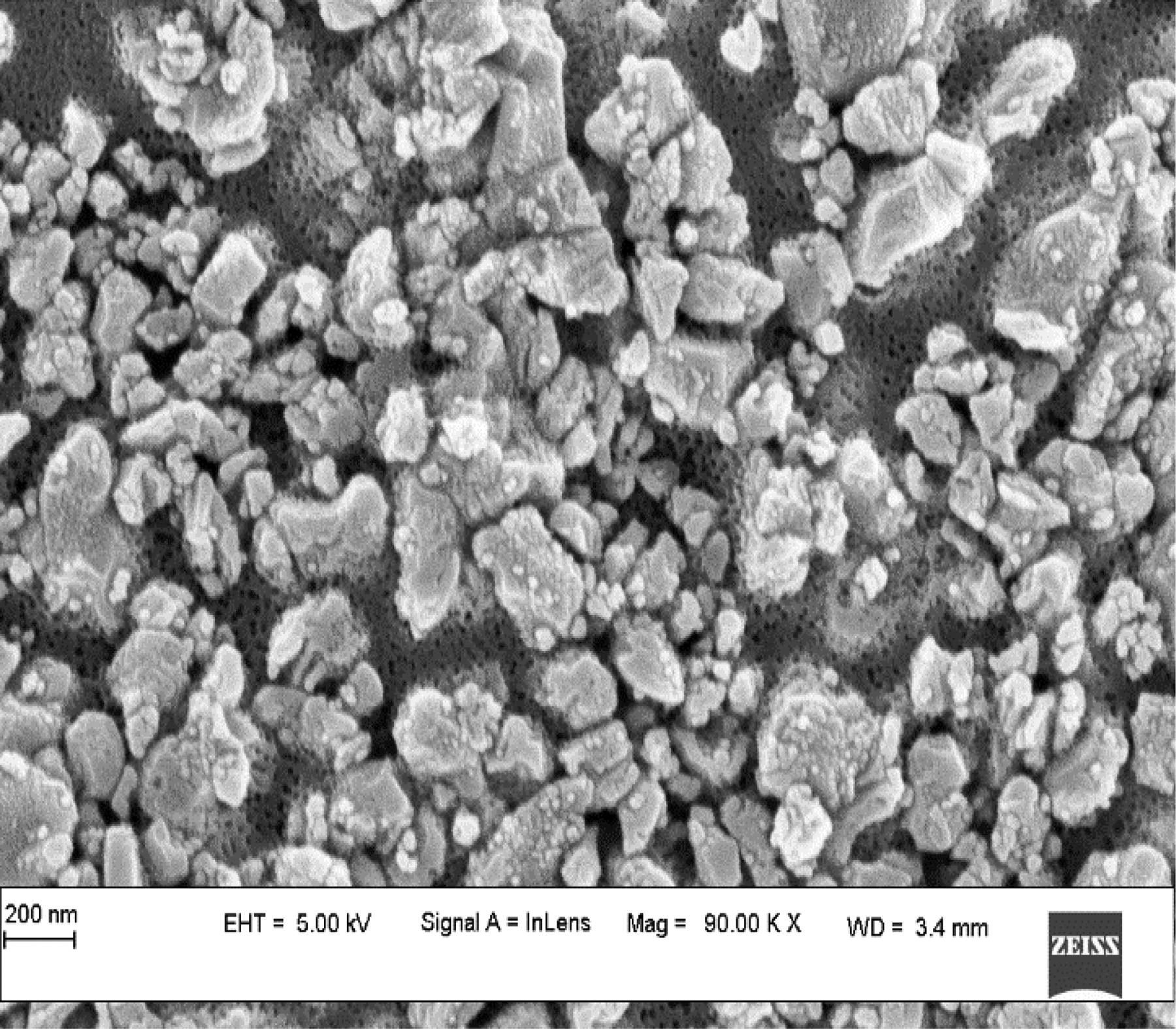
FESEM image of the quercetin/sulfobutyl-ether-β-cyclodextrin-loaded chitosan nanoparticle (QCD-NP). FESEM, field emission scanning electron microscopy.
Structural characterization of QCD-NP
Differential scanning calorimetry
Quercetin thermogram showed a broad endothermic peak at 126°C indicating the evaporation of water molecules strongly held in the crystal lattice, thus requiring a higher temperature. The second endothermic peak at 322°C shows the melting and decomposition of quercetin (Figure 4a). The thermogram of HMW chitosan exhibited a broad endothermic band at 93°C, which depicted evaporation of surface-bound water, and an exothermic peak at 310°C due to decomposition. The SBE-β-CD thermogram showed a broad endothermic band at around 90°C, which also corresponded to the liberation of crystal water; moreover, the peak at 270°C was attributable to sample decomposition. The thermogram of the QCD IC revealed that the characteristic peaks of pure quercetin and SBE-β-CD disappeared, as complexation occurred by replacing water with the guest molecule. 30 The thermogram of the unloaded NP represents no interaction with just the superimposition of individual components. However, the DSC thermogram of QCD-NP showed an endothermic peak (128°C) with less intensity in comparison with pure quercetin, which might be due to drug dispersion in chitosan NPs. 51,52

DSC thermograms

Attenuated total reflectance–Fourier transform infrared spectroscopy
The ATR-FTIR spectra of quercetin, SBE-β-CD, HMW chitosan, QCD IC, unloaded NP, and QCD-NPs are shown in Figure 4b. The IR molecular band and peaks obtained for quercetin include the following: 3,412–3,201 cm−1 (phenolic O-H stretching), 1,665 cm−1 (C=O), 1,602 cm−1 (C=C stretching of aromatic ring), 1,375 cm−1 (C-OH), and 1,257 cm−1 (C=O stretching vibration) . 47,53 The HMW chitosan showed an absorption spectrum at 3,405 cm−1 because of OH and NH stretching, 1,666 cm−1 (C=O; amide-I stretching vibration), 1,515 cm−1 (-NH2; amide II bending vibration), and 1,375 cm−1 (C-N; amide III stretching vibration). 54 The IR spectra of SBE-β-CD showed an intense and broad band: 3,740–3,010 cm−1 (OH stretching vibrations), 3,000–2,800 cm−1 (CH), 1,646 cm−1 (H-OH bending of water molecules linked to SBE-β-CD), 1,187 cm−1 (CH), and 1,041 cm−1 (O=S=O stretching vibrations). 29 For the QCD IC, many of the distinctive bands of quercetin (1,602, 1,375, and 1,257 cm−1) disappeared completely. Furthermore, the absorption intensity of the C=O band (1,665 cm−1 for quercetin) shifted to a lower frequency, that is, 1,646 cm−1 for the QCD IC, which suggests that complexation of quercetin with SBE-β-CD involved oxygen atom of the carbonyl group. In addition, there was an increase in peak intensity at 1,012 cm−1 and a slight change in band shapes and widths in comparison with quercetin and SBE-β-CD, suggesting an interaction between quercetin and SBE-β-CD. 47
The IR spectrum of the unloaded NP showed superimposition of the spectra of both SBE-β-CD and HMW chitosan. In the case of QCD-NP, the characteristic peaks of quercetin at 3,412 cm−1 because of the vibration of phenolic and hydroxy groups (3,400 cm−1) and absorption peaks assigned to aromatic bending and stretching (C=O, C=C, C-OH) around 1,600, 1,650, and 1,300 cm−1 disappeared. The disappearance of characteristic peaks of quercetin in the spectrum of QCD-NP revealed polymerization of the QCD IC in chitosan NPs, which probably occurred by the formation of hydrogen bonds or hydrophobic interactions. 55
Preparation and Characterization of In Situ Gel (QCD-NP-ISG)
The composition, gelation temperatures, and gelation time of the various trials taken are shown in Table 3. The poloxamer molecules in the solution exhibited a zigzag configuration initially, which transformed into a close-packed configuration and then to a viscous gel because of the increasing temperature. Formulations 6 and 7 behaved as a mobile viscous liquid at room temperature and transformed into a semisolid transparent gel at body temperature (37°C). In comparison with the G7 formulation, the G6 formulation took more time to form a gel as the temperature was slowly increased to 37°C. The gel transition temperature of the G7 formulation containing 23% poloxamer 407, and 3% poloxamer 188 was the most appropriate for preparing an in situ gel compared with the other trials conducted for the given application. 56 The prepared gel was white, homogeneous, and smooth with the consistency of a semisolid.
Composition of the In Situ Gel
QCD-NP, quercetin–cyclodextrin-loaded chitosan nanoparticles.
Drug content, gelation temperature, and gelation time
The drug content of the optimized QCD IC-loaded chitosan NP in situ gel (QCD-NP-ISG) was found to be 108.6 µg/mL. The temperature at which the liquid phase transforms into a gel is known as the gelation temperature. A vaginal in situ gel requires a gelation temperature of 37°C. If the formulation’s gelation temperature is <37°C, the gel may form at room temperature, posing manufacturing and administration challenges. If the gelation temperature exceeds 37°C, the vaginal gel remains fluid at body temperature and does not offer sustained release in the body cavity, resulting in a shorter retention time. 20 A major facet in the production of a desirable in situ hydrogel is the gelation time. This is important because it prevents drainage from the application site, allowing the active material to stay on the mucosal tissue for longer. Table 3 displays the gelation temperature and gelation time of the prepared formulations. The optimized formulation’s gelation temperatures and time were found to be 37°C and 65 s, respectively.
Determination of pH
The pH of the formulation is an important factor for vaginal compatibility. The vaginal pH is usually found between 3.5 and 4.5; however, this may vary on several changing conditions, including the presence of semen and fungal and bacterial infections (pH 5–8). 35 The pH of the optimized formulation was found to be 4.08 at room temperature (25 ± 2°C), which, in turn, shows that the prepared gel is biocompatible and suitable for vaginal application.
Viscosity measurement
The viscosity of the final formulation (G7) was found to be 158.9 cps at room temperature (25 ± 2°C), for shear stress (22.6 dyne/cm2) and shear rate (14.2/s), which indicated that the gel was in a fluid state, convenient for injection into the vagina. However, the viscosity of the formulation increased in SVF, at 37°C, which was desirable for resisting the vaginal cleansing action and achieving prolonged residence in body cavities such as the vagina. The poloxamer 407/poloxamer 188 concentration was finely tuned to prepare a thermosensitive in situ gel with the desired sol–gel transition temperature and mucoadhesive properties.
In vitro drug release study
The diffusion study for the optimized QCD-NP-ISG was carried out and compared with pure quercetin and QCD-NP using the Franz diffusion apparatus, and the results are shown in Figure 5b. The cumulative drug release of QCD-NP was found to be 168.11 µg (76.69 ± 3%) at the end of 48 h, and therefore, sustained release was achieved. The possible reason for the initial burst release followed by a slow and continuous release could be attributed to the dissociation of quercetin adhered to the surface of QCD-NP and drug diffusion through the polymer matrix. However, the cumulative release of QCD-NP from ISG at the end of 48 h was further less, 145.2 µg (66.85 ± 2.2%), because of the crosslinked matrix of the gel, enabling a further slow and prolonged release of quercetin from QCD-NP-ISG. Although in vitro studies of QCD-NP showed a better release pattern in comparison with QCD-NP-ISG, the amount of quercetin available at the administration site (vagina) for prolonged release from QCD-NP, in vivo, will be less because of the strong self-cleansing ability of the vagina leading to the expulsion of QCD-NP, which is a liquid formulation. Therefore, to achieve high local concentration and a longer retention time, NPs are incorporated into an in situ gel as it is bioadhesive and crucial for the treatment of vulvovaginitis. 57 On the contrary, the cumulative release of pure quercetin was only 61.7 µg (12.3 ± 2.5%) at the end of 48 h, because of poor solubility of the drug in the aqueous phase, which caused precipitation of quercetin on the dialysis membrane. 58
In vitro mucoadhesion test
The mucoadhesion test for quercetin, QCD-NP, and QCD-NP-ISG was carried out in vitro, following the procedure reported by Berginc K et al., 32 and the results are presented in Table 4. The percentage of quercetin adhered to the vagina for quercetin, QCD-NP, and QCD-NP-ISG was found to be 9.79, 75.32, and 90.23%, respectively. The mucoadhesive strength of QCD-NP was improved in comparison with pure quercetin, owing to the increased binding of HMW chitosan’s positively charged groups with negatively charged groups of mucin. 59 It is important to highlight the fact that for good mucoadhesion property, the polymer chain and its polar functional groups should have greater flexibility. In addition, the chitosan interacts with the epithelial junction of the vaginal mucosa, enhancing drug permeability. 60 The enhanced mucoadhesive strength of QCD-NP-ISG could be due to the cumulative effect of chitosan and poloxamer. Higher bioadhesive strength is due to increased electrostatic interaction, hydrogen bonding, and surface energy properties of the poloxamer along with chitosan, which increases the viscosity of formulation. 59 As a result, the formulation will be able to withstand the physiological clearance process in vivo.
In Vitro Mucoadhesion of Quercetin, Nanoparticles, and In Situ Gel
QCD-NP-ISG, in situ gel incorporated with QCD-NP.
HET-CAM assay
HET-CAM assay was carried out by identifying the irritant reactions (hemorrhage, lysis, and coagulation) that occurred upon contact of formulations with the chorioallantoic membrane of the fertilized hen’s egg, as shown in Figure 5c. It was found that the negative control, NaCl (0.9% w/v), showed no irritancy, whereas the positive control, NaOH (0.1 M), had an irritation score of 15.36, indicating that they were extremely irritant. No irritant potential was observed for the formulations pure drug (Q), QCD, QCD-NP, BCD-NP, QCD-NP-ISG, and blank ISG, as shown in Table 5. These results are considered within the acceptable range according to the Interagency Coordinating Committee on the Validation of Alternative Methods protocol, 61 as there were no lysis, hemorrhage, and coagulation for the tested formulations. Therefore, the developed formulation was nonirritant and suitable for vaginal application.
Irritant Score and Extent of Damage in Hen’s Egg Chorioallantoic Membrane Test Assay
Irritant score:
Antimicrobial assay
The antibacterial tests were performed against two major bacterial strains responsible for vulvovaginitis by the agar well diffusion method, and the results are shown in Table 6. The zone of inhibition (ZI) observed for quercetin at 5, 10, 15, 20, 25, and 30 µg/mL against S. aureus and P. aeruginosa is shown in Figure 6a and b.
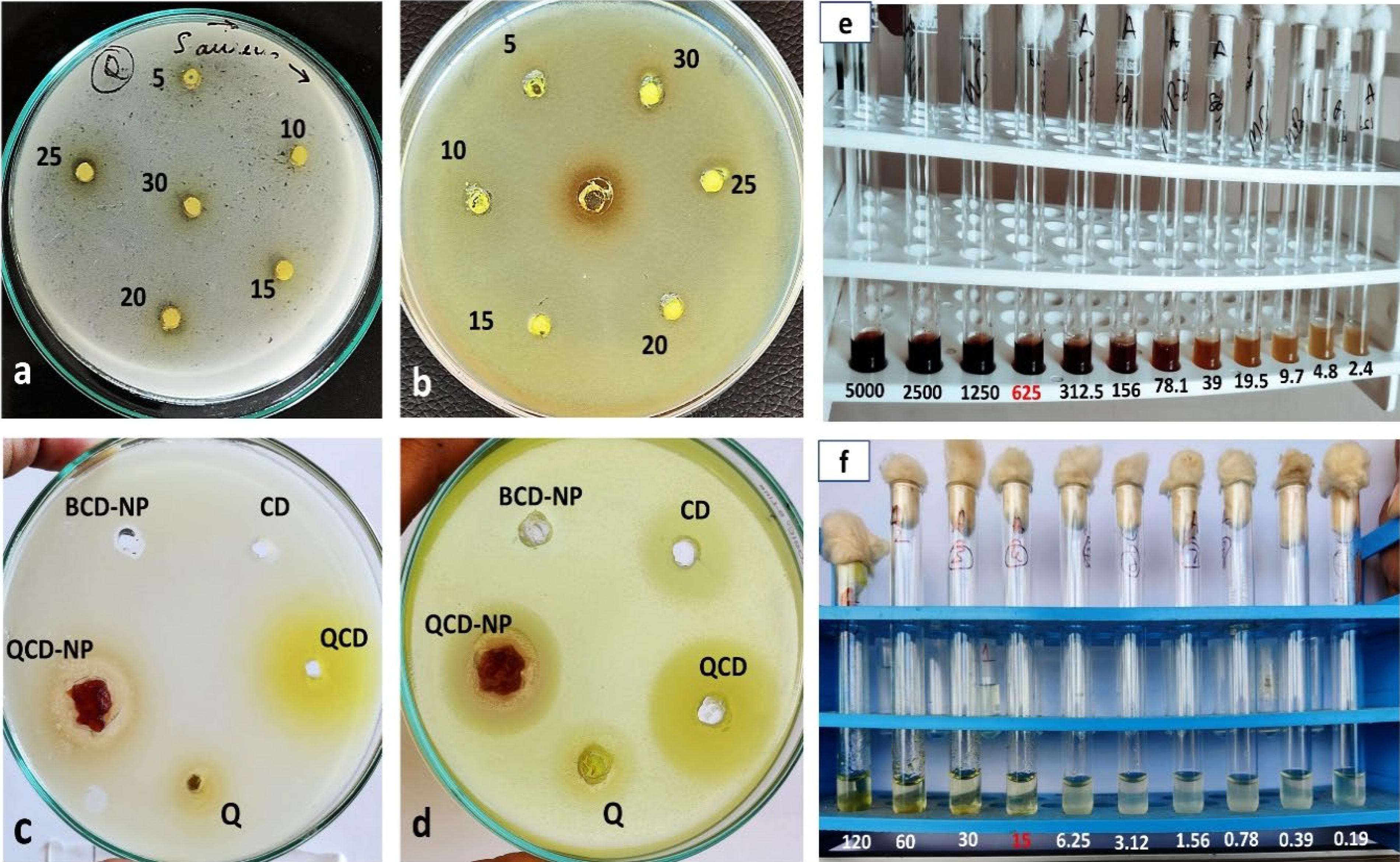
Antibacterial activity of quercetin at 5, 10, 15, 20, 25, and 30 µg/mL against Staphylococcus aureus
Antibacterial Efficacy of the Formulations Against Staphylococcus aureus and Pseudomonas aeruginosa
ZI, zone of inhibition.
The QCD-NP showed better antibacterial activity against S. aureus (Clear ZI: 1.3 cm, Partial ZI: 2.1 cm) and P. aeruginosa (Clear ZI: 1.8 cm, Partial ZI: 2.3 cm) compared with pure quercetin (Clear ZI: 0.6 cm for S. aureus, Partial ZI: 1.6 cm for P. aeruginosa) despite its thick consistency making it less available for well diffusion (Figure 6c and d). 62 It has to be noted that more than 50% of QCD-NP remained as such in the well at the end of 24 h of incubation because of the high viscosity of the formulation. The formulation QCD-NP might have shown much higher antibacterial activity and ZI in vivo than what was observed in vitro. To account for the difference, MIC studies were carried out as shown in Figure 6e and f, and the MIC of quercetin and QCD-NP against S. aureus was found to be 625 µg/mL and 15 µg/mL (42-fold better antibacterial activity for NP formulation), respectively. Furthermore, not only the QCD-NP but also the QCD IC (Clear ZI: 2.3 cm, Partial ZI: 2.7 cm) showed higher antibacterial activity against P. aeruginosa in comparison with pure quercetin. This could be due to the slow and sustained release of quercetin from QCD IC. The higher antibacterial efficacy of QCD in comparison with QCD-NP against P. aeruginosa could be due to the thin consistency of QCD solution in the well, making it freely available for diffusion. The BCD and BCD-NP were considered negative controls. The chitosan NPs (QCD-NP) showed antibacterial activity more effectively because of the cationic groups present in chitosan that interact with negatively charged bacterial surfaces and the presence of quercetin that synergistically diminished bacterial cell regeneration. Quercetin, mainly composed of phenolic constituents, is well known to increase membrane permeability by suppressing the potassium and calcium transport across the bacterial membrane, inhibiting nucleic acid synthesis, and reducing enzyme activities. Besides, based on the ZI, it was found that our formulation (QCD-NP) was more effective in inhibiting P. aeruginosa compared with S. aureus, which could be due to the high resistance of the S. aureus strain used and its thick peptidoglycan layer, making it less penetrable for our nanoformulation. 63,64
CONCLUSION
A mucoadhesive thermosensitive gel incorporated with QCD IC-loaded chitosan NPs, for the treatment of vulvovaginitis, was successfully prepared. In this study, we aimed to design chitosan NPs for better mucoadhesive property, which was further incorporated in an in situ gel to improve the release, permeability, and residence time in the vagina. The mucoadhesion test showed that the formulations prepared with polymers (QCD-NP and QCD-NP-ISG) showed better mucoadhesive nature compared with the pure drug, quercetin. Furthermore, the in vitro antibacterial studies revealed the QCD-NP to have better antibacterial efficacy against the most common bacterial strains, S. aureus and P. aeruginosa, known to cause vulvovaginitis. The efficacy of the formulations can be further studied by in vivo and clinical experiments. Considering that conventional therapy for vulvovaginitis lacks efficacy owing to lesser residence time, high toxicity, and resistance, the favorable antibacterial results obtained suggest the QCD-NP-ISG formulation to be a promising alternative to treat vulvovaginitis.
Footnotes
ACKNOWLEDGMENT
The authors thank Manipal College of Pharmaceutical Sciences and Manipal Academy of Higher Education, Manipal, India, for providing adequate facilities for the research.
AUTHORS’ CONTRIBUTIONS
A.M. and S.L. contributed to the conceptualization of this work. A.M., P.M., K.S., and M.P. conducted the experiments, contributed to the scientific discussion, and drafted the article. The final article was revised by S.L.
CONSENT ON PUBLICATION
All authors agree on the publication.
AUTHOR DISCLOSURE STATEMENT
The authors declare no conflicts of interest.
FUNDING INFORMATION
The authors are grateful to the All-India Council for Technical Education, New Delhi, India, for providing financial assistance in the form of the National Doctoral Fellowship to A.M. (
SUPPLEMENTARY MATERIAL
Supplementary Figure S1
Supplementary Figure S2
Supplementary Figure S3
Supplementary Table S1