




Abstract
Introduction:
Abamectin (ABA) is an insecticide that is commonly used in agricultural spraying. Although it is known to have neurotoxic effects on living organisms in the ecosystem and humans, the details of these effects, including the duration and amount of exposure, are not fully understood. The aim of this study is to evaluate the effect of ABA exposure on human microglia clone 3 (HMC3) cells. These cells were chosen because they are suitable for modeling immunological functions such as cytokine production, cell migration, and inflammation response.
Materials and Methods:
The impact of ABA on the viability of HMC3 cells was evaluated using the 3-(4,5-dimethylthiazol-2-yl)-2,5-diphenyltetrazolium bromide assay, while the effect of ABA on Interleukin-18 (IL-18) levels was assessed through enzyme-linked immunosorbent assay. Furthermore, the influence of ABA on apoptosis parameters was determined through qRT-PCR.
Results:
Increasing concentrations of ABA were observed to decrease the viability of HMC3 cells. The IC50 dose was determined to be 25.71 μg/mL. IC50: 25.71 μg/mL and half of IC50 doses (IC50/2: 12.85 μg/mL) were applied to the cells. The results of the dose applications were compared with the results of the control groups. There was a significant increase in IL-18 levels in cells treated with 25.71 μg/mL ABA (p < 0.001). There was a significant increase in p53 and BAD expression levels in cells treated with 25.71 μg/mL ABA (p < 0.05). There was a significant decrease in BCL2L1 expression levels in cell treated with 25.71 and 12.85 μg/mL ABA (p < 0.05).
Conclusions:
These results clearly demonstrate that it can be concluded that ABA exposure caused a decrease in the survival of HMC3 cells and induced apoptosis. Long-term exposure to ABA may increase the risk of neurodegenerative diseases as a result of inflammation and decreased cell viability.
Introduction
Avermectins are metabolites produced by the fermentation of Streptomyces avermitilis. These metabolites belong to the family of 16-member macrocyclic lactone compounds. Abamectin (ABA) is a mixture of avermectins. 1 It contains a minimum of 80% B1a and a maximum of 20% B1b. 2 The chemical difference between Avermectin B1a and B1b is due to the presence of either a methylene group or an ethylene group at C-26. 3 The use of ABA as an anthelmintic and antiparasitic agent for animals is a common practice. Furthermore, it is an active ingredient in nematicides and insecticides for agricultural purposes.3,4 The insecticidal properties of ABA are well documented, and the substance may also be harmful to mammals. 5 The mechanism of action of ABA involves effects on the gamma-aminobutyric acid (GABA) system and chloride channels. GABA receptors are found in the nerve cells of the central nervous system and regulate the neural basal tone of the brain. Laboratory animals have exhibited vomiting, convulsions, pupil dilation, and coma as a result of ABA poisoning. Furthermore, ABA has been reported to have genotoxic effects in some studies. 3
ABA is a respiratory chain inhibitor that has no effect on the activity of the enzymes NADH dehydrogenase or succinate dehydrogenase. The mechanism of action is similar to that of oligomycin and carboxyatractyloside. This similarity suggests that it has a direct effect on FoF1-ATPase and/or adenine nucleotide translocator (ANT). 5 The FoF1-ATPase is an enzyme that is located in the inner membrane of the mitochondrion. It is responsible for the synthesis of Adenosine triphosphate (ATP), which is driven by the electrochemical gradient of protons generated in the respiratory chain. The enzyme consists of two main components: Fo, which is an integral membrane protein that functions as a proton channel, and F1, which is a hydrophilic moiety that contains the catalytic and regulatory sites. ANT is another crucial component of the mitochondrial mechanism responsible for ATP synthesis. It plays a role in both pathological and physiological mitochondrial events, making it a primary target for drug-induced toxicity.3,5
The ABA has been demonstrated to be involved in the induction of oxidative stress, Nrf2-Keap1, NF-κB pathway activation, and mitochondrial pathway-mediated apoptosis induction. 6 Reactive oxygen species are the by-products of oxidative metabolism. This includes normal mitochondrial respiration. Nevertheless, these conditions are also the result of pathological processes, such as inflammation. Oxidative stress leads to the deterioration and loss of mitochondria. Neurodegenerative diseases can result from such mitochondrial damage and loss. 7
A significant number of pesticides have the potential to induce functional changes in the immune system through both humoral and cellular immune responses. For the assessment of the immune response, cytokines are important indicators. 8 The immune system plays a crucial role in neurodegenerative diseases, with a number of studies indicating a link between the immune system and brain structure, vascular pathology, and cognitive function. Microglia and astrocytes are the major resident glial cells in the brain. They mediate neuroinflammation by producing chemokines, cytokines, and other neuroinflammatory molecules. 9 Increased levels of IL-18 can cause long-term consequences of traumatic brain injury and neurodegeneration. 10
The objective of this study was to examine the toxic effects of ABA, a chemical commonly utilized in agricultural settings, on human microglia clone 3 (HMC3) cells, a crucial component of the human central nervous system’s active immune defense. Additionally, the study sought to ascertain the influence of ABA on cytokine levels and apoptosis mechanisms.
Materials and Methods
Cell culture
The HMC3 cell line was obtained from the American Type Culture Collection. The cells were cultured in phenol-free Dulbecco’s modified Eagle’s medium (DMEM; Capricorn, Germany) was supplemented with 100 IU/mL penicillin, 10 μg/mL streptomycin (Sigma-Aldrich, USA), and 10% fetal bovine serum (Sigma-Aldrich, USA) and maintained in a humidified incubator at 37°C, 5% CO2. Upon reaching 80% confluence, the cells were collected, washed with phosphate-buffered saline (PBS; Capricorn, Germany), counted, and prepared for seeding in each well.
Cell toxicity
The cells were seeded in 96-well plates at 1 × 10⁴ cells/well and incubated overnight prior to ABA (Syngenta, Switzerland) treatment. The cells were then treated with ABA at concentrations of 15.62 to 250 μg/mL for 24 hours. The final concentration of 3-(4,5-dimethylthiazol-2-yl)-2,5-diphenyltetrazolium bromide (MTT; Sigma, USA) was adjusted to 0.5 mg/mL. Following a 4-hour incubation period at a CO2 incubator, the resulting formazan crystals were dissolved in DMSO. A multiscan plate reader (Synergy HTX BioTek, USA) was employed to quantify cell viability at 570 nm. The percentage of cell viability was calculated according to the following formula: viability % = (ODtreated cells/ODcontrol cells) × 100.
Enzyme-linked immunosorbent assay for IL-18
Overall, 1 × 10⁴ cells/well were seeded in a 96-well plate and incubated overnight to allow cells to attach. The following day, the media and detached cells were removed. The cell layer was washed three times in PBS and once in serum-free DMEM. Cells were incubated in DMEM for 4 hours prior to treatment with ABA. In order to gain a deeper understanding of the impact of ABA on HMC3 cells and to facilitate the interpretation of the results, doses of IC50 = 25.71 μg/mL and half of this dose (IC50/2) were then applied for 24 hours.
The cells were homogenized with an ultrasonic homogenizer following washing with PBS. Following the centrifugation, the supernatants were transferred to Eppendorf tubes for analysis and stored at −80°C until analysis. 11 The IL-18 (MyBioSource, USA) studies were conducted in accordance with the manufacturer’s instructions.
Real-time polymerase chain reaction analysis
IC50 = 25.71 μg/mL and half of this dose (IC50/2) doses were applied to HMC3 cells for 24 hours. The total RNA isolation, measurement of RNA concentration and purity, translation to cDNA, and real-time PCR applications were conducted in accordance with the methodology previously described in our study and in accordance with the manufacturer’s instructions. 12 On the website of the US National Center for Biotechnology Information, specific mRNA sequences were identified. Potential primer sequences were then tested. ACTB F: CATGTACGTTGCTATCCAGGC R: CTCCTTAATGTCACGCACGAT, p53 F: TCTACAAGCAGTCACAGCACAT R: CAACCTCAGGCGGCTCATAG, BCL2L1 F: GCATATCAGAGCTTTGAACAGG R: GAAGGAGAAAAAGGCCACAATG, BAD F: GAGTCGCCACAGCTCCTAC R: GGAGTCCACAAACTCGTCACT. The expression of the ACTB gene was employed to normalize the results. The Cq values were subsequently normalized and calculated utilizing the formula 2−ΔΔ Cq.
Statistical analysis
Unless otherwise stated, the experiments in this study were repeated three times (n:3)13,14 In the statistical analysis, enzyme-linked immunosorbent assay and expression results were compared between the groups. The normal distribution of the numerical parameters was analyzed using the Shapiro–Wilk W test. One-way Analysis of variance (ANOVA) with the Bonferroni test using GraphPad Prism v. 5 was used for statistical analysis (GraphPad Software, Inc., La Jolla, CA, USA). Additionally, IC50 was determined using GraphPad Prism 5 software. The level of significance was set at p < 0.05.
Results
Cell toxicity and viability
The results of the MTT assay demonstrate that the effect of ABA on the viability of HMC3 cells is shown in Figure 1. It is observed that cell viability decreases with increasing concentration. In our study, the IC50 was found to be 25.71 μg/mL (R2: 0.9845).
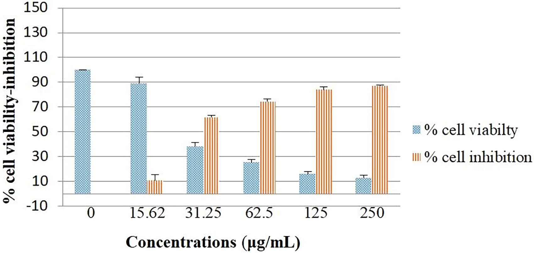
Cell viability and inhibition depending on ABA concentrations.
IL-18 results
The results demonstrated that the IL-18 level increased in a dose-dependent manner, with the highest levels observed in the IC50 and IC50/2 groups compared to the controls (p < 0.001; Fig. 2).
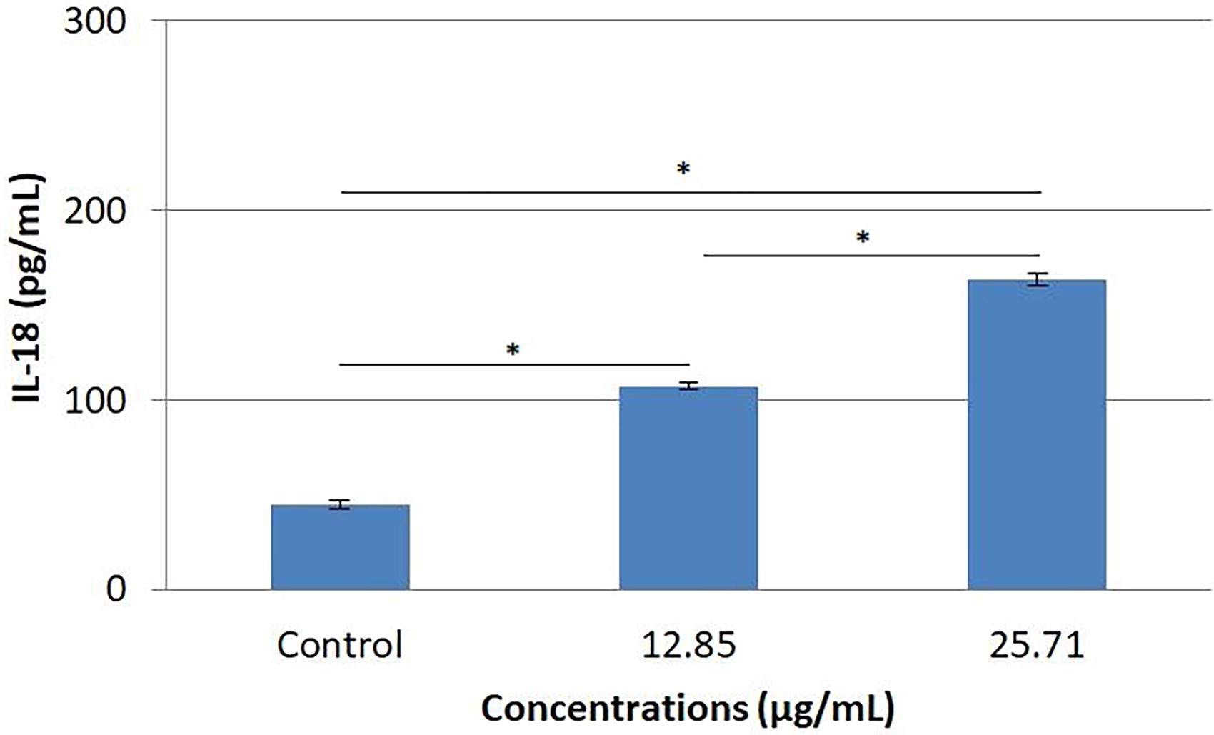
IL-18 levels. Values are presented as means ± standard deviatıon (SD). *p < 0.001.
Expression results
The expression of apoptotic genes (p53 and BAD) was found to be elevated in the ABA group (at IC50 dose) in comparison to the control group, while the expression of the antiapoptotic gene (BCL2L1) was observed (IC50/2 dose) to be reduced. Figure 3 illustrates this.

Relative mRNA expression results and statistical comparison between groups. Values are presented as means ± SD. *p < 0.05. ACTB, actin beta; BAD, BCL2 associated agonist of cell death; BCL2L1, BCL2 like 1.
Discussion
Pesticides are widely used to protect crops from insects. 15 The global demand for avermectin is about 5000 tons per year, mainly produced by Chinese companies. 16 Undegradable pesticide residues can enter the food chain through air, water, and soil. 17 Depending on the amount and route of exposure, they are potentially toxic to humans and can cause acute and chronic health problems. 18 Toxicological and epidemiological studies have identified potential health risks such as cancer, genetic malformations, neurodevelopmental disorders, and damage to the immune system. 17
In recent years, ABA has become one of the most widely used insecticides in agriculture. While studies have improved our understanding of its effects on other living organisms, research continues to understand the mechanism of adverse effects on human health.
ABA has been reported to inhibit the development of aquatic organisms, cause behavioral disorders and neurotoxicity, 19 induce oxidative stress, inflammation, and apoptosis. 20 ABA induces oxidative stress by decreasing antioxidant enzyme activities and increasing Malondialdehyde (MDA) levels. It activates inflammation by increasing Inducible nitric oxide synthase (INOS) levels and pro-inflammatory transcription. 20 ABA exposure induces oxidative stress by causing the production of reactive oxygen species and activating the Nrf2-Keap1 pathway. It causes inflammation by activating the NF-κB pathway and apoptosis in carp through the mitochondrial pathway. 6 In the central nervous system, microglia and astrocytes play a pivotal role in regulating inflammatory responses. 21 Oxidative stress may act as a mediator of neuroinflammation.22,23 Neuroinflammation is a key factor in the development of neurodegenerative diseases, including Alzheimer’s, Parkinson’s and amyotrophic lateral sclerosis. 24 Neuroinflammation causes the release of products that can have neurotoxic effects, such as pro-inflammatory cytokines. 25 Pro-inflammatory cytokines were released at the site of neurodegeneration in the brain, which in turn stimulated the release of damage-associated molecular patterns (DAMPs). DAMPs are commonly found in the neurodegenerative brain of Alzheimer’s disease (AD) patients. 26 Inflammatory cytokines derived from microglia can trigger neuronal degeneration and promote the plaque formation typical of AD. There is increasing evidence that IL-18 may play a role in this scenario. 27 Cultivation and activation of HMC-3 microglial cells in a medium containing LPS and ATP resulted in decreased cell viability and increased release of IL-1β and IL-18 compared with nonactivated cells. 28 Chlorpyrifos, a pesticide, increased Nitric oxide (NO), MDA, and O2 parameters and upregulated the expression of IL-1β and NLRP3 genes in BV-2 cells. 29 Avermectin has been demonstrated to induce neurotoxicity in carp by disrupting the blood-brain barrier structure and inducing oxidative stress, inflammation (pro-inflammatory TNF-α, IL-6, IL-1β), and apoptosis. Furthermore, the NF-κB and PI3K/Akt pathways have been identified as being involved in this process. 30
In summary, immune dysregulation has been identified as a significant underlying condition of diseases. The initial protective anti-inflammatory phase is thought to involve microglia in the CNS and Treg and Th-2 cells in the periphery. Preclinical data suggest that this protective response occurs at the onset of motor neuron and neuromuscular junction degeneration. As motor neurons continue to degenerate and accumulate, this response shifts from anti-inflammatory to pro-inflammatory. Microglia become pro-inflammatory and induce the release of neurotoxic factors from astrocytes. These factors can kill motor neurons. 31
The results of our experiments on HMC3 cells showed that ABA caused a decrease in cell viability, an increase in IL-18 levels (p < 0.001; IC50=25.71 μg/mL and IC50/2 doses), an increase in the expression levels of apoptotic genes (BAD and p53; p < 0.05; IC50=25.71 μg/mL), and a decrease in the expression level of anti-apoptotic gene (BCL2L1; p < 0.05; IC50=25.71 μg/mL and IC50/2 doses). The protein–protein interaction between IL-18, BAD, p53, and BCL2L1 is shown in Figure 4.
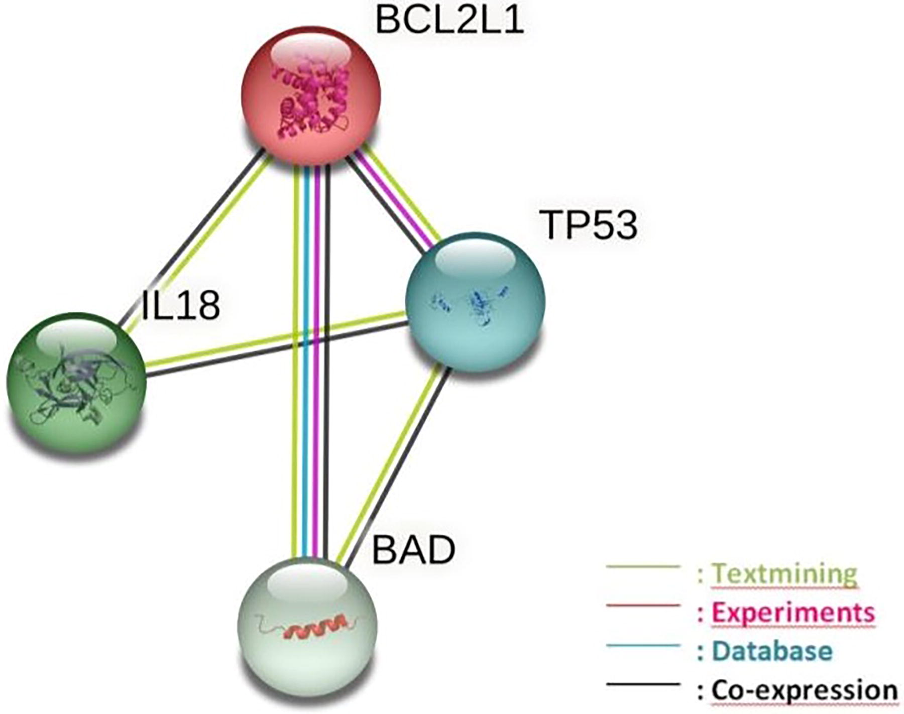
The STRING network view. Colored lines between the proteins indicate different types of interaction evidence.
Extensive research into the effects of pesticide use on humans is just beginning to gain momentum. ABA increased the level of inflammation, induced an apoptosis mechanism, and decreased cell viability by causing an increase in oxidative stress in HMC3 cells, one of the elements of the human nervous system. There is a paucity of studies in the literature on the toxic effects of ABA in human HMC3 cells. Demonstrating this in this study will add to the literature on the effects of our research on human cells.
Conclusions
The results of our study suggest that given the continuity and magnitude of ABA exposure, ABA may induce pro-inflammatory effects and apoptosis, which play a promoting role in the pathogenesis and development of neurodegenerative diseases. We suggest that our findings may contribute to further molecular-based research on neuroinflammation and neurological and neurodegenerative diseases.
Footnotes
Authors’ Contributions
O.S.: Data curation, formal analysis, methodology, software, validation, visualization, and writing—original draft. F.N.C.: Formal analysis and writing—review and editing. U.Ş. and M.Y.T.: Data curation, methodology, validation, and writing—review and editing. F.İ.Ö. and M.A.: Visualization and writing—original draft. All authors have read and approved the final article.
Author Disclosure Statement
The authors have stated that they have no potential conflicts of interest with this article.
Funding Information
There was no funding for this study.