
Abstract
Significance:
A key role of the tumor microenvironment (TME) in cancer progression, treatment resistance, and as a target for therapeutic intervention is increasingly appreciated. Among important physiological components of the TME are tissue hypoxia, acidosis, high reducing capacity, elevated concentrations of intracellular glutathione (GSH), and interstitial inorganic phosphate (Pi). Noninvasive in vivo pO2, pH, GSH, Pi, and redox assessment provide unique insights into biological processes in the TME, and may serve as a tool for preclinical screening of anticancer drugs and optimizing TME-targeted therapeutic strategies.
Recent Advances:
A reasonable radiofrequency penetration depth in living tissues and progress in development of functional paramagnetic probes make low-field electron paramagnetic resonance (EPR)-based spectroscopy and imaging the most appropriate approaches for noninvasive assessment of the TME parameters.
Critical Issues:
Here we overview the current status of EPR approaches used in combination with functional paramagnetic probes that provide quantitative information on chemical TME and redox (pO2, pH, redox status, Pi, and GSH). In particular, an application of a recently developed dual-function pH and redox nitroxide probe and multifunctional trityl probe provides unsurpassed opportunity for in vivo concurrent measurements of several TME parameters in preclinical studies. The measurements of several parameters using a single probe allow for their correlation analyses independent of probe distribution and time of measurements.
Future Directions:
The recent progress in clinical EPR instrumentation and development of biocompatible paramagnetic probes for in vivo multifunctional TME profiling eventually will make possible translation of these EPR techniques into clinical settings to improve prediction power of early diagnostics for the malignant transition and for future rational design of TME-targeted anticancer therapeutics. Antioxid. Redox Signal. 28, 1365–1377.
Introduction
T
Individual cancer cells generated by causal genetic insults behave differently depending on their specific tissue microenvironment. Numerous therapeutic strategies elaborated in cellular systems in vitro in the absence of TME factors are not reproduced in further studies in animals and humans. There is approaching scientific consensus that in vivo TME profiling opens opportunities for preclinical screening of anticancer drugs and predicting therapeutic effectiveness, as well as developing new diagnostics and noninvasive guided therapeutic strategies based on TME manipulation.
A reasonable radiofrequency penetration depth in tissues makes magnetic resonance imaging (MRI) and low-field electron paramagnetic resonance (EPR)-based techniques the most appropriate approaches for noninvasive assessment of the chemical TME parameters such as pO2, extracellular pH (pHe), phosphate (Pi), GSH, and redox. MRI relies largely on imaging of water protons and is widely used in clinical settings providing anatomical resolution while lacking functional sensitivity. Potentially attractive 31P NMR measurements of extracellular [Pi] and pH based on a signal from endogenous phosphate are normally masked by several-fold higher intracellular Pi concentrations (36, 37). In contrary to the NMR applications, EPR measurements rely on spectroscopy and imaging of specially designed paramagnetic probes providing functional sensitivity. Note that exogenous EPR probes have advantages over exogenous NMR probes due to a much higher intrinsic sensitivity of EPR and absence of endogenous background EPR signals.
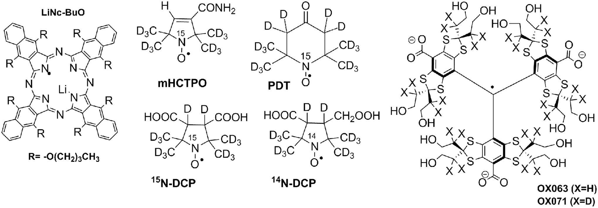
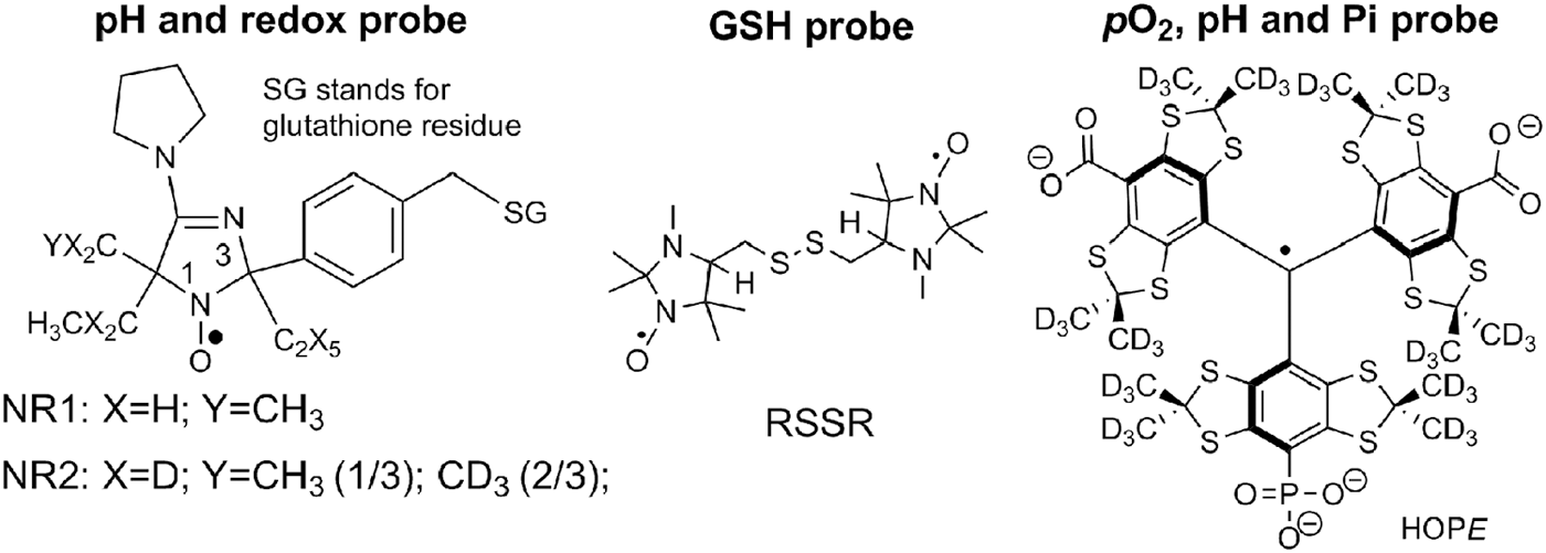
In vivo EPR applications rely on use of both particulate and soluble paramagnetic probes (see Fig. 1 for the exemplified structures). Advantages of particulate probes used for EPR oximetry are high functional sensitivity, stability in living tissue, and minimal toxicity (11, 34, 76, 90). The particulate probe may remain in the place of implantation for long periods of time, enabling repeated measurements of tissue pO2 for up to weeks and months after implantation. On the contrary, soluble nitroxide (NR) and triarylmethyl (TAM) probes may possess functional sensitivity to various parameters of TME such as pO2, pH, Pi, redox, and GSH (9, 10, 20, 93) and allow for spatially resolved measurements using EPR-based imaging techniques (13, 29, 66, 70, 94). The developments of a dual-function pH and redox nitroxide probes, NR1 and NR2 (10, 94), GSH probe (59, 93), and multifunctional pH, pO2, and Pi trityl probe (20), shown in Figure 2, provide unsurpassed opportunities for in vivo concurrent measurements of several TME parameters and their correlation analyses independent of probe distribution and time of measurement (9). Here we overview the recent advances of in vivo molecular EPR-based spectroscopy and imaging of TME and redox using functional paramagnetic probes, and applications of these approaches in various animal models of cancer.
In vivo EPR oximetry of TME
Significant oxygen deficiency has been reported for many tumors, which is associated with the tumor-increased resistance toward radiation and chemotherapy (19, 47, 104). The hypoxic TME promotes processes driving malignant progression, such as angiogenesis, genetic instability, and metastasis (46, 83). Therefore, noninvasive and real-time pO2 measurements using EPR oximetry are of importance in preclinical research and have the potential for clinical applications (100).
EPR oximetry is predominantly based on the physical phenomenon of Heisenberg spin exchange between the molecules of paramagnetic probe and oxygen, which does not interfere with oxygen metabolism, therefore providing basis for noninvasive EPR oxygen measurements in biological systems. Spin-exchange of the radical probe with comparatively long relaxation time and oxygen diradical molecule with short relaxation time results in shortening both the longitudinal (T1) and transverse (T2) relaxation times of the probe in inverse proportionality to the rate of encounter of probe and oxygen molecules, which in turn is proportional to oxygen concentration and oxygen partial pressure (27, 49). NRs were the first paramagnetic probes used for EPR oximetry (2, 3, 41, 71). Backer et al. (3) pioneered applications of continuous waves (CW) EPR T2 oximetry method based on oxygen-induced line broadening of the NR probe (for Lorentzian lineshape EPR linewidth,
The NRs were the first probes explored for mapping oxygen environment using EPR imaging (EPRI) (68) and dynamic nuclear polarization imaging (40) (also termed proton/electron double-resonance imaging, PEDRI, or Overhauser -enhanced MRI, OMRI). However, comparatively broad NR spectral lines complicate their oximetric imaging application, particularly for pulsed EPRI (50). Further progress in EPR oximetry was enabled by the development of the trityl radicals or TAMs by Nycomed Innovation (1). TAM probes show a single narrow EPR line with the width of about 100 mG or lower [160 mG for OX063 and 80 mG for OX071 (27), see Fig. 1 for the structures]. The oxygen-induced line broadening of the TAMs in aqueous solutions is about 500 mG/mM of oxygen (1, 6), similar to that for the NRs, while the concentration broadening is about 10 mG/mM (1, 82), which is one order of magnitude less than that for the NRs. These factors make TAMs preferable EPR oximetric soluble probes with oxygen sensitivity approaching 1 mmHg of pO2.
TAM probes were first proposed as advanced oximetric probes for PEDRI (66) due to their long relaxation times allowing for easy saturation of EPR transitions. The presence of oxygen shortens the relaxation times and consequently results in the decreased transfer of polarization from electron to nuclear spins during the EPR irradiation. In vivo oximetry is one of the most straightforward and useful applications of PEDRI (1, 66) overviewed by Kishimoto et al. in a separate review of this Forum (61).
Elas et al. (23) demonstrated a utility of CW EPRI using TAM probes for pO2 mapping and identifying hypoxic areas in TME. Spectral–spatial CW EPRI provided spatial resolution of (1 mm)3 and pO2 accuracy of about 3–4 mmHg with acquisition time of about 20 min (23). Long relaxation times of the TAM probes open opportunity to apply pulsed EPR techniques further advancing pO2 mapping. In addition to elimination of object motion artifacts, pulsed EPRI decreases acquisition times. Time-domain pulsed EPR measurements of free induction decay allow for imaging the decay time, (
Recently, Halpern and colleagues (27, 28, 30) demonstrated that spin-lattice relaxation (T1)-based EPRI oximetry using TAM probes has significant advantages compared to (T2)-based analogs. Both longitudinal and transverse relaxation rates, R1 and R2, or the corresponding inverse relaxation times, 1/T1 and 1/T2, of TAMs are linearly proportional to pO2 with a similar sensitivity. However, T1 is about one order of magnitude less sensitive to probe concentration self-relaxation compared with that for T2. This facilitates high-precision measurements and imaging of the oxygen tension using spin-lattice relaxation EPR in live animal tissues with 1 mmHg pO2 resolution and 1 mm spatial resolution, and acquisition time of 10 min or less. An exemplified three-dimensional oxygen map of fibrosarcoma tumor obtained with T1-based EPRI is shown in Figure 3 demonstrating high pO2 heterogeneity of TME and clearly identifying hypoxic areas (29).

Authors suggested (29) that that EPR images of tumor regions with low oxygenation can be used as a tool to guide radiation therapy. The first preliminary data obtained in a fibrosarcoma mouse model with targeting small hypoxic areas of tumor using conformal radiation therapy demonstrate the promising enhancement of cancer radiation treatment. Current development of the EPR imaging oximetry using TAM probes such asOX071 (29) opens the path to human applications but their potential clinical translation hinges on achieving regulatory approval for the use of these imaging agents in human subjects, requiring significant investment.
Particulate probes have certain advantages for potential clinical translation. In addition to already mentioned high functional sensitivity, stability in living tissue, minimal toxicity, and capacity for repetitive measurements during months after implantation (11, 34, 76, 90, 99, 100, 107), they can more readily be approved for use in human subjects. Of the particulate oximetric probes, India ink has been previously approved for clinical use as an anatomical marker, and it has been the first probe used for tissue pO2 measurements in humans by Swartz and his colleagues (99, 107). Alternative probes such as LiPc derivatives (76, 89, 91) have an advantage over India ink in oxygen sensitivity. Recently, Kuppusamy and his colleagues encapsulated LiNc-BuO probe (Fig. 1) in an oxygen-permeable polymer polydimethylsiloxane (84, 85), a biocompatible silicone material used in a wide range of medical applications. The authors termed the obtained oxygen sensor as OxyChip and demonstrated its high oxygen sensitivity and safety in animal models (84, 85). Recently, the Phase I clinical trial on oxygen measurements in subcutaneous tumors by EPR oximetry using OxyChip has been sponsored by the Dartmouth-Hitchcock Medical Center and National Cancer Institute (17).
In vivo EPR assessment of reducing capacity and acidosis of TME
Tumor reliance on glycolysis is known to generate significant alterations in the TME acidity and redox status. Areas of hypoxia and acidosis are common features of TME in solid tumors. Cells that exist in such adverse TME conditions can significantly alter the tumor response to cytotoxic anticancer therapies. The acidic extracellular pH in tumors has a number of important consequences, playing a role in tumor initiation, progression, and therapy (37). pHe has been identified as a significant prognostic factor both in experimental transplantable tumor models and in spontaneous tumors (79). In its turn, high reducing capacity of TME is an important determinant in the response of the tumor to certain chemotherapeutic agents, radiation, and bioreductive cell cytotoxins (18).
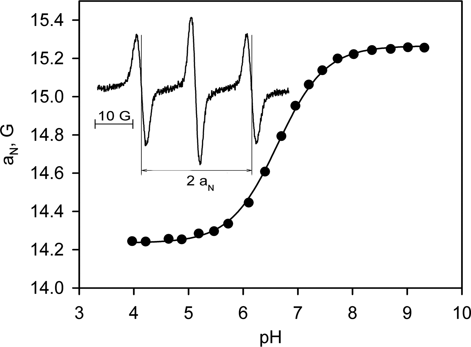
NRs and TAMs containing ionizable group with an equilibrium constant Ka , exist in aqueous solution in protonated, RH+, and unprotonated, R, forms. EPR measurements of the ratio of their concentrations allow for calculation of the pH values based on the Henderson–Hasselbalch equation, [RH+]/[R] = [H+]/Ka . Among the various NR types, imidazoline and imidazolidine nitroxides have been used in most EPR spectroscopy and imaging pH metric applications (53, 56) due to the significant changes of the EPR spectral parameters on protonation of nitrogen atom N-3 (ΔaN ≈ 1 G and Δg ≈ 0.0002) (58). Recently, pH-sensitive trityl radicals were also synthesized (7, 20). One of the great strengths of the EPR pH probes is that the method is ratiometric, the pH measurement being independent of the probe concentration but dependent on the ratio, [RH+]/[R]. Spectral simulation or spectral parameters sensitive to [RH+]/[R] ratio can be used for accurate pH determination. The spectral parameters used as pH markers include the ratio of peak intensities when RH+ and R signals are resolved, and the observed hyperfine splitting, aN, measured as a distance between the EPR lines of the triplet when RH+ and R signals are overlapped. Sensitivity of the observed aN value to pH depends on the spectrometer frequency and settings such as modulation amplitude (57).
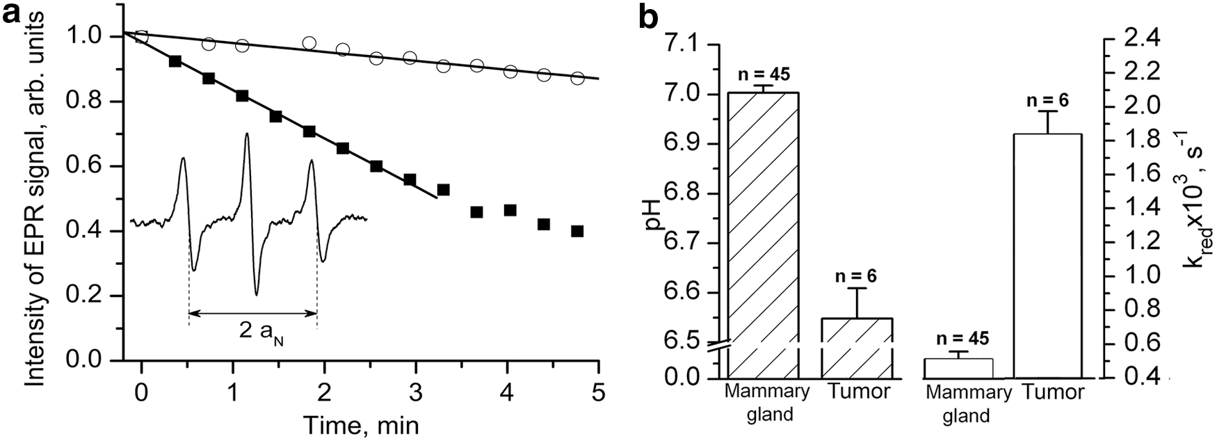
Figure 4 shows the observed aN dependence of the NR1 radical on pH described by a standard titration curve. Note that the structure of NR1 probe has been specially designed for the pH measurements in extracellular TME. First, value of the pKa of the NR1 radical, 6.6 at 37°C (10), allows for in vivo pH measurements in the range from 5.6 to 7.6 (accuracy, 0.05 pH units), which covers the range of acidic pH in solid tumors. Binding of cell-impermeable GSH tripeptide to the NR1 restricts its penetration into the cells ensuring EPR measurements of extracellular pH values (108). The bulky ethyl groups at carbons C2 and C5 provide steric hindrance around the radical fragment significantly decreasing the rate of reduction (60) in a highly reducing microenvironment.
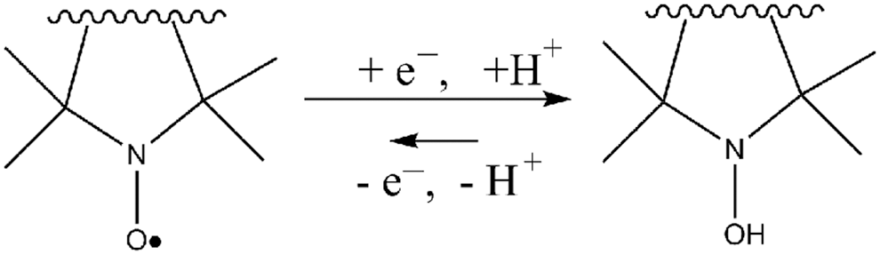
The EPR signal decay due to NR reduction to the corresponding hydroxylamines (Fig. 5) in living tissues is often considered as a limiting factor in EPR applications of the NR probes. On the positive side, the rates of the nitroxide decay allow for EPR evaluation of the tissue reducing capacity in various physiological and pathophysiological states (10, 70, 88). In its turn, cyclic hydroxylamines can be oxidized by biological oxidants into the corresponding EPR-measured NRs and provide useful information on oxidative stress and reactive oxygen species (ROS) generation. In contrast to the EPR spin-trapping approach (4, 43), oxidation of hydroxylamines is not oxidant specific and their use to measure specific ROS requires additional controls. In vitro and in vivo applications of cyclic hydroxylamine are reviewed in this Forum by Dikalov et al. (21).
An application of cell-permeable NRs allows for EPR assessment of intracellular redox status (69, 70), while application of cell-impermeable NRs allows for assessment of the reducing capacity of extracellular matrix. Note that in case of pH-sensitive NRs, redox-sensitive marker, signal amplitude, and pH-sensitive marker, aN, are independent spectral parameters, therefore making these NRs dual-function pH and redox probes. Figure 6 exemplifies the pH and redox measurements in mouse model of breast cancer (10) using NR1 probe. The data obtained for the group of female FVB/N MMTV-PyMT mice in tumors and normal mammary glands support extracellular acidosis and high reducing capacity of TME.
Note that tumor tissue is characterized by high heterogeneity, and therefore, spatially resolved functional measurements of TME are critically important, for example, allowing identification of the area with compromised redox and pH homeostasis. Both EPR imaging (13, 54, 70) and PEDRI (65, 94) were explored for redox and pH mapping. In case of PEDRI, the deuterated analog of the NR1 probe, NR2 (Fig. 2), has been used for pH mapping of TME in mouse model of breast cancer (61, 94). The narrow signal of the deuterated NR2 probe is easily saturated by EPR irradiation, which results in high PEDRI signal enhancement and allows for shorter acquisition time. PEDRI applications for pH and redox mapping of TME are discussed in a separate article of this Forum (61).
Figures 7 and 8 illustrate the applications of EPRI for pH (38) and redox (70) mapping of the TME in living mice using nitroxide probes. The three-dimensional EPR map of extracellular pH in TME of squamous cell carcinoma in right hind leg of C3H mouse was measured after i.v. injection of cell-impermeable NR1 probe (Fig. 7a). Figure 7b shows a significantly more heterogeneous and acidic distribution of pHe values in TME compared to normal tissue.
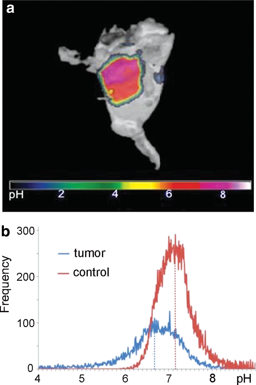
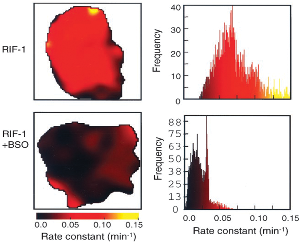
Figure 8 demonstrates the EPRI of TME reducing capacity measured in RIF-1 tumor-bearing mice using cell-permeable 3-carbamoyl-proxyl nitroxide probe (70). An observation of significant decrease in the rates of the NR signal decay for the mice treated with glutathione biosynthesis inhibitor, L-buthionine-S,R-sulfoximine, supports an important role of GSH as an intracellular redox buffer.
In vivo EPR assessment of intracellular GSH
A ubiquitous thiol-containing tripeptide, GSH, presents in virtually all mammalian tissues and plays a central role in cell biology. The intracellular GSH concentrations in mammalian cells are in the range between 1 and 10 mM and are close to half of the total thiols/disulfides in the cells. The redox couple of reduced GSH form and its oxidized disulfide form, GSSG, is a major component of intracellular redox buffer (92, 95). A metabolic reprogramming in proliferating cancer cells results in enhanced NADPH formation via the pentose phosphate pathway interconnected with glycolysis followed by facilitation of reduced GSH synthesis. High concentrations of GSH and low negative GSH redox potentials have been found in cancer cells of various tumor types (10, 33, 48, 103, 105). Furthermore, it has been demonstrated that tumor redox status can be modified by tissue glutathione level, in vivo (70). An increased level of GSH has been found in whole blood samples from patients with nonsmall-cell lung cancer (35) apparently reflecting high GSH concentration and low redox potential characteristic for proliferating malignant cells of distant malignant tissue.
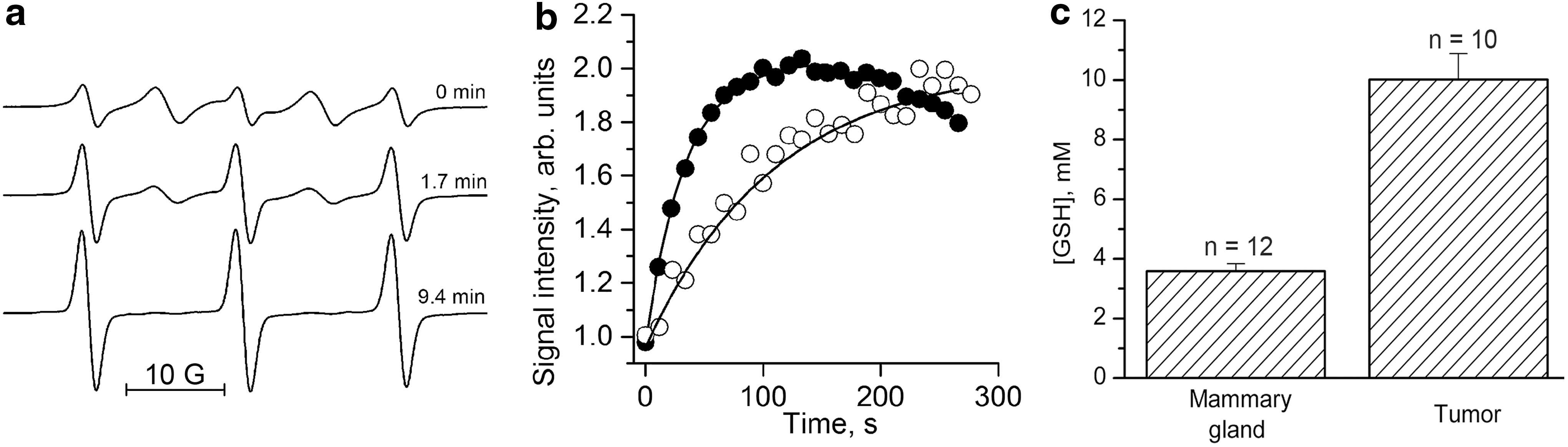
The EPR approach allowing for assessment of glutathione redox status in vivo using GSH-sensitive probes might provide a useful indicator of cancer progression and aggressiveness. Currently, EPR spectroscopy, in combination with the disulfide biradicals of the imidazolidine type (59) (see Fig. 2 for the RSSR structure) and its isotopically substituted analogs (93), represents the only method that allows for noninvasive quantitative [GSH] measurements in living subjects in preclinical applications (10, 93). The RSSR probe represents a paramagnetic analog of disulfide Ellman's reagent (24, 25), which on the reaction of thiol/disulfide exchange with GSH splits its disulfide bond resulting in formation of two monoradicals and cancelation of intramolecular spin-exchange between the monoradical fragments. Figure 9a illustrates the effect of reaction of the RSSR probe with glutathione on the EPR spectra resulting in disappearance of the “biradical” spectral components and a corresponding increase of the intensity of the monoradical components. The rate of the increase of the amplitude of the monoradical component is proportional to the GSH concentration and is a convenient GSH-sensitive EPR spectral parameter. The observed rate constant of the reaction between GSH and RSSR, kobs = 2.8 ± 0.2 M −1s−1 (T = 34°C, pH 7.2) (10). This corresponds to the time constants of exponential kinetics in the range from 0.6 to 6 min for the physiologically relevant intracellular GSH concentration from 1 to 10 mM providing an experimentally convenient time window for the EPR detection. Note that the RSSR disulfide biradical being a small lipophilic molecule easily crosses cellular membranes where it reacts with intracellular GSH providing a reliable approach for determination of GSH, in vivo (10, 93). This EPR approach relies on a major contribution of the intracellular glutathione in the pool of the thiols easily accessible for the reaction with the disulfide biradicals. Figure 9b exemplifies the kinetics of the monoradical spectral peak intensity change measured in mammary tumor (●) and normal mammary gland (○) using L-band EPR. The solid lines are the fits of the initial part of the kinetics by the monoexponent yielding [GSH] = 10.7 and 3.3 mM for the tumor and normal mammary gland, correspondingly.
Recently, new disulfide paramagnetic reagents, including disulfide biradicals of pyrrolidine type (73), trityl-nitroxide and trityl-trityl disulfide biradicals (78), were synthesized. Trityl disulfides are cell impermeable (78) and do not allow assessment of intracellular GSH. The use of pyrrolidine disulfides for EPR assessment of thiols was demonstrated in vitro (22, 73) and then in vivo for mapping thiol redox status in tumor-bearing mice (31). Further comparative studies of the rates of the penetration of the pyrrolidine disulfide nitroxides into the cells, their reaction with GSH, and reduction by intracellular reductants are required to determine the capacity of these probes for quantitative in vivo EPR measurement of intracellular glutathione concentrations.
In vivo concurrent EPR measurements of tissue pO2, pHe, and Pi
Concurrent in vivo EPR measurements of the multiple TME parameters using a single paramagnetic probe allow for their correlation analysis independent of probe distribution, time of probe delivery, and spectrum acquisition, therefore providing unique insight into the relationships between these parameters in solid tumors (45). Recently, we designed a trityl paramagnetic probe, HOPE, with unique spectral properties (8, 20) that allowed for simultaneous in vivo monitoring of tissue pH, pO2, and [Pi] in extracellular microenvironment in various animal models of cancer (9, 20). Note that 31P NMR of endogenous phosphate predominantly measures intracellular phosphate while EPR detects the HOPE probe in interstitial extracellular space: bulky hydrophilic HOPE probe does not cross cellular membrane and its EPR signal from the blood is not observed due to probe complexation with plasma albumin (96). Figure 10 illustrates functional sensitivities of the spectral parameters of the HOPE probe.

Figure 11 illustrates multifunctional measurements performed in FVB/N wild-type mammary glands and in the TME of MMTV-PyMT transgenic mice, which spontaneously develop breast cancer and emulate human tumor staging (74). The comparison of the mean values of pO2 in tumors and in normal tissues (50 ± 3 mmHg vs. 58 ± 3 mmHg, correspondingly) and pHe (6.99 ± 0.03 in TME vs. 7.1 ± 0.03 in normal tissue) supports an appearance of hypoxic and acidic regions in the tumor. The most significant several-fold differences were observed for interstitial [Pi] (1.8 ± 0.2 mM in TME vs. 0.84 ± 0.07 mM in normal tissue), indicating a potential role of interstitial Pi as TME marker of tumor progression. Figure 11e–h shows the correlation between the individual values of the TME parameters measured with the HOPE probe. The observed positive correlation between pO2 and pHe in normal tissue versus the absence of correlation between these parameters in tumors supports tumor reliance on glycolytic metabolism independent of oxygen availability—the latter exemplifies in vivo manifestation of Warburg effect. Figure 11g and h shows negative correlation between pO2 and [Pi] both in normal tissue and TME supporting association of increase in [Pi] with changes in bioenergetic status on insufficient oxygen delivery.
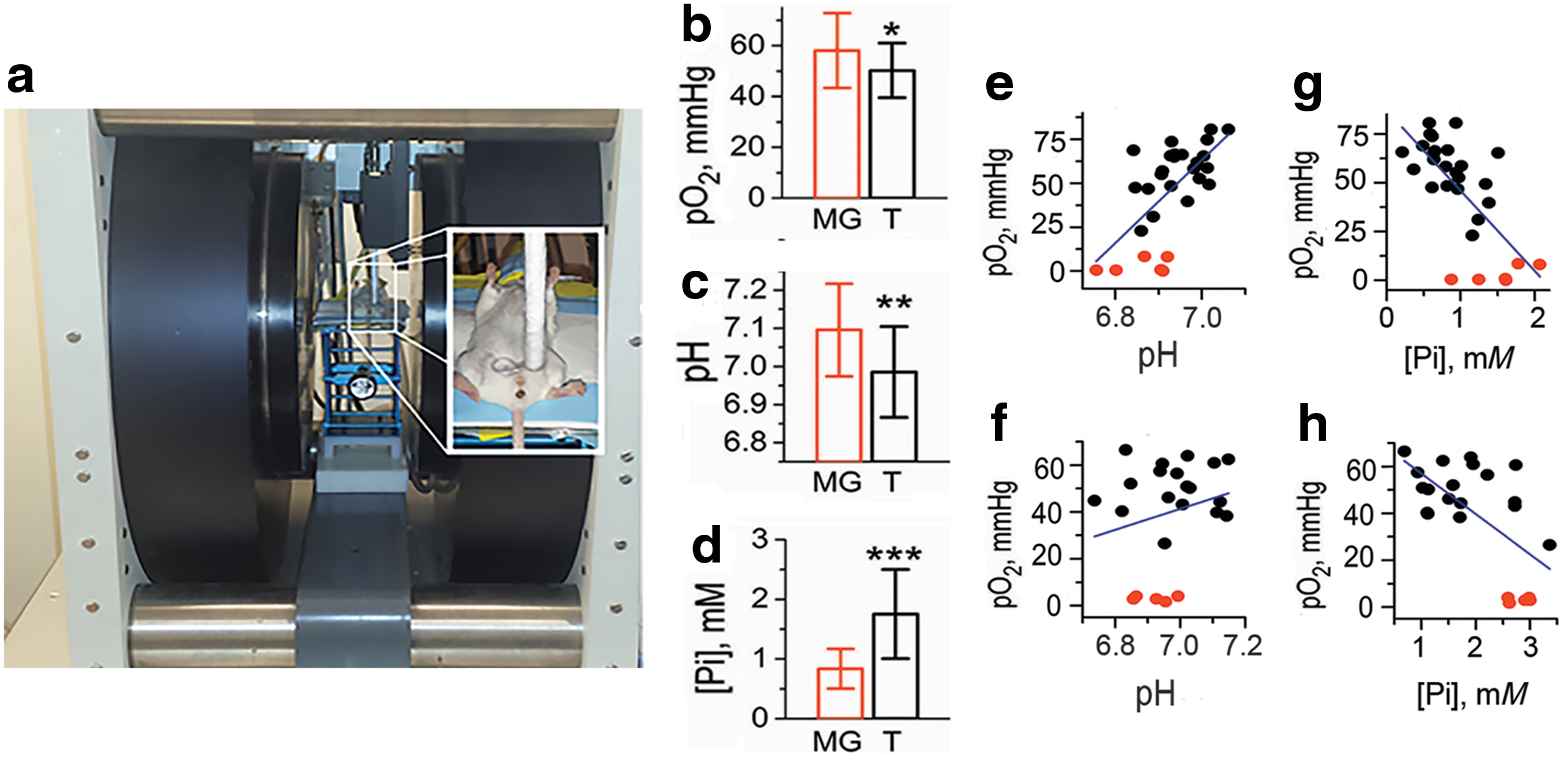
The EPR profiling of TME of nonmetastatic PC14 and metastatic PC14HM tumor xenografts using HOPE probe showed that pO2 and pHe values do not significantly differ for these two xenografts (9). On the contrary, extracellular inorganic phosphate concentrations allowed for discriminating between nonmetastatic and metastatic tumors—the measured levels of [Pi] were significantly higher in the metastatic PC14HM xenografts (9). The associate of high interstitial Pi concentrations with alterations in tumor metabolism (98, 102), Pi contribution in buffer-facilitated proton transport (39), and a high demand in phosphorus supply for the rapid growth according to the “growth rate hypothesis” (26) may underline the potential contribution in tumor progression and aggressiveness [see Ref. (9) for detailed discussion]. Further studies are required to evaluate physiological significance of the observed high TME phosphate concentrations and the amplification in highly metastatic tumors, and whether it may provide additional opportunities for TME-targeted therapy.
Janus-faced TME and EPR profiling of normalization during anticancer treatment
The increased rates of ROS generation (5, 75, 80, 87) and lipid peroxidation (32, 72) have been measured in many cancers. We hypothesized (55) that specific TME parameters deviate from the corresponding counterparts in normal tissues facilitating extracellular ROS production and exposing cancer and nontransformed cells to a highly oxidizing environment. The cancer cells are well equipped with overexpressed antioxidant defense to survive in oxidative TME. On the other side, an exposure of the normal cells to a highly oxidizing environment may result in unavoidable and fatal toxicity. This “Janus-faced” character of the TME facilitates cancer growth at the expense of normal cells. Normalizing parameters of the tissue microenvironment may decrease selection pressure for malignant phenotype. Therefore, EPR monitoring of TME parameters during anticancer treatment may provide a tool for optimization of therapy efficiency and contribute to the development of TME-targeted therapeutic approaches.
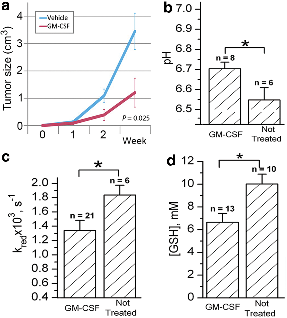
Figure 12 illustrates an example of in vivo EPR profiling of TME using functional probes, NR1 and RSSR, during the treatment with anticancer drug (10). It has been reported a significant decrease in tumor growth (Fig. 12a), angiogenesis, metastases, and pO2 level on treatment with granulocyte macrophage colony-stimulating factor (GM-CSF) in a PyMT model of breast cancer (34). Figure 12b–d shows EPR-measured TME parameters of PyMT mice bearing breast cancer tumors not treated and treated with GM-CSF. Note significant normalization of TME parameters on treatment with GM-CSF, namely, an increase of pHe, decrease of tissue reducing capacity, and GSH concentration.
Conclusions
The recent progress in EPR instrumentation designed to be used in humans (100) and the development of biocompatible paramagnetic probes with spectral sensitivity to the parameters of local chemical microenvironment eventually will make possible the translation of EPR techniques for multifunctional profiling of TME into clinical settings. These techniques will allow improving prediction power of early diagnostics for malignant transition and for future rational design of TME-targeted anticancer therapeutics.
Footnotes
Acknowledgments
This work was partially supported by NIH grants CA194013, CA192064, and U54GM104942. The WVCTSI is acknowledged for start-up to V.V.K. The content is solely the responsibility of the author and does not necessarily represent the official views of the NIH.
Author Disclosure Statement
No competing financial interests exist.