




Abstract
Significance:
Modulation of nuclear factor (erythroid-derived 2)-like 2 (Nrf2)-mediated antioxidant response is a key aspect in the onset of diabetes-related cardiovascular complications. With this review, we provide an overview of the recent advances made in the development of Nrf2-targeting strategies for the treatment of diabetes, with particular attention toward the activation of Nrf2 by natural antioxidant compounds, nanoparticles, and oxidative stress-modulating biocompatible scaffolds.
Recent Advances:
In the past 30 years, studies addressing the use of antioxidant therapies to treat diabetes have grown exponentially, showing promising but yet inconclusive results. Animal studies and clinical trials on the Nrf2 pathway have shown promising results, suggesting that its activation can delay or reverse some of the cardiovascular impairments in diabetes.
Critical Issues:
Hyperglycemia- and oscillating glucose levels-induced reactive oxygen species (ROS) accumulation is progressively emerging as a central factor in the onset and progression of diabetes-related cardiovascular complications, including endothelial dysfunction, retinopathy, heart failure, stroke, critical limb ischemia, ulcers, and delayed wound healing. In this context, accumulating evidence suggests a central role for Nrf2-mediated antioxidant response, one of the most studied cellular defensive mechanisms against ROS accumulation.
Future Directions:
Innovative approaches such as tissue engineering and nanotechnology are converging toward targeting oxidative stress in diabetes. Antioxid. Redox Signal. 36, 707–728.
Introduction
Frederick Benting, father of insulin, a discovery turning its first century this year, was quoted to say “Insulin belongs to the world, not me” when asked about his choice to symbolically sell the rights for his fresh discovery to the University of Toronto for only $1.00. Before that discovery, patients diagnosed with diabetes mellitus (DM) could only be assigned to a strict diet for the few years they had left to live. In 2019, the International Diabetes Federation estimated that 463 million people were living with DM worldwide, and expected that number to reach 700 million by 2045 (138). Currently available treatments that focused on glycemic control significantly improved patients' quality of life and life expectancy, thanks to the steady flow of technological advances in support of glycemic control maintenance.
These relatively recent achievements mainly include the use of glucose- and pH-responsive platforms to achieve glycemic control. However, artificial glycemic control in diabetic patients can only delay the onset of diabetes complications. Indeed, hardly controllable blood glucose fluctuations, as well as hyperglycemia, lead to the production of advanced glycation end products (AGEs), which in turn generate intracellular reactive oxygen species (ROS) through the activation of several receptor for advanced glycation end products (RAGE)-dependent signaling pathways, ultimately leading to insulin resistance (152).
Moreover, AGEs play a role in the onset of the so-called metabolic memory: the mechanism through which hyperglycemia and glucose fluctuations trigger the epigenetic modification of endothelial, cardiac, and vascular cells, activating a vicious circle in which oxidative stress becomes self-supporting (160).
Consequently, endothelial cells (ECs) antioxidant systems become overwhelmed, triggering the modification of a plethora of signaling cascades, including among others the downregulation of protein kinaes A and B (Akt), as well as peroxisome proliferator-activated receptor (PPAR) (157). As a consequence, the balance between ROS accumulation and endothelial nitric oxide synthase (eNOS) activity is impaired (44). These events lead to the onset of endothelial dysfunction (ED): a complex metabolic condition characterized by impaired responsiveness to the angiogenic stimuli and by disturbed cross talk between ECs and pericytes (43). Systemic ED represents one of the earliest events in the onset of most diabetes-related complications (150).
Heart failure (HF) is one of the most problematic cardiac complications of diabetes (163). HF can arise as a consequence of the fibrotic regenerative response affecting the necrotic cardiac tissue immediately after acute ischemic events such as myocardial infarction (reparative fibrosis), or, as is the case for DM, as a result of cumulative damage originating from ROS- and AGE-driven cardiomyocytes (CM) apoptosis (reactive fibrosis) (121). Both scenarios involve macrophage invasion of the necrotic area and their transition toward the M1 proinflammatory phenotype.
Wound healing is also defective in diabetic patients on account of the disturbances in the cross talk between ECs and pericytes, which creates the conditions for a diminished angiogenic potential and a reduced peripheral partial oxygen tension, ultimately affecting the regenerative potential of peripheral tissues (122).
Research on nuclear factor (erythroid-derived 2)-like 2 (Nrf2) activation-based therapeutic strategies in diabetes is recently gaining momentum since this transcription factor has been recognized as a major switch in the enzymatic antioxidant response to oxidative stress (101). Moreover, its AGE-RAGE-dependent inactivation makes it an interesting target for diabetes management (Fig. 1). Indeed, Nrf2-mediated antioxidant responses are dysfunctional in diabetes (10), and dysregulation of the Nrf2 redox pathway affects the healing of diabetic wounds (130). In line with the consolidation of oxidative stress as a central player within diabetes progression, a great effort was made to find suitable treatments to stimulate Nrf2 signaling and modulate ROS production.

Several natural antioxidant compounds, micronutrients, as well as metallic nanoparticles (NPs) have been associated with a pro-Nrf2 activity (81). Inorganic, metallic NPs gathered remarkable attention in several therapeutic applications, including, among others, wound healing (167) and diabetes (21). Thanks to their impressive versatility, to their intrinsic antioxidant potential, and to the possibility to finely tune their surface characteristics, NPs can be engineered to target specific tissues or metabolic milieux (121), such as chronically inflamed tissues in diabetic patients, and therefore might represent a valuable tool for finalizing the therapeutic potential demonstrated by natural antioxidant compounds.
Moreover, tissue engineering (TE) currently offers a growing number of suitable platforms for the treatment of diabetes-related complications, with a particular focus on vascular restoration and chronic diabetic wound healing (15). Particular interest was reserved to the possibility for scaffolds to regulate the equilibrium between oxidant and antioxidant processes (30), and to their intriguing potential role as ROS-responsive carriers of antioxidant compounds (191).
Hence, in this review, we aim to offer a comprehensive and up-to-date overview of the synergy that is emerging between TE, metallic NPs, and antioxidant therapy in the quest for an effective treatment of diabetes progression, with a particular focus on cardiovascular-related complications (Fig. 2). The narrative will first contextualize Nrf2 activation within the scope of diabetic cardiovascular complications; second, the well-studied effect of natural antioxidant compounds on Nrf2 activation will be addressed; finally, the more recently emerging approaches involving NPs and TE will be discussed.

Nrf2 Signaling in Diabetes and Its Cardiovascular Complications
Nrf2-mediated antioxidant response: an overview
Nrf2 is a master gene regulating the antioxidant response to ROS accumulation and is expressed in several human tissues (31, 200). Regulation of its activity relies almost entirely on its interaction with Kelch-like ECH-associated protein 1 (Keap1), a 624 amino acid, homodimeric protein, which in turn is bound to cullin3–ring box1 (Rbx1) cytoplasmic, E3 ubiquitin ligase complex (84). In the absence of oxidative stress, the Keap1-cullin3-Rbx1 complex keeps Nrf2 within the cytoplasm, consequently inhibiting its transcriptional activity and inducing its proteasome 26S-driven digestion.
Upon high glucose–induced oxidative stress, key Keap1 cysteine residues are oxidized by upstream antioxidant response elements (AREs) activators (e.g., pro-oxidants; electrophiles including ROS, reactive nitrogen species [RNS], and antioxidant compounds; and phase-2 enzyme inducing-compounds) triggering the release of Nrf2 from the E3 ubiquitinase complex and its casein kinase (CK2) phosphorylation-dependent translocation into the nuclear compartment (68, 125). Through its heterodimeric interaction with small Maf protein (sMaf), Nrf2 is able to bind with ARE DNA sequences, leading to the transcriptional activation of several genes involved in the antioxidant response (Fig. 3).
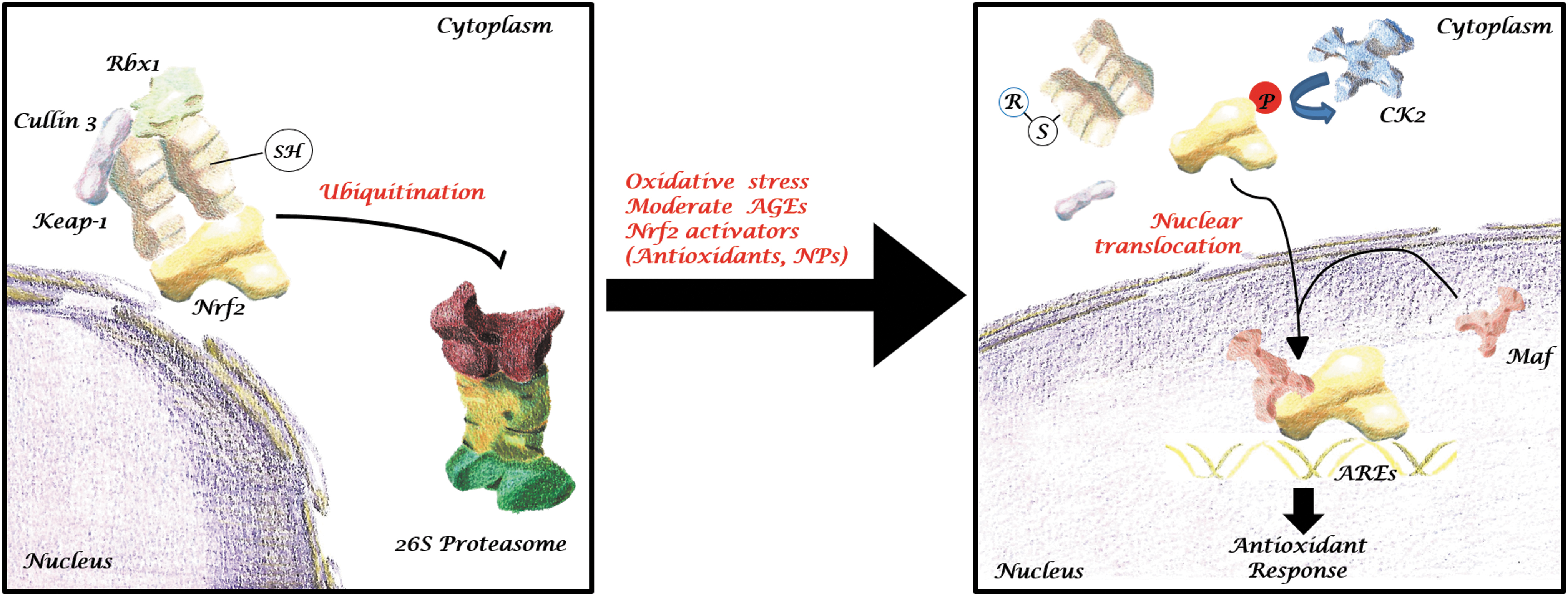
Keap1 exists as a homodimer in the cytoplasm and, at a minor extent, in the nucleus and endoplasmic reticulum (176). Each monomer is composed of three functional domains, namely broad-complex, tramtrack, and bric a brac domain (BTB), intervening portion (IVP), and Kelch domain (36). BTB is responsible for Keap1 homodimerization, interaction with cullin3 and Rbx1, and, importantly, it hosts C151, one of the 27 reactive cysteine residues that confer to Keap1 the ability to detect redox fluctuations (48). The inner domain, IVP, is the major responsible for Keap1 sensitivity to electrophiles as it bears the remaining 26 reactive cysteines. Finally, each dimer contains two Kelch domains, responsible for Keap1 interaction with Nrf2 and several other competitive targets.
Nrf2 activation in diabetes
Diabetes can affect Keap1-Nrf2 interaction at several levels, which encompass their transcription, post-translational modification, and degradation. Indeed, similar to what is observed with aging, DM is associated with a progressive epigenetic deregulation of the KEAP1 genomic region, which causes a concomitant increase in its expression, thwarting antioxidant response potential (106). KEAP1 is constitutively expressed, although its half-life, normally ≈13 h, largely depends on the extent of its p62\SQSTM1-dependent degradation, which can be accelerated by electrophiles such as tert-butylhydroquinone or 1,2-naphthoquinone, and is accelerated by ubiquitination (155).
p62\SQSTM1 is a component of the macroautophagic machinery, the constitutive process through which the cell clears itself from damaged organelles and recycles its building blocks. Specifically, p62\SQSTM1 binds to Keap1 Kelch-like domain in an Nrf2-competitive way and targets it to inclusion bodies, which content is degraded during selective autophagy (71). A number of studies demonstrated that autophagosomes accumulate in CM of diabetic mice (105, 118, 137) due to both increased production and reduced degradation (18, 105). p62 was shown to accumulate in the cytoplasm as a consequence of diabetes-related autophagy impairment (41). Therefore, p62/Keap1/Nrf2 might play a role in the initial defense mechanism against DM-induced ROS accumulation.
Moreover, the prolonged exposition to high levels of insulin eventually leads to the constitutive activation of the mTOR/p70S6K pathway, causing insulin resistance through insulin receptor substrate 1 (IRS1) serine phosphorylation (165). The resulting inhibition of the IRS1/PI3K/Akt pathway impairs both autophagy and, through the depletion of Akt-induced increase in Nrf2 expression and nuclear translocation, antioxidant response (68, 75). However, deregulation of autophagy is associated with cytoplasmic accumulation of p62\SQSTM1 (171), which in turn sequestrates Keap1, amplifying ROS-driven Nrf2 nuclear translocation (177). Importantly, also physical exercise was proven to inhibit Nrf2 degradation through inhibition of its interaction with Keap1 in mice diabetic model (110, 131) as well as in a rat model (193).
Nrf2 activity in the context of diabetic cardiovascular complications
Transient activation of Nrf2, followed by its downregulation, was observed during the onset of diabetic ED. ED is the initial event in macrovascular complications such as coronary artery disease, peripheral arterial disease, stroke, and microvascular complications leading to nephropathy, neuropathy, wound healing impairment, and retinopathy. The etiology of ED is different between type 1 diabetes mellitus (T1DM) and type 2 diabetes mellitus (T2DM). Indeed, while the insurgence of ED in T1DM is mainly triggered by hyperglycemia alone, in T2DM also insulin resistance and circulating fatty acids play a role (54, 74). Considering the effects of hyperglycemia on the endothelium at the molecular level, one of the first steps toward the development of ED is represented by the inhibition of the enzyme glyceraldehyde 3-phosphate dehydrogenase.
This event causes the accumulation of glycolytic metabolites, leading to the activation of numerous metabolic pathways, such as protein kinase C, a known activator of Nrf2, sorbitol, but also methylglyoxal (MG) and AGEs, therefore increasing oxidative stress and apoptosis (35, 64).
However, insulin resistance prevents glucose transporter GLUT4 plasma membrane translocation in endothelial as well as skeletal muscle and adipose cells, reducing nitric oxide production through the downregulation of eNOS and further increasing vasoconstriction (82). Moreover, hyperinsulinemia exacerbates the shift toward a proinflammatory state of the endothelium, orchestrating the overexpression of adhesion molecules such as E-selectin and VCAM1, together with the vasoconstrictor endothelin-1, which increases oxidative stress through the activation of NAD(P)H oxidase (32, 40).
ROS accumulation has been reported in ED models also on account of xanthine-, aldehyde- and glucose-oxidase hyperactivation (46, 112, 162). A major source of ROS is beyond doubt the mitochondrion: ROS accumulation in such organelles is associated with altered calcium equilibrium, which is at the basis of several DM cardiovascular complications (51, 154). Importantly, an excessively oxidant microenvironment triggers the oxidation of eNOS cofactor BH4 to BH2, hindering eNOS homodimerization. Monomeric eNOS is unable to synthesize NO, and synthesizes O2- instead, exacerbating oxidative stress (47).
Therefore, amelioration of oxidative stress conditions into the EC milieu is vital to alleviate the onset of ED in diabetic patients. At the same time, a protective effect was described for Nrf2-free Keap1 against RNS and ROS accumulation. Indeed, Keap1 Kelch domain, when freed from Neh2, its target domain on Nrf2, can interact and create a complex with GAPDH and eNOS. When this complex is formed, C151 on Keap1 BTB domain acts as a sensor for RNS, triggering antiapoptotic post-translational modifications of key factors (86).
A significant reduction in Nrf2 binding activity to AREs was observed in the peripheral blood mononuclear cells (PBMCs) of diabetic and prediabetic patients compared with nondiabetic control. PBMCs are a heterogeneous cell population originating from the bone marrow, including lymphocytes, natural killer, and several subclasses of proangiogenic and hematopoietic progenitors, all of which are pivotal for several aspects of peripheral inflammation and tissue regeneration. Interestingly, diabetic patients without medical supervision showed significantly lower Nrf2 nuclear localization in PBMCs than prediabetic subjects and diabetic patients with medical supervision, suggesting that glycemic control is vital to maintain Nrf2 levels (72).
Nrf2 inactivation has also been observed in a model of hind limb ischemia, where uncontrolled oxidative stress damaged ECs function, resulting in impaired angiogenetic potential (45). Critical limb ischemia is considered the end stage of diabetic ED in the scope of peripheral circulation characterized by chronic ischemic pain, skin ulcerations, and gangrene, resulting from ED-dependent angiogenic deficit aggravated by the insurgence of neuropathy and ischemic events (150, 151). Nrf2 is upregulated in keratinocytes in the early phases of wound healing, triggering the secretion of Ccl2, a macrophage chemotactic agent, which in turn induces epidermal growth factor (EGF) secretion by macrophages, therefore stimulating keratinocytes proliferation and wound closure (168).
Consistent with the deleterious effects of diabetes and CLI on wound healing, a reduced amount of Nrf2 mRNA and protein was reported in diabetic patients' skin (91). Moreover, pharmacological stimulation of Nrf2 into diabetic mice wounds significantly shortened wound closure time (129).
In the context of HF, Nrf2 nuclear translocation deficiency and the consequent increase in ROS signaling are associated with increased fibrosis and reduced CM contractility and viability (24, 200). In diabetic human left ventricles obtained from autopsied hearts, Nrf2 nuclear translocation was significantly reduced compared with control hearts and was associated with ROS-dependent development of insulin resistance (156). Moreover, the same team demonstrated how after an initial upregulation, Nrf2 expression drops during diabetes progression as a consequence of antioxidant response impairment (94).
As can be expected while appreciating the protective role of Nrf2, its overactivation in several cancers in vivo and in vitro has been described, raising a warning concerning the pursuit of its overstimulation for therapeutic purposes (92). Notably, a large number of malignancies were found to bear mutations in Nrf2 and/or Keap1 (34). Concern about Nrf2 uncontrolled overexpression should not keep us from pursuing the stabilization of the innate antioxidant response as a therapeutic approach for the treatment of diabetes-related cardiovascular complications but should instead encourage a cautious approach (31).
Phytochemical Antioxidants Targeting Nrf2 in Diabetes and Its Cardiovascular Complications
Phytochemical compounds targeting Nrf2 in diabetes
Since the discovery of Nrf2 as a central player in oxidative stress response, several studies addressed the application of phytochemical compounds able to enhance its activity in the management of diabetes and its complications (108, 149, 175). A pivotal role in the onset of diabetes, especially type 1 diabetes, is played by hyperglycemia-induced apoptosis in pancreatic β-islets. Nrf2 activation by phytotherapy antioxidants exerts a protective function on insulin-producing cells (8, 22, 79, 90, 132, 149, 199).
Treatment of insulinoma cell lines with naringenin, an antioxidant compound isolated from citrus fruit (132), or with sulforaphane, an isothiocyanate abundant in cruciferous vegetables (149), was found to increase Nrf2 expression and nuclear translocation in a dose-dependent manner. Sulforaphane and quercetin can also induce miR-let7 expression, which positively regulates Nrf2 signaling (7).
In streptozotocin (STZ)-treated mouse MIN6 insulinoma cell line, oral administration of naringenin induced Nrf2 nuclear translocation and triggered the upregulation of Nqo-1 and glutathione S transferase gene expression, leading to a partial recovery in their viability and ROS production (132). Notably, the effective concentrations of naringenin alone caused a mild reduction in MIN6 cell viability and a slight increase in ROS production compared with the negative control, which might represent the triggering mechanism by which Nrf2 expression is increased by naringenin.
Subcutaneous injection of sulforaphane-mediated Nrf2 nuclear translocation exerting a protective effect from NF-κB-mediated cytokine damage through the activation of phase 2 antioxidant enzymes (149, 175), restoring insulin production in RINm5F rat insulinoma pancreatic β cells exposed to high glucose damage (149).
The administration of naringenin (132), withaferin A (159), or sulforaphane (149, 199) to STZ-induced diabetic mice resulted in an increased expression of Nrf2 and a partial recovery of insulin (132, 159) and glucose blood levels (132, 159, 199). Similar results were obtained in a rat model: the administration of aspalathin, a polyphenolic compound present in Aspalathus linearis extract, to STZ-treated β-pancreatic cells reduced apoptosis and partially recovered viability loss in a dose-dependent manner (108). Moreover, in the same study, aspalathin induced Nrf2 nuclear translocation, increased the expression of antioxidant response genes Nqo-1, superoxide dismutase 1 (SOD1), and heme oxygenase-1 (HO-1), and significantly reduced the negative effect of H2O2 and high glucose insults on insulinoma β cell line 1E (INS1E) cell viability.
Interestingly, aspalathin induced the accumulation of p62, which resulted in Keap1 sequestration and constitutive Nrf2 signaling activation, thus providing a hypothetical mechanism of action for this antioxidant compound. Glycemic control amelioration through the activation of the canonical Nrf2 pathway was also observed in MG- (22, 90), STZ- (8, 119), and Goto-Kakizaki (79) rat diabetic models. Upon oral administration of the antioxidant and PPARγ-agonists ankaflavin (90) and scopoletin (22) to the MG diabetic rat model, Nrf2 phosphorylation on serine 40 was also associated with reduced serum, pancreatic, and hepatic levels of AGEs. In addition, ankaflavin treatment significantly reduced the MG-triggered increase of RAGE expression in PBMCs (90).
Targeting of Nrf2 activity by phytochemical compounds also proved effective in increasing glucose uptake (26, 95, 159, 174, 190). Indeed, resveratrol (26) and 7-hydroxycoumarin (95) markedly ameliorated insulin resistance and glucose uptake in Hep G2 human hepatoma cell line through Nrf2 expression enhancement (95) and activation (95, 174). Notably, resveratrol-induced Nrf2 phosphorylation was found to be triggered by extracellular signal-regulated kinases (ERK) signaling and not through p38 (26). Similarly, dietary oleanolic acid (174) and Lycium barbarum polysaccharide (190) were found to reduce hepatic insulin resistance in mice diabetic models through an Nrf2-mediated reduction of ROS production, resulting in increased glucose uptake, reduced glycogen lysis, increased glycogen synthesis, reduced gluconeogenesis, and improvement in total body and liver weight profile.
Phytochemical antioxidants targeting Nrf2 in diabetic cardiovascular complications
A growing number of studies report positive effects on diabetic nephropathy and neuropathy achieved through Nrf2 activation by natural antioxidants (8, 62, 79, 109, 111, 119, 195, 196, 199). However, the most critical aspect of diabetes progression is the onset of cardiovascular complications. Loss of balancing between ROS production and antioxidant response leads to chronic inflammation, which in turn enhances local ROS production, installing a self-perpetuating vicious circle deleterious for the vasculature and for the peripheral tissues (43, 160). Therefore, great effort has been dedicated to the study of the Nrf2 pathway activation by phytochemical antioxidants in this scope (37, 134, 141, 164, 170, 187, 197).
Nrf2 activation by sulforaphane (187), aspalathin (37), or diallyl trisulfide (DATS) (164) supplementation in cell culture media was observed in hyperglycemic conditions and was shown to improve antioxidant response in vitro on a model of hyperglycemia-induced ED (187), in addition to preventing apoptosis in rat CM (37, 164). Moreover, human microvascular endothelial cells treated with Barleria lupulina hot water extract-derived Nrf2 activators, 4-ethylcatechol, 4-vinylcatechol, or 4-methylcatechol, displayed reduced stress fibers accumulation and increased expression of the cell junction molecule claudin-5, in line with an improved endothelial barrier functionality (141). Particularly interesting data were harvested from mice diabetic models of diabetic cardiomyopathy (37, 170, 197).
Both subcutaneous administration of sulforaphane (197) and oral administration of resveratrol (170) or aspalathin (37) to diabetic mice models resulted in an NrF2-mediated protective effect against cardiac complications. In particular, sulforaphane and resveratrol administration resulted in the Nrf2-mediated reversion of diabetes-induced 4-HNE overexpression (170, 197). As a result, diabetes-induced cardiac remodeling, adipose tissue accumulation, and inflammation were reduced (170, 197). In the rat model of diabetic cardiomyopathy, dietary DATS (164) and
Despite the wide amount of experimental data in support of the validity of natural antioxidant compounds as therapeutic agents against the progression of diabetes and its complications, clinical interventions failed to deliver conclusive results to date (142). One possible explanation for this so-called “antioxidant paradox” may be found in the mutually triggering nature of oxidative stress and inflammation, and in the failure of the explored candidate antioxidant drugs to efficiently counteract at the same time both conditions (19).
NPs as Nrf2 Activators in Diabetes and Its Cardiovascular Complications
Several classes of NPs have been shown to interact with Ngf2-dependent antioxidant response, including bare metallic NPs, functionalized metallic NPs, and biologically originating NPs such as exosomes and microvesicles. The following sections will discuss the interplay between these classes of NPs and their downstream Nrf2 pathway-related molecular targets.
Metallic NPs targeting Nrf2 and ROS accumulation in diabetes
Metallic NPs are emerging as a novel and promising tool for the regulation of ROS production and inflammation (Fig. 4), both through Nrf2-dependent and Nrf2-independent mechanisms. Gold nanoparticles (AuNPs) were shown to induce Nrf2 nuclear translocation in human vascular ECs (89), macrophages, and keratinocytes (49), whereas silver nanoparticles (AgNPs) triggered Nrf2 activation in Caco-2 cells (Caco2). Aortic ECs showed a marked increase in Nrf2 protein expression in rats treated intratracheally with CeO2 (cerium oxide) nanoparticles (CONPs) (115). Metallic NP-mediated activation of the Nrf2 signaling pathway was suggested to take place through three different mechanisms: (i) direct interaction with Keap1 thiol groups, (ii) mild direct generation of ROS, and (iii) indirect ROS generation by binding to the antioxidant cytoplasmic molecule GSH (49, 83, 182).
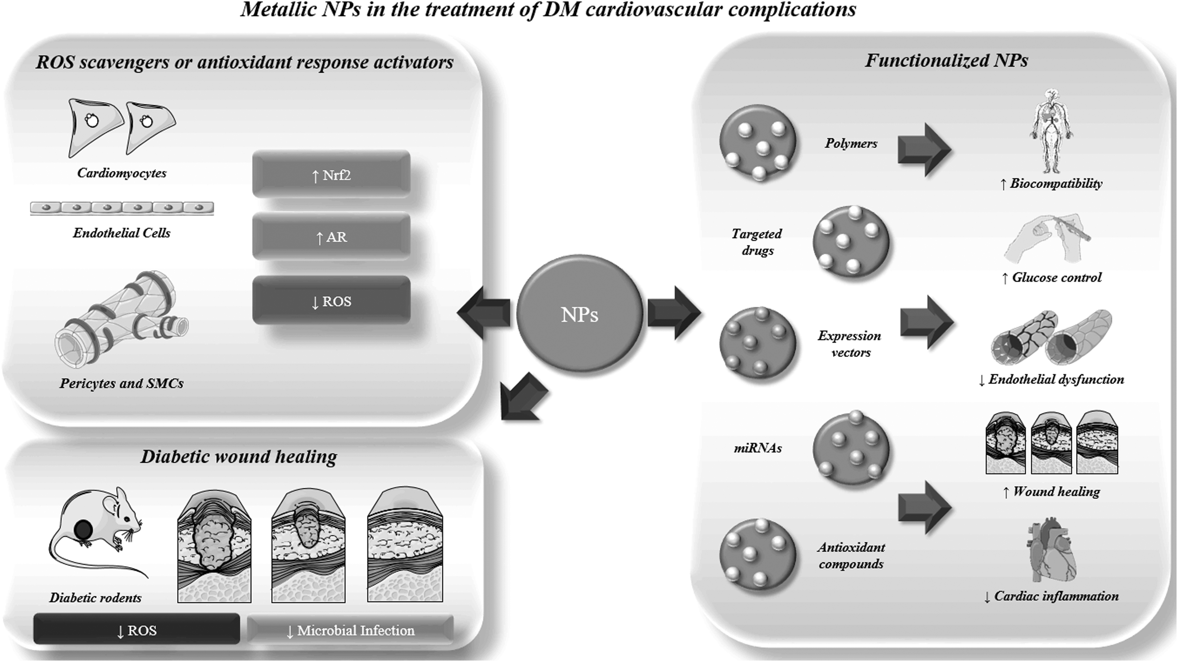
Moreover, NP-mediated Nrf2 nuclear translocation was associated with p65-NF-κB pathway inhibition (81). Although NPs are often used as ROS inducers, as is the case in the treatment of cancer or bacterial infections, a growing number of studies report their successful application as ROS modulators in the treatment of diabetes and its cardiovascular complications (167, 189). Indeed, as a rule of thumb, NPs show a biphasic effect when it comes to ROS-scavenging/stimulation effects (100, 131). Whereas low concentrations of NPs trigger the activity of antioxidant enzymes such as catalase and SOD, as it is desirable when dealing with diabetes and its complications, high concentrations of NPs can overwhelm such antioxidant systems, making a reliable and target-specific weapon for cancer treatment.
This latter behavior comes as a result of (i) NP accumulation to the target tissue through enhanced permeability and retention (EPR) and\or cancer environment-specific molecular targeting, and (ii) NP-triggered apoptosis of target cancer cells through mitochondrial membrane depolarization and consequent oxidation of key molecules in the electron transport chain such as NADPH and NADPH oxidase (50, 100). Indeed, excessive NP concentrations are associated with cytotoxicity in vitro (16, 87) and neutrophils activation, which leads to an additional ROS production in vivo (100).
Physicochemical properties at the basis of such behaviors all reside in NP surface to volume ratio, size, and chemical composition: a higher surface on volume ratio is associated with higher reactivity, whereas the chemical composition of the surface, as well as the electron density and distribution, determine the NP interactome (1).
Recently, a growing number of nanomaterials showed antioxidant properties and are often referred to as “nanoantioxidants.” Nanoantioxidants can be sorted into two categories based on their chemical composition: NPs bound to antioxidant drugs and NPs with intrinsic antioxidant properties. The latter category includes several metallic NPs, namely CONPs, YONPs (yttrium oxide nanoparticles), CuNPs (copper nanoparticles), vanadium pentoxide, manganese dioxide, ZONPs (zinc oxide nanoparticles), iron oxide, cobalt oxide, AuNPs, AgNPs, and platinum. Metallic NPs, in general, have shown promising results in vitro and in animal diabetic models (Drosophila melanogaster, mice, and rat) concerning their Nrf2-mediated ROS modulating activity (3, 9, 59).
Metallic NPs targeting Nrf2 in diabetic cardiovascular complications
Glycemic control is fundamental to avoid AGE-mediated onset of ED, at the onset of diabetic cardiovascular complications. In vitro models of oxidative stress-mediated inhibition of insulin secretion demonstrated that metallic NPs pretreatment exerted a protective effect on pancreatic β cells (61, 127, 158). Pretreatment in a culture of isolated β cells (158) or explanted β-pancreatic islets (61, 127) with CONPs and YONPs alone or in combination reduced the negative effect of H2O2 on cell viability (61, 127), restoring insulin secretion (158). CONPs (77, 79, 81), YONPs (80), superparamagnetic iron oxide NPs (4), and AgNPs (5, 77) administration to pharmacologically induced diabetic rats improves glycemic control by increasing the antioxidant response (77, 80, 81) and reducing AGEs (5).
In STZ-treated rodents, an almost complete recovery to normal plasma glucose levels was reported after 14 days of oral AgNP and CONP administration (77). Similar results were obtained with STZ-treated pregnant mice in a model of gestational diabetes (166). In mice, AuNP intraperitoneal injections restrained STZ-induced hyperglycemia through phosphatidylinositol 3 phosphate kinase (PI3K)–Akt-mediated Nrf2 activation (17, 102).
Metallic NPs have been proven a valuable tool in the treatment of several cardiovascular complications of DM (11, 12, 16, 25, 134, 138, 145, 183, 184, 192) (Table 1). Indeed, in vitro studies demonstrated that CONPs and AgNPs could reduce ROS production in vitro in CM (11, 192) and human umbilical vein endothelial cells (134), whereas AuNPs reduced ROS production in rat aortic smooth muscle cells by Nrf2-mediated HO-1 transcription enhancement (83). An in vitro study conducted on human vascular ECs reported an ROS generation-independent increase in Nrf2 protein levels and nuclear translocation upon AuNP administration. Chromatin immunoprecipitation analysis revealed that following nuclear translocation, Nrf2 was bound to the HO-1 promoter, enhancing its expression (89).
Pure Metallic Nanoparticles Tested in Preclinical Studies on Diabetes-Related Cardiovascular Complications to Date and Relative Outcomes
AgNPs, silver nanoparticles; ALA, α-lipoic acid; AuNPs, gold nanoparticles; AuNRs, gold nanorods; CM, cardiomyocytes; CONPs, cerium oxide nanoparticles; CuNPs, copper nanoparticles; Cx-43, connexin-43; EGCG, epigallocatechin gallate; ER, endoplasmic reticulum; HMECs, human microvascular endothelial cells; HUVECs, human umbilical vein endothelial cells; MCPIP, monocyte chemotactic protein-induced protein; MHC, myosin heavy chain; PHBV, poly(3-hydroxybutyrate-co-3-hydroxyvalerate); RAGE, receptor for advanced glycation end products; ROS, reactive oxygen species; STZ, streptozotocin.
Moreover, CONP-mediated reduction of ROS production increased CM viability in response to high glucose (192), reduced autophagy and aberrant tube formation in an in vitro model of diabetic retinopathy (134), and increased human mammary EC adhesion and proliferation on synthetic wound dressing membranes (12). AgNPs helped to restore cell size and lipid peroxidation in H9c2 cardiomyoblasts subjected to high glucose-induced oxidative stress (11). Recent observations point to a pleiotropic effect on ROS production by CONPs, involving both direct ROS scavenging and a mild Nrf2 pathway preactivation (67, 135).
Metallic NPs are also gathering great attention in the treatment of chronic diabetic wounds on behalf of their angiogenic (11, 161, 175, 176) and antimicrobial (16, 138, 167) activities. For example, CuNPs show antibacterial properties against many bacteria (16, 138) making them an attractive tool for the dressing of diabetic wounds and ulcers. The antimicrobial properties of CuNPs reside in the release of Cu2+ ions, which are associated with a concentration-, formulation- and release dynamics-dependent degree of cytotoxicity (183, 184).
Integration of copper ions into folic acid-containing metal–organic framework (MOF) (183, 184), resulting in Cu-MOF NPs, or in polycaprolactone (PCL) films (16), significantly reduced toxicity. Moreover, Cu-MOF NPs enhanced cell migration in vitro (183, 184) and significantly enhanced angiogenesis in a mice model of diabetic chronic wound healing, alone or in combination with a hydrogel carrier, when compared with vehicle control (183). Similarly, high but not low concentrations of AgNPs were associated with increased cardiac ROS production in ex vivo perfused hearts of diabetic rats (133).
However, incorporation of AgNPs within cellulose nanocrystals enhanced wound healing in a mice diabetic model through transforming growth factor-β (TGF-β) and platelet-derived growth factor β-driven stimulation of resident cells (140). AgNPs also proved helpful in reducing AGE-induced retinal ED derived edema in an in vitro rat diabetic model (143). Moreover, in vivo preclinical studies already confirmed metallic NPs as valuable tools in wound healing of mice diabetic models, through stimulation of keratinocytes and fibroblasts proliferation and migration (25, 99, 125, 183, 184).
Functionalized antioxidant metallic NPs in diabetic cardiovascular complications
Functionalization of metallic NPs have been explored to ameliorate their targeting efficiency, biocompatibility, and to boost their therapeutic potential. Polyethylene glycol (PEG) binding to AuNPs, or PEGylation, ameliorated NPs biocompatibility by reducing AuNP-induced platelet aggregation (140). Aluminum nanoparticles (AlNPs) allowed improving the short half-life (13 h) of liraglutide, a recently approved drug-targeting glycemic control in diabetes that should normally be assumed through daily injections (57). Complexing of liraglutide with tannic acid and AlNPs, indeed, resulted in the prolonged release of the drug, achieving extended glucose control for up to 5 days with a single injection and, importantly, reducing the incidence of cardiovascular complications in a diabetic mice model (57).
Functionalized antioxidant NPs in ED
Functionalized metallic NPs have been tested in the treatment of ED (38, 66). Oral administration of AgNPs functionalized with docosahexaenoic acid to STZ-induced diabetic rats significantly ameliorated systemic ED by lowering red blood cells membrane cholesterol and triglycerides, while improving plasma nitric oxide content (66). In proliferative diabetic retinopathy, ED is characterized by the inhibition of pericytes regulatory activity on ECs, resulting in their hyperproliferation, which leads to the formation of a dense network of highly fenestrated capillaries and edema (53).
Oral administration of AuNPs functionalized with resveratrol, an Nrf2 activator, reduced edema and hyperproliferation of ECs in an STZ-induced diabetic rat model through the inhibition of ERK and NF-κB signaling (38). These results were confirmed by further observations concerning tumor microenvironments, where tannic acid-functionalized AuNPs normalized the tumoral angiogenesis by inhibition of smad 2/3 fibrotic signaling, enhancing pericytes coverage and increasing tight junctions between ECs (65).
Functionalized antioxidant metallic NPs in chronic wound healing
The healing of chronic wounds is also inefficient in diabetic patients as a consequence of ED-related angiogenic deficit (52). Topical administration of ultra-small (7 nm in diameter) AuNPs conjugated with antibacterial peptide LL37 and with vascular endothelial growth factor (VEGF) plasmid vector onto diabetic mice wound models resulted in a synergistic antibacterial effect between AuNPs and the antimicrobic peptide, accompanied by an increase in angiogenesis due to enhanced efficiency in the transfer of VEGF expression vector, eventually leading to faster wound closure rates (171).
AuNPs were also found to decrease RAGE expression in mouse fibroblasts when administered in combination with EGCG and ALA (AuEA) (26). Topical administration of AuEA on diabetic mice wounds caused an increased expression of proangiogenic factors VEGF and angiopoietin 2, significantly accelerating the wound healing process (26).
Functionalized antioxidant NPs in HF
Accumulation of therapeutic NPs to the microenvironment of the failing heart can be achieved through either active or passive targeting. Passive targeting, alternatively described as EPR, which capitalizes on the increased permeability observed in the fibrotic tissue of the failing heart, proved quite effective for metallic antioxidant NPs by virtue of the high level of ROS in the inflamed cardiac wall, especially for imaging purposes (58). Anyway, active targeting of specific molecular targets through the functionalization of a wide array of NP species is also gathering attention since it allows for higher specificity and for the possibility to achieve additional therapeutic effects (148). Through the reaction with cationic gold salts, AuNPs can be modified into various shapes, including cylindrical nanorods (NRs) (33).
In an in vitro examination, gold nanorods (AuNRs) were functionalized with polyelectrolytes and polystyrene sulfonate and integrated into a three-dimensional (3D) collagen matrix seeded with cardiac fibroblasts (147). In this setup, AuNRs enabled the modulation of cardiac fibroblasts switch to myofibroblast phenotype. When AuNRs were integrated into gelatin methacrylate hydrogels, a fourfold increase was generated in rat CM adhesion after 24 h, with positive effects also observed on CM viability and beating synchrony at high rates (114). Results were mostly attributed to the measured change in Young's modulus and porosity of the scaffold. AuNRs were also integrated into collagen scaffolds seeded with rat CM, producing an increase in cell adhesion and intercalated disk formation detected by connexin-43 staining (127).
The study also revealed that the increased formation of intercalated disks was dependent on the integrin β1\pAKT\GATA4 signaling pathway, suggesting a role for the observed increase in stiffness at the nanoscale upon AuNR integration in the scaffolds. Peptide-modified NPs can be obtained with the advantage of a stronger interaction with hydrogels or scaffolds for their delivery and retention, conferring to such scaffolds additional antioxidant and/or antimicrobial properties. It is the case for AuNPs and AgNPs that, upon CGS-LL37 peptide functionalization and integration in an injectable collagen hydrogel, were shown to induce M2 macrophage polarization in vitro (60).
A novel NP containing 25Mg isotope and porphyrin MC16 as a nanocarrier proved beneficial in a rat model of pharmacologically induced HF, resulting in an amelioration of the oxidative stress profile accompanied by a restoration of heart rate and blood pressure (13). In a mice model of diabetic cardiomyopathy worsened by ovariectomy-induced estrogen deficiency, tail injection of AuNPs functionalized with an antagonist of the M1 promoter miR-155 (antago-miR155) increased M2/M1 macrophage ratio while reducing CM apoptosis and ameliorating heart function (69).
Biological NPs targeting Nrf2 in diabetic cardiovascular complications
The Nrf2 signaling pathway is also influenced by extracellular vesicle (EV)-mediated paracrine signaling (93). EVs are a heterogenic group of cell-derived vesicles including exosomes (ø 40–150 μm) and microvesicles (ø > 150 μm) (37).
While microvesicles are shed directly from the plasma membrane, exosomes originate within the endosome from the invagination of the endosomal membrane and are released in the extracellular milieu upon fusion of the endosomal membrane with the plasma membrane. EVs can deliver messages in the form of signaling molecules including proteins and several classes of coding and noncoding RNAs to distant recipient cells and have been shown to play an important role in several pathological and physiological processes. For instance, adipocyte-derived stem cell exosomes exert a beneficial effect on T1DM through several pathways, including modulating the immune cell response (116).
A growing number of miRNAs is being associated with the regulation of Keap1-Nrf2 signaling axis (27). In a rat model of HF, it was shown that cardiac cells from the left ventricle secreted large numbers of exosomes enriched in miRNAs associated with Nrf2 pathway dysregulation upon TNF-α proinflammatory stimulation (161). However, miR-200a has been found to ameliorate diabetic retinopathy and nephropathy through direct TGF-β2 and Keap1 modulation, respectively (180, 186). miR-7 has Keap1 among its targets (76), is present in serum exosomes, and its elevated serum levels have been associated with an increased incidence of T2DM microvascular complications, suggesting that it could play a role in the antioxidant response taking place during the onset of diabetes-induced ED (169).
miR-27 is responsible for the indirect inhibition of Nrf2 signaling by targeting SPRY2: downregulation of SPRY2 results in an acceleration in EGF receptor degradation, reducing ERK phosphorylation and therefore inhibiting Nrf2 expression. Exosomal miR-27 was studied in the peculiar context of ovarian hyperstimulation syndrome (OHSS), where baseline levels of ROS production are reduced compared with healthy controls (98). In that study, miR-27 was found to be downregulated in exosomes isolated from the follicular fluid of OHSS patients, resulting in decreased ROS levels.
Moreover, of direct interest to the onset of diabetic cardiovascular complications, its delivery by adipose-derived stem cell (ADSC)-derived exosomes triggered insulin resistance in skeletal muscle cells through PPARγ downregulation in vitro and in vivo (194). Production of miR-28-containing exosomes by Schwann cells is reportedly increased in high glucose-stimulated Schwann cells. Such exosomes, in turn, reduce axonal growth in distal neurons in vitro, therefore promoting the development of diabetic neuropathy (70), which is associated with the progression of ED toward the onset of critical limb ischemia.
These data highlight the important role of EVs in orchestrating Nrf2 signaling and ROS accumulation in diabetic cardiovascular complications. Some studies are beginning to harness the potential of such signaling networks toward the development of novel therapeutic strategies in the scope of diabetic cardiovascular complications. Through activation of the Nrf2 antioxidant response program, administration of hESC-derived exosomes containing miR-200a restored vascular ECs angiogenic potential in a model of ulcer healing in aging mice through downregulation of Keap1 (23).
Exosomes derived from ADSCs overexpressing Nrf2 significantly improved wound healing in a rat diabetic model compared with exosomes from wild-type ADSCs, which in turn significantly improved wound healing compared with nontreated animals, through balancing of ROS accumulation (96). Treatment of mesenchymal stem cells with a potent Nrf2 activator resulted in an increased secretion of exosomes capable of erasing the delay in wound closure in diabetic mice compared with healthy controls.
Hydrogels and Solid Scaffolds to Target Nrf2 Signaling in Diabetic Cardiovascular Complications
The capability to mimic the three dimensionality of the natural tissue plays a fundamental role in determining the correct function of cells in vivo as well as in vitro. As a direct consequence, scaffold-based TE approach gained a central role in diabetes research, aiming for the recreation of complex microenvironments with adequate stimuli to support cell proliferation, interaction, and signaling. As the centrality of Nrf2 role in the development of diabetes and its complications as a regulator of both oxidative stress and inflammation became more and more evident, increasing efforts have been dedicated to the design of a suitable platform for the delivery of Nrf2-activating cues.
Natural scaffolds such as collagen, gelatin, chitosan, agarose, and decellularized matrices hold the advantage to reduce the undesirable inflammatory response, to be fully biocompatible and biodegradable (123). However, biodegradable synthetic polymers, such as polyurethane, poly(lactic-co-glycolic acid) (PLGA), polyvinyl alcohol (PVA), poly(
Hydrogels are a class of 3D, highly biocompatible scaffolds that can be obtained from synthetic or biological hydrophilic polymers or a composition of both (20). By definition, they form a 3D macromolecular network, able to retain large amounts of water. Hydrogels are easy to produce in various sizes, shapes, and forms [e.g., injectable (97, 172), spreadable (42, 103), or sprayable (28)]. Moreover, their functionalization with drugs, ROS scavengers, or cells is easily achievable. Of particular interest for the ROS-related cardiovascular complications of diabetes, hydrogels can be designed so that the release of the therapeutic load is triggered by particular environmental stimuli, such as high levels of ROS (ROS-responsive), elevated temperature, pH fluctuations, or following the application of electric fields (20) (Fig. 5).

Hydrogels also offer remarkable advantages in terms of their biocompatibility. Indeed, the timing of their degradation can be regulated by finely tuning their macromolecular composition and structure. Due to their high biocompatibility, their porous structures (both similar to the native extracellular matrix [ECM]), and their unique biochemical properties, hydrogels have drawn considerable attention in different biomedical fields, including ROS-related diabetic cardiovascular complications.
Nrf2 activating hydrogels for the treatment of diabetic cardiovascular complications
Hydrogel formulations brought significant improvement to research in DM-related complications, for example, nonhealing diabetic wounds (42, 103, 180, 198) and cardiovascular diseases (CVDs) (85, 97, 172) (Table 2). As previously mentioned, hyperglycemia-induced ROS accumulation in diabetic subjects plays a central role in the onset and progress of CVDs; therefore, ROS-responsive hydrogels intuitively represent an effective tool against such process. Indeed, ROS-responsive hydrogels have gathered considerable attention due to their unique features: they can exploit their beneficial function either by directly scavenging ROS (101, 172) or by ROS-induced drug release (97, 172, 198). Therefore, several applications have been explored for ROS-responsive hydrogels in chronic diabetic wound healing (42, 103, 180, 198) and CVD research areas (85, 97, 173).
Antioxidant Hydrogels in Diabetic Cardiovascular Complications
bFGF, basic fibroblast growth factor; CVDs, cardiovascular diseases; GM-CSF, granulocyte–macrophage colony-stimulating factor; HB-PBAE, hyperbranched ROS-sensitive macromer; PDA, polydopamine; PEAX,
Antioxidant hydrogels in the treatment of diabetic HF
Antioxidant hydrogels have been applied to the treatment of diabetic HF models. Li et al. (97) synthesized an injectable ROS-responsive PVA hydrogel able to enhance myocardial repair in a rat model of ischemia–reperfusion injury through the release of basic fibroblast growth factor upon injection into the pericardial cavity (97). The hydrogel inhibited the apoptosis of CM and promoted their proliferation, protected cardiac functions, and reduced fibrosis, enhancing angiomyogenesis. Similarly, Wang and colleagues (172) have developed an injectable ROS-responsive hydrogel composed of hyperbranched ROS-sensitive macromer (HB-PBAE) combined with multiacrylate end groups, thiolate-modified hyaluronic acid (HA-SH), and polydopamine deposited on tanshinone IIA (TIIA) NPs (172).
TIIA, an active component of Salvia miltiorrhiza root (Danshen) extract with cardioprotective, antioxidant, and anti-inflammatory properties, is released as a consequence of the ROS-dependent hydrogel degradation. Studies in rats demonstrated that the hydrogel decreased infarction size, improved heart functions, and decreased inflammation by inhibition of IL-6, Il-1β, and TNF-α expression. An additional hydrogel (85) used to repair infarcted cardiac tissues is
Antioxidant hydrogels in the treatment of diabetic chronic wound healing
Antioxidant hydrogels were also proven effective in the treatment of nonhealing diabetic wounds (42, 103, 180, 198). An example of such an approach can be found in the ROS-scavenging hydrogel developed by Fan et al. (42). This hydrogel, based on alginate and positively charged Eudragit® NPs containing edaravone, exploits edaravone's Nrf2 activating (56) function to improve diabetic wound healing.
Furthermore, alginate reduces the possibility of bacterial infections while keeping the environment moist, supporting re-epithelization and decreasing scar formation (42). In diabetic mice, the topical application of this hydrogel promoted wound healing in a dose-dependent manner: while a low dose of edaravone accelerated diabetic wound healing, a high dose leads to an excessive ROS-scavenging activity, paradoxically inhibiting wound healing (42).
These results can be explained in light of the well-known biphasic effect of ROS in wound healing (42). In a recent study, Zhao and colleagues (198) developed a ROS-responsive PVA/N1-(4-boronobenzyl)-N3-(4-boronophenyl)-N1,N1,N3,N3-tetramethylpropane-1,3-diaminium (TPA)-based hydrogel with antioxidant and antibacterial properties. Due to the ROS-scavenging activity of TPA, this hydrogel promoted wound healing by downregulating proinflammatory cytokines, upregulating M2 phenotype macrophages, and improving angiogenesis and collagen production. In addition, loading of the hydrogel with the antibiotic mupirocin and granulocyte–macrophage colony stimulating factor helped to accelerate the healing process (198).
A chitosan hydrogel containing a mixture of flavonoids extracted from Passiflora positively stimulated the antioxidant response in terms of an increased expression of Nrf2-controlled glutathione peroxidase (GPx) in a rat model of diabetic wound healing, while reducing wound closure time. In accordance with the observations on the Nrf2-activating effect of COS, chitosan hydrogel alone enhanced wound closure rate and GPx expression similarly to the flavonoid-containing counterpart (56). In a rat model of diabetic erectile dysfunction, a diabetes complication strictly dependent on ED, chitosan hydrogels strongly enhanced tissue retention of incapsulated ADSCs while acting in synergy with them to improve endothelial function and reduce EC apoptosis (188).
Recently, hydrogels have also been studied as carriers for the targeted delivery of antioxidant metallic NPs to the chronic wound site (103, 180).
In an example of this highly innovative strategy, Wu and colleagues successfully restored angiogenesis and wound healing in diabetic rats by using a topically applicable hydrogel loaded with CONPs and antagomiR-26a, an inhibitor of the recently identified key negative regulator of angiogenesis in the diabetic miR-26a (180). Similarly, Masood et al. (103) applied topical ROS-responsive chitosan-PEG hydrogel loaded with AgNPs to nonhealing diabetic wounds in rabbit models. Due to AgNPs strong antioxidant and antibacterial activities, AgNP-loaded chitosan-PEG hydrogels significantly accelerated wound healing (103). Moreover, chitosan-encapsulated insulin alleviated mesenteric ED through the modulation of inflammation in diabetic rats (185).
Antioxidant scaffolds for diabetic wound healing
The ideal scaffold for diabetic wound healing should support target tissues through a positive impact on ROS-generated ED while harnessing host cells' regenerative potential. In such a perspective, various attempts have been made to exploit the endogenous antioxidant system to improve diabetic wound healing (Fig. 6).

PLGA and PCL-based scaffolds have been used to deliver Nrf2 activator and ROS scavenger curcumin (29, 106). The capability of controlling the release of curcumin and lactic acid from the curcumin-laden PLGA was shown, with the therapeutic effect of accelerating the wound closure by downregulating the inflammatory response, improving the granulation tissue formation, and boosting the re-epithelialization (29). PCL nanofibers loaded with curcumin were developed in the study of Merrel et al., where they exhibited a cytoprotective effect toward human fibroblasts under oxidative stress conditions in vitro, and an increased wound healing rate in vivo (106).
In a recent work, poly(propylene sulfide) (PPS) microspheres, besides showing a direct ROS-scavenging activity (117), were shown to promote site-specific, ROS-responsive release of Nrf2-activating curcumin, exerting a powerful antioxidant and anti-inflammatory action against chronic inflammation (73). Anyway, PPS-based systems are not responsive to superoxide, which represents the most reactive ROS. To overcome this limitation, a novel PPS-PEG structure was conjugated with SOD (63). Aligned electrospun PLLA was used as a carrier for the delivery of asiatic acid, an Akt/Nrf2 activator with antioxidant, anti-inflammatory, and antibacterial properties, resulting in excellent re-epithelization, angiogenesis, and ECM formation in a diabetic mice model of wound healing (55).
The electrospinning technique was also used to generate chitosan/PVA nanofibers loaded with ZONPs, in an attempt to exploit the aforementioned antibacterial activity of metallic NPs in parallel with Nrf2-activating properties of chitosan (2). Notably, ZONP-loaded chitosan/PVA scaffolds implanted in diabetic rabbits exhibited increased wound contraction compared with the nonloaded ones. Improved, scarless wound healing was also achieved by the implantation of a silver-catechin nanocomposite tethered collagen scaffold with angiogenic and antibacterial properties (78). The scaffold formulation enabled the modulation of TGF-β1 and TGF-β3 expression, promoting the scarless wound healing in the infected, severe burn wounds.
With the aim to simultaneously provide antioxidant properties, supply oxygen, and induce angiogenesis, a novel exosome-laden hybrid scaffold (OxOBand) was developed, which promoted wound closure and skin regeneration in diabetic wounds (144). The scaffold is composed of multiple components with specific tasks: antioxidant polyurethane to alleviate the oxidative stress; ascorbic acid, combined with a calcium peroxide cryogel, to release oxygen for up to 10 days in a hypoxic environment; exosomes from ADSCs to modulate cellular migration, proliferation, and collagen synthesis. The OxOBand scaffold proved able to attenuate oxidative stress, induce vascularization, and enhance collagen turnover in a murine diabetic model.
Conclusions
Glycemic control and ROS production are critical determinants for the outcomes of diabetes progression. Achieving glycemic control alone undoubtedly allowed for a massive improvement in the quality of life and life expectancy of diabetic patients. However, the diabetic condition is still associated with particularly poor outcomes in wound healing and CVDs, not to mention with shorter life expectancy. Therefore, modulation of the increase in ROS production associated with the diabetic condition may represent the next frontier in diabetes management. Several attempts have been made at using antioxidant strategies to treat DM, showing promising but yet inconclusive results.
Metallic NPs offer an intriguing option to target oxidative stress in the endothelium within the hyperglycemic diabetic milieu, especially if administered in combination with scaffold platforms, which enable their slow release directly in target tissues. However, biologically originating NPs such as exosomes performed very effectively as upregulators of the Nrf2-dependent antioxidant response in the scope of diabetes-related CVDs. The key for the next step in the management of diabetes and its cardiovascular complications might therefore reside in the definition of specific platforms, responsive to the peculiar microenvironment of ED, for the delivery of antioxidant, Nrf2-activating compounds.
Footnotes
Authors' Contributions
All listed authors provided substantial contributions to the conception of the work. All listed authors participated in the drafting and/or critical revising of the intellectual content, provided a final approval before the submission of the article, and agree to be accountable for all aspects of the work in ensuring that questions related to the accuracy or integrity of any part of the work are appropriately investigated and resolved.
Author Disclosure Statement
No competing financial interests exist.
Funding Information
Funding/financial support was obtained from the Italian Ministry of Health, Ricerca Corrente to the IRCCS MultiMedica.