
Abstract
Abstract
Hematemesis in a healthy newborn is most often caused by swallowed maternal blood. Maternal blood due to fibrocystic breast disease in human milk has not previously been reported in the literature. We report here a newborn case with hematemesis in which the mother had fibrocystic breast disease, and we want to emphasize this rare entity. Physicians should be aware of this rare condition, and fibrocystic breast disease of the mother should be included in the differential diagnosis of newborns with hematemesis.
Introduction
Fibrocystic breast disease (FBD) is a condition of breast tissue affecting an estimated 30–60% of women and at least 50% of women of childbearing age. It is characterized by noncancerous breast lumps in the breast, which can sometimes cause discomfort, often periodically related to hormonal influences from the menstrual cycle. 5 To the best of our knowledge, no complication related to breastfeeding has been reported in the literature. Histologically, FBD may present as simple cystic glandular hyperplasia, adenosis, chronic cystic mastitis with apocrine metaplasia, and ductal papillomatosis. 6 It can be unilateral or bilateral. Chronic cystic mastitis with apocrine metaplasia, as well as ductal papillomatosis, can cause bloody milk. It is not common but is reasonable as a cause of bloody galactorrhea. We here report a newborn case with hematemesis in which the mother had FBD, and thus hemorrhagic breastmilk due to this condition.
Case Report
A 3-day-old, 3,400-g female newborn was hospitalized after a 1-day history of hematemesis. She was born at term by cesarean section and was discharged home 48 hours after the delivery. She was the second child of a healthy 28-year-old mother without any significant medical history. The mother exclusively breastfed her up to postnatal day 3.
On postnatal day 4, the patient had multiple episodes of hematemesis. Her parent reported three bouts of hematemesis on the following day. The infant was admitted to the emergency department and subsequently transferred to our hospital for further evaluation. The family history was negative for bleeding disorders. Physical examination revealed a well-appearing afebrile newborn with normal vital signs and systemic findings. Laboratory tests revealed normal complete blood count (hemoglobin, 18.2 g/dL; platelet count, 332,000/mm3). The prothrombin and partial thromboplastin times were 14 and 32.8 seconds, respectively. Digital rectal examination revealed soft stool in the vault that tested positive for occult blood. Gastric lavage revealed digested blood. The result of the Apt–Downey test on the patient's specimen proved that the blood was of maternal origin.
The mother had neither nipple discharge before the delivery nor sore or cracked nipples at that time. She did not report any pain during suckling. The mother could not stay with her baby because she had another child for whom to care. Although she was asked for expressed breastmilk, she did not, and our patient had to be fed with formula. No further hematemesis or melena was observed after admission.
The next day, expression of unilateral hemorrhagic breastmilk was observed (Fig. 1). Although the mother had no pain, detailed breast ultrasonography revealed cystic breast disease on the side with bloody milk. The mother was reassured that her newborn was not bleeding internally and was referred to her primary care physician for follow-up.
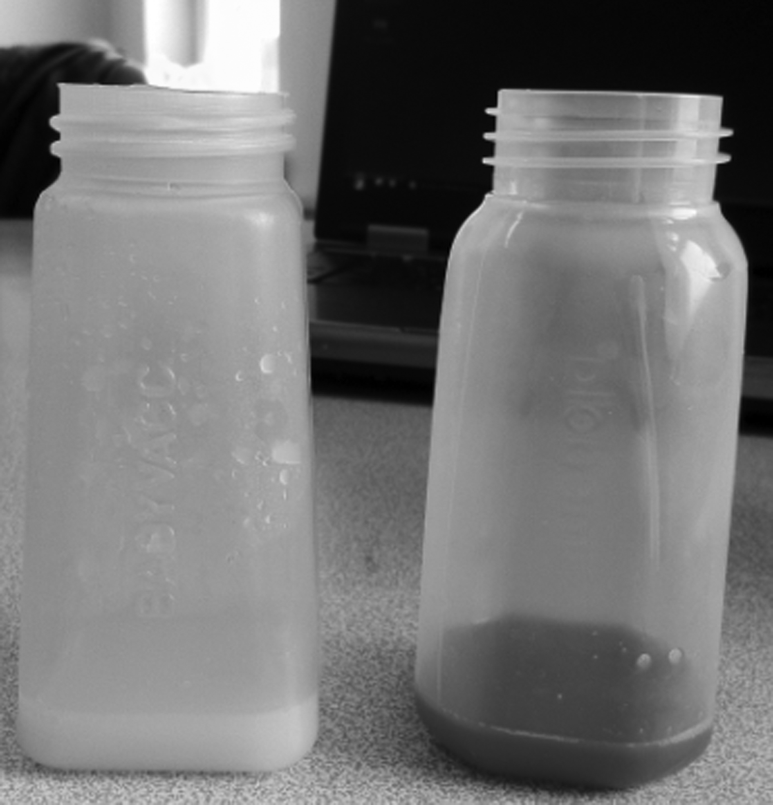
Expressed breastmilk samples from the right and left breasts demonstrate unilateral involvement with blood from the left breast.
After a few days, the neonate was discharged well on day 6 of life. The breastmilk remained hemorrhagic during the following several months but had disappeared on follow-up. Cystic changes of the breast also decreased in size on follow-up.
Discussion
Hematemesis may occur in a newborn who has had no previous signs of illness or distress, and prompt evaluation is required. The various possible causes are swallowed maternal blood, blood from cracked nipples, acute gastritis, stress ulcers and erosions, drugs, coagulopathy, vascular malformations, milk protein intolerance, and rusty-pipe syndrome. FBD of the mother has not previously been reported as a cause of hematemesis in a newborn.
Vitamin K-deficient bleeding (hemorrhagic disease of the newborn) should be considered in neonates who were not given vitamin K prophylaxis at birth. Stress gastritis or ulcers are generally associated with critical illness. Congenital anomalies, including intestinal duplications or vascular anomalies, may also present with gastrointestinal hemorrhage. Coagulopathy in a newborn may also be caused by infection, liver failure, or a congenital coagulation factor deficiency. Our patient's coagulation tests were all normal, and she had received vitamin K prophylaxis at birth. Milk protein intolerance may present with upper gastrointestinal tract bleeding, although lower gastrointestinal tract bleeding is much more common. True upper gastrointestinal tract bleeding in a neonate must be distinguished from swallowed maternal blood. The Apt–Downey test is not ordered frequently by emergency department physicians, but it may be very useful to rule out the infant as the source of the bleeding. In our patient the result of the Apt–Downey test on the patient's specimen proved that the blood was of maternal origin.
Maternal causes of hematemesis in the newborn are swallowed maternal blood, at the time of delivery or from cracked nipples during breastfeeding, and rusty-pipe syndrome. Lafreniere 7 estimated that 15% of lactating women had blood in their early secretions. Changes in the color of breastmilk can be of concern for nursing mothers. When malignant causes are appropriately ruled out, brown-colored breastmilk usually results from an obvious or subtle hemorrhage of the areola or nipples, both of which cause pain during suckling. However, there is another distinctly uncommon condition termed “rusty-pipe syndrome.” It is commonly bilateral, and most cases begin at birth or in early lactation. The condition is painless and remains unnoticed unless the mother is expressing the milk. It is thought to be due to the delicate network of capillaries, which are easily traumatized, which results in blood leaking into breast secretions. To continue breastfeeding is recommended as the condition is self-limited, and most cases clear within the first week. 8 The mother of our patient did not have crackled nipples and pain. When she was observed during milking, the blood directly originated from the breast, not from the nipple. We have moved away from the diagnosis of rusty-pipe syndrome because of unilateral involvement and the presence of a more logical cause.7–9
In the mammary gland of nonpregnant females, a large fatty pad exists. As pregnancy progresses, the adipose cells of the pad are gradually replaced by ducts, alveoli, blood vessels, and connective tissue. Alveoli are not formed before pregnancy is established. The breasts go through further changes throughout pregnancy and postpartum in order to sustain lactation. Breast changes begin following conception, and the weight of the breast increases approximately 12 ounces during pregnancy. In addition, blood flow to the breast doubles. After birth, milk is formed, and it is released into the milk-producing cells and ductal system. The two most critical hormones involved in milk production are prolactin and oxytocin, and as long as milk is withdrawn frequently from the breast, the alveolar cells will continue secreting milk for an indeterminate period of time. Over the first few months, the high baseline prolactin level that is the norm in the early weeks gradually decreases to the lower baseline that is the norm for later lactation. 10
FBD is a cumulative process, unilateral or bilateral, caused partly by the normal hormonal variation during a woman's monthly cycle. The most important of these hormones are estrogen, progesterone, and prolactin. During pregnancy, the mammary tissue has estrogen receptors and progesterone receptors. During lactation the mammary gland has estrogen and prolactin receptors, but not progesterone receptors. Histologically, FBD may present as simple cystic glandular hyperplasia, adenosis, chronic cystic mastitis with apocrine metaplasia, and ductal papillomatosis. Chronic cystic mastitis with apocrine metaplasia, as well as ductal papillomatosis, can cause bloody human milk. It is not common but is reasonable as a cause of bloody galactorrhea. 6 The ultrasonographic examination of our patient's mother revealed FBD, and there was no other reason of bloody human milk. We think that the mother had FBD that exacerbated at pregnancy, anastomosed to the ducts, and emerged during lactation by the breast changes (e.g., size, blood flow, replacement of adipose tissue by ducts, and hormone levels and responses) and after a few months resolved by normalization of the prolactin level.
In conclusion, this case report represents hematemesis in a healthy newborn that was found to be the result of swallowed maternal blood arising from FBD of her mother. We suggest that FBD should be kept in mind and may be confirmed by breast ultrasonography in case of bloody breastmilk.
Footnotes
Disclosure Statement
No competing financial interests exist.