Society of General Physiologists Symposium on “Ion Channels and Transporters in Immunity,Inflammation and Antitumor Immunity” (held online on September 11,2020)
Available accessMeeting reportFirst published online December, 2020
Society of General Physiologists Symposium on “Ion Channels and Transporters in Immunity,Inflammation and Antitumor Immunity” (held online on September 11,2020)
Ion channels and ionic transporters are expressed in every cell in the body, including the cells of the immune system. This international meeting was dedicated to some of the latest advances in the field and covered a range of topics in seven presentations. Zinc transport by the ZIP7 transporter expressed in the endoplasmic reticulum, transporting Zn2+ into the cytoplasm, was shown to be essential for B-cell development and functioning. Consequently, complete loss of ZIP7 was likely to be fatal (Sophie Hambleton). The protein kinase domain of TRPM7 cation channel was involved in proinflammatory T-cell differentiation (Susanna Zierler). A particularly novel development focused on ‘optogenetic immunotherapy’ involving photo-switchable Ca2+ (STIM1/ORAI) signaling in T-cells enabling spatially and temporally distinct immune signaling including transcriptional reprogramming (Yubin Zhou). Starting with genetic screening and functional genomics, T-cells were shown to express a volume-regulated anion (Cl−) channel (with LRRC8C and LRRC8A as essential components). The channel's activity controlled production of inflammatory cytokines and autoimmunity in the murine spinal cord (Axel Concepcion). Ca2+ signaling induced by CRAC/ORAI in TH17 lymphocytes in neuroinflammatory spinal cord was studied further by confocal imaging. Treg cells were found to inhibit TH17 functioning, in part, by suppressing Ca2+ signaling. This approach could open up a novel immunotherapy method (Michael Cahalan). S1PR (a GPCR that mediates S1P signaling) and SPNS2, a lymphatic S1P transporter, controlling T-cell trafficking in lymph nodes were characterized by Susan Schwab. Finally, pathogenic/damaged cytosolic DNA binding to the cyclic GMP-AMP synthase protein to enable synthesis of the secondary messenger cyclic GMP-AMP, leading to immune response involving production of cytokines and activation of natural killer cells was demonstrated by David Raulet. Thus, the meeting highlighted how studies of cellular ion channels and ionic transporters could elucidate both the understanding of immune cell functioning and, ultimately, improve its clinical management.
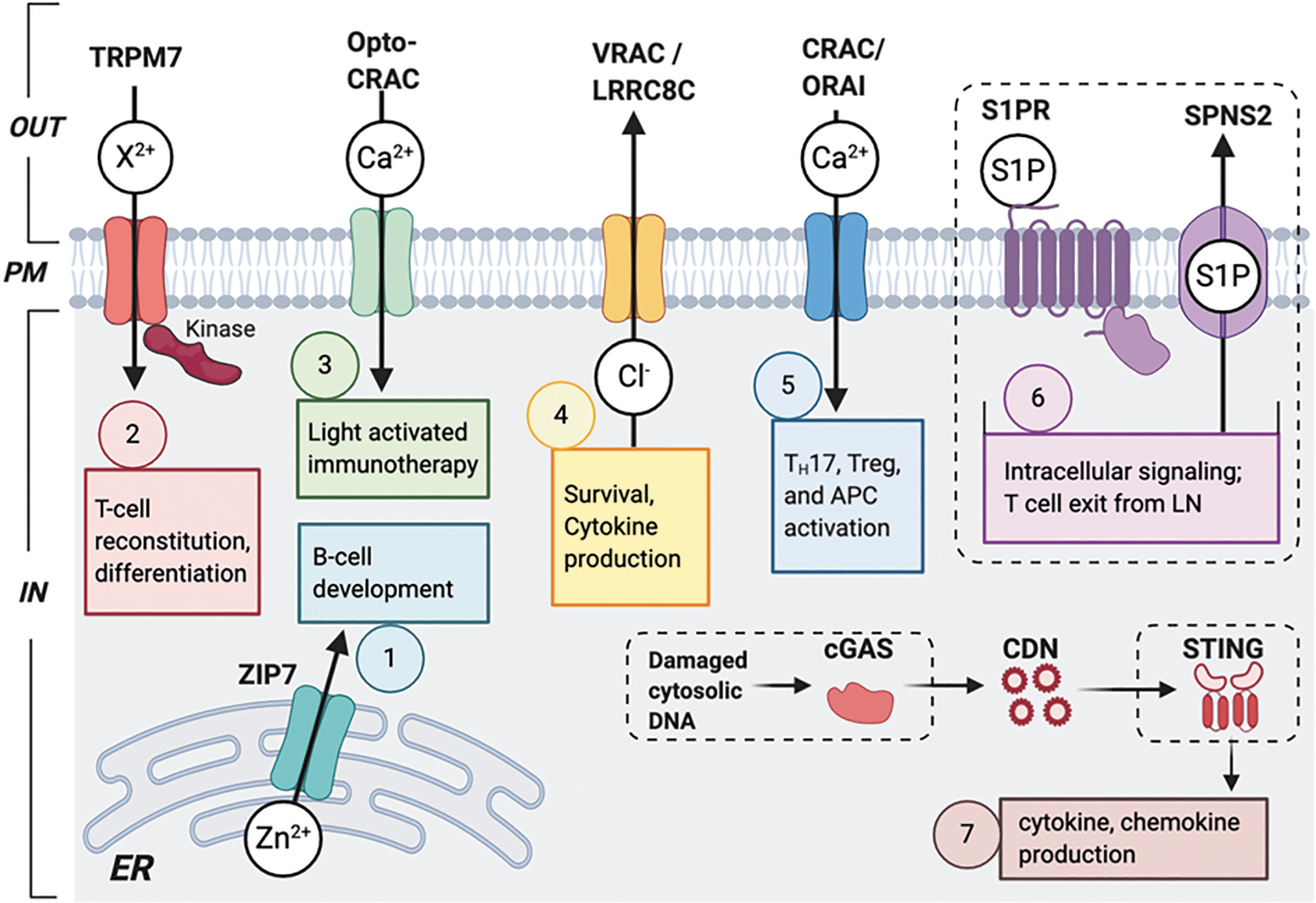
Ion channels, first conceived by Hodgkin and Huxley in the 1950s, have been scrutinized with increasing intensity ever since. The classic work was done on giant nerves and muscles as model systems. The field developed rapidly, both conceptually and technically, and as smaller and smaller cells became accessible, we now know that every cell in the body possesses some combination of ion channels and uses these in a plethora of functions from the most fundamental (e.g., transcription) to whole cell (e.g., migration). Expression of the ion channels is frequently associated with “transporters” either closely or remotely. Not surprisingly, therefore, genetic and/or epigenetic modification of ion channels and transporters (ICTs) can give rise to a diversity of pathologies. One of the true surprises of this field has been the discovery of functional ICTs in all major classes of cells in the immune system, not only the plentiful lymphocytes but also natural killer cells (NKCs), dendritic cells, and macrophages (Fig. 1).1 Thus, ICTs are intimately involved in the physiology and pathophysiology of innate and adaptive immunity. So, this SGP symposium organized by Bimal Desai, PhD, and Stefan Feske, MD, PhD, was greatly welcome and dedicated to some of the latest advances in the field (Fig. 2).2
An overview of ICTs in immune cells. ICTs can be located in the plasma membrane and/or intracellular organelles and permeate a variety of monovalent and divalent ions. In this figure, the ICTs are grouped by the ions they conduct. Some ICTs have been demonstrated in multiple immune cell types, including T cells, B cells, NKCs, and mast cells. Ca2+-permeant channels include the ATP-gated P2RX7 receptor, the TRP channels TRPM2 and TRMP7, TRPV1-2, voltage-gated calcium channels (Cav), CRAC (formed by ORAI proteins), and IP3R. Mg2+ influx in immune cells is mediated by the Mg2+ transporters MagT1 and the Mg2+-permeable, but nonselective, cation channel TRPM7. Zn2+ homeostasis is maintained by ZIPs (Zrt-/Irt-like proteins) and Zn2+ transporters (ZnT). The influx of divalent cations, especially Ca2+, requires a negative membrane potential, which, in T lymphocytes and other immune cells, is maintained by the voltage-gated K+ channels Kv1.3 and KCa3.1, and the Na+ channel TRPM4. ICTs associated with ion channelopathies in human patients are indicated by an asterisk (*). Reproduced from Feske et al.1 with permission. CRAC, calcium release-activated channel; ICT, ion channels and transporter; NKC, natural killer cells; TRP, transient receptor potential.
ICTs, and their associated basic mechanisms discussed in the symposium. The topics covered are numbered in order of the presentations. (1) ZIP7 is a zinc transporter expressed on the ER membrane and transports Zn2+ from the ER stores into the cytoplasm (Sophie Hambleton). (2) TRPM7 channel, fused to an enzymatic α-protein kinase domain, conducting divalent cations (Ca2+, Mg2+, and Zn2+) into T cells (Susanna Zierler). (3) Optogenetic immunotherapy. Opto-CRAC channel in immune cells enabling photoswitchable STIM1/ORAI signaling. A second photoactivatable protein LiCAR engineered into T cells, which was discussed, is not shown for clarity (Yubin Zhou).2 (4) VRAC expressed in the plasma membrane, mediating efflux of Cl– to control RVD in T cells. LRRC8C and LRRC8A are essential components of the VRAC channel (Axel Concepcion). (5) CRAC/ORAI-induced Ca2+ signaling in TH17 cells in the spinal cord in MS (Michael Cahalan). (6) S1PR (a GPCR that mediates S1P signaling) and SPNS2, a lymphatic S1P transporter, controlling T cell trafficking in LNs (Susan Schwab). (7) Pathogenic/damaged cytosolic DNA binding to the cGAS protein to enable synthesis of cGAMP, leading to immune response involving production of cytokines and activation of NK cells (David Raulet). cGAS, cyclic GMP-AMP synthase; ER, endoplasmic reticulum; GPCR, G protein-coupled receptor; LiCAR, chimeric antigen receptor; LN, lymph node; PM, plasma membrane; VRAC, volume-regulated anion channel.
The meeting kicked off with a presentation by Sophie Hambleton, MD (University of Newcastle, United Kingdom), entitled ZIP7: An unexpected player in B-cell development. Dietary zinc deficiency is responsible for over 800,000 deaths annually, mostly in the developing world. Zinc has profound impacts on cells as a structural component of many proteins and a co-factor in enzymatic reactions. The concentration of Zn2+ in the cytoplasm is managed by ZIP transporters, most of which lie in the plasma membrane. ZIP7 is one of these transporters regulating the level of intracellular Zn2+ in many different cell types, including immune and cancer cells. Interestingly, ZIP7 is found on the membranes of the endoplasmic reticulum (ER) where it transports Zn2+ from its vast stores into the cytoplasm. A mutation (P198A) in the gene coding for ZIP7 (SLC39A7) was first identified in twin patients of agammaglobulinemia (absence of B cells and platelets in peripheral circulation with consequent inability to produce antibodies). Using whole-exosome sequencing, the disease-causing biallelic mutation P198A was shown to cluster around the Zn2+ binding pocket inside the ZIP7 protein near the cytoplasmic region. To assess the Zn2+ transporting functions of ZIP7 proteins, each variant was expressed individually in Xenopus oocytes or human embryonic kidney cells. A Zn2+-sensitive dye (zinquin) was used to measure the level of intracellular Zn2+. The transfections showed that wild-type (WT) proteins, as expected, caused an increase in the concentration of cytoplasmic Zn2+, while mutant proteins did not. A “knock-in” murine model with a ZIP7 allelic series was developed to further elucidate the mechanisms of the disease. Animals that possessed mutations autologous to the one described in the twin patient (P198A) were small and died soon after weaning. These mice were born without a normal population of B cells (although the bone marrow remained relatively intact), confirming a conserved function of the SLC39A7 gene in B lymphocyte development. RNAseq revealed that many genes essential for B cell development were differentially expressed between WT and homozygous mutant animals. Measuring the impact of the ZIP7P198A mutation on the Zn2+ content of developing B cells (using fluorescence resonance energy transfer-based probes) affirmed that cytoplasmic Zn2+, but not ER Zn2+, was reduced significantly in the diseased cells. Furthermore, the lowered concentrations of cytoplasmic Zn2+ increased phosphatase activity in developing ZIP7-deficient B cells. Consequently, B cell receptor signaling and key downstream developmental and prosurvival mechanisms were suppressed. The latter included the recombination-activating gene and B cell-activating factor receptor pathways. It was concluded that ZIP7 deficiency was a novel genetic cause of agammaglobulinemia with absent B cells, and the complete loss of this protein was likely to be fatal. Much remains to be done, however, to fully understand the fundamentals of Zn2+ in B cells (and other cell types), associated signaling mechanisms, and their functional role in immune cells.
One of the relatively new and exciting properties of ion channels is their ability to display functional duality as both ionic permeators and enzymes, the physiological consequences of which are not yet fully understood in the immune system. This was the area investigated by Susanna Zierler, PhD (LMU Munich, Germany), in a presentation entitled TRPM7 channel-kinase function—from cellular signaling to immune system homeostasis. Dr Zierler's laboratory focuses on studying immune cell signaling at the ionic level and aims to identify novel pharmacological targets for proinflammatory diseases and leukemia. This led to work on the “transient receptor potential” (TRP) channels, especially TRPM7, which conducts divalent cations (Ca2+, Mg2+, and Zn2+). Importantly, TRPM7 is fused to an enzymatic α-protein kinase domain. Its conditional genetic inactivation (TRPM7−/−) would disrupt thymopoiesis (T lymphocyte selection) and alter chemokine and cytokine production profiles. Heterozygous TRPM7 mutants (TRPM+/ΔK), animals without a functional kinase, survived, but developed hypomagnesemia-driven allergic hypersensitivity, which could be reversed with a high-magnesium diet. Heterozygous TRPM+/ΔK mast cells released excessive amounts of histamine upon IgE stimulation. Through single-point mutation (TRPM7R/R) studies, this phenotype was shown to be due to the kinase domain of TRPM7 regulating mast cell exocytosis. In contrast, the kinase did not interfere with T lymphocyte selection. Further studies of TRPM7R/R mice revealed a diversity of signaling defects and lineage effects in regulatory T cells (Treg cells) and TH17 lymphocytes (CD4+ T lymphocytes secreting substantial amounts of interleukin 17A). Interestingly, TRPM7R/R T cells could differentiate into Treg cells, but not into TH17 cells. SMAD2 phosphorylation in TRPM7R/R mutants was significantly reduced upon TGF-β1 stimulation, suggesting that TRPM7 kinase also controlled SMAD2 signaling. Proximal ligation assays revealed that SMAD2 was indeed a novel substrate of TRPM7 kinase. Finally, the basic findings were questioned in the context of leukemia patients who often undergo bone marrow transplants. TRPM7 kinase inhibition was found to prevent “graft versus host disease” (GvHD), an immunological complication whereby transplanted cells attack the host. Thus, modulations of TRPM7 kinase would inhibit T cell reconstitution, proinflammatory T cell differentiation, and GvHD in mice. Future work will aim at replicating these effects in humans. Clinical applications could be facilitated by the development of novel modulators of TRPM7 such as waixenicin A, a bioactive component of Hawaiian soft corals.
No modern ion channel meeting would be complete without the mention of “optogenetics.” Yubin Zhou, PhD (Texas A&M, USA), did this in his presentation entitled Optogenetic regulation of CRAC channel function and signaling in immune cells. Optogenetics combines genetics with optics to control the membrane potential and downstream behavior of specific cells. In this study, optogenetic methods were applied to immune cells in two ways, by engineering (1) an Opto-calcium release-activated channel (CRAC) channel enabling either photoswitchable STIM1/ORAI signaling or (2) a photoswitchable “chimeric antigen receptor” (LiCAR) into T cells, thereby creating LiCAR T cells.3 CRAC channel-based optogenetics could be incorporated into immune cells through different approaches. First, LOV2, a photosensory protein, could be used as an “optical switch” and engineered to replace the CC1 component of the SOAR region of soluble STIM1 protein. Thus, optically induced conformational modification of LOV2 would expose the SOAR domain of STIM1 and enable it to engage and activate the ORAI channel liberally. Second, another photosensitive protein, CRY2, was engineered into CC1-SOAR to enable light-dependent oligomerization. Subsequently, “remote” photic stimulation of the cells could elicit spatially and temporally distinct signaling and immune responses, including transcriptional reprogramming. A major challenge in the application of optogenetics, however, is the delivery of light to deep tissues in vivo. This has been overcome by the use of “wireless optogenetics,” whereby a unique class of optical nanomaterials called “upconversion nanoparticles” (UCNPs) can transform and transmit low-energy photons into high-energy photons or transform near-infrared (NIR) light into blue emission. UCNPs are also small enough to circulate in blood and be manipulated so as to target immune cells. Thus, the workers succeeded in using UCNPs and NIR light control of Opto-CRAC to successfully aid tumor killing in a mouse model of melanoma. Further applications were being aimed at “nano-optogenetic immunotherapy” enabling time- and site-specific modulation of T cells, mitigating the side effects associated with conventional cell-based immunotherapy.
Axel Concepcion, PhD (New York University, USA), presented a talk entitled Novel ion channel regulators of T-cell function. By compiling information from the human genome, in a heroic effort, it was possible to find over 700 ICT genes. This set the scene for the elucidation of functional role of these ICTs using genetic screens (based on gene expression) and functional genomics (using CRISPR/Cas9 sgRNA screening). Some of the ICT genes were expressed specifically in T cells and coded for LRRC8s, a protein family comprising 5 members (LRRC8A-E) that constitute the pore region of the “volume-regulated anion channel” (VRAC). LRRC8 proteins were expressed in the plasma membrane and mediate the efflux of Cl– and organic osmolytes to facilitate regulatory volume decrease (RVD) upon osmotic swelling. Further studies showed that LRRC8A knockout abolished VRAC currents. Interestingly, targeting different combinations of LRRC8 members could change the inactivation kinetics of VRAC without affecting the channel current. It was concluded that LRRC8A, along with at least one additional LRRC8 homolog, was required to constitute a functional VRAC in T cells. qPCR analysis of CD4+ T cells established LRRC8C as the predominant LRRC8 homolog expressed in both mice and human T lymphocytes, comprising over 60% of the total VRAC composition at the transcriptional level. Functionally, LRRC8C deletion in T cells abolished osmotically activated VRAC currents and caused the collapse of RVD. Furthermore, LRRC8C-deficient (LRRC8C−/−) T cells demonstrated increased proliferative activity upon T-cell receptor (TCR) stimulation in vitro. LRRC8C−/− T cells also exhibited enhanced survival rates (decreased apoptosis) and increased production of cytokines (interleukin-2 and interferon-gamma) upon phorbol myristate acetate (PMA)/ionomycin stimulation (PMA/ionomycin bypass TCR stimulation and cause T cell activation). In addition, LRRC8C deficiency enhanced CNS inflammation in the “experimental autoimmune encephalomyelitis (EAE) murine model of multiple sclerosis (MS). This occurred through increased numbers of CD4+ T cells in the spinal cord and increased production of inflammatory cytokines. LRRC8C deficiency enhanced T cell-mediated antiviral immunity to influenza (confirmed by the decrease in mRNA of viral titers in the lung). Clearly, the next step seemed to exploit LRRC8C as an essential component of the VRAC channel in T cells as a novel immunotherapeutic drug target.
Michael Cahalan, PhD (UC Irvine, USA), a pioneer of the field of ITCs in immune cells, presented his recent work on Regulatory T-cells as suppressors of TH17 cell Ca2+ signaling in the spinal cord during murine autoimmune neuroinflammation. Again, the EAE murine model of MS was used and Ca2+ signaling in T cells in the spinal cord was visualized during disease progression. Pathogenic TH17 cells cause aggravated symptoms by attacking the myelin sheaths. Two-photon microscopy enabled the visualizing of spatial organization of endogenous Treg, TH17, antigen-presenting cells (APCs), and axons throughout the disease progression in the spinal cord. At the onset, pathogenic TH17 cells solely congregated at the caudal end of the cord. At peak disease, the population and spread of the TH17 cells increased, and Treg cells infiltrated the entire cord. APCs could also be seen to infiltrate the cord and co-localize with Treg cells. During the chronic disease phase, clearance of pathogenic TH17 cells and APCs, but the maintenance of Treg cells, were observed to be associated with partial remission. A genetically encoded ratiometric Ca2+ indicator (Salsa6f) was incorporated into transgenic EAE mice to image cytosolic Ca2+ in TH17 cells in 3D. The cells exhibited high levels of Ca2+ signaling at the onset of EAE, but this decreased at peak disease. Interestingly, TCR-induced Ca2+ signaling in TH17 cells was suppressed by the presence of Treg cells. It was concluded that Treg cells would inhibit TH17 functioning by reducing APC density, by limiting TH17 access to APCs, and by suppressing Ca2+ signaling in TH17 cells. Accordingly, it was suggested that a novel immunotherapy method could involve increased influx of engineered Treg cells into sites of neuroinflammation. Also, TH17 suppression would lead to a remission of symptoms in this model of MS and the ICTs responsible for the Ca2+ signaling (CRAC/ORAI1, Kv1.3, KCa3.1, etc.) could be pharmacologically targeted to reduce TH17 activation and thus ameliorate symptoms of the disease.
The presentation of Susan Schwab, PhD (NYU School of Medicine, USA), entitled Exit strategies: S1P and T-cell migration, focused on elucidating the mechanisms of immune cell migration and understanding the establishment of chemotactic gradients of sphingosine-1-phosphate (S1P) and their effects on immune cells. The emergence of T cells from lymph nodes (LNs) is driven by gradients of S1P, high in circulatory fluids and low in lymphoid tissues. There are five subtypes of S1P receptor (S1PR1-5). These are G protein-coupled receptors and targeting their signaling mechanisms has already shown clinical promise, in fact, approved by the FDA for the treatment of relapsing-remitting MS. Numerous other S1PR drugs for autoimmune disorders are currently being developed. One of the main effects of SP1R drugs is to block S1P signaling to trap T lymphocytes in lymphoid organs and prevent them from entering the circulation and reaching sites of inflammation. Unfortunately, however, S1P drugs have multiple “off-target” side effects on the cardiovascular system that also expressed S1PRs. The novel S1P transporter, SPNS2, was found to be essential for the supply of lymph S1P from lymphatic endothelial cells, but not for the supply of blood S1P, from red blood cells. To explore roles of SPNS2 in vivo, an SPNS2-deficient (SPNS2−/−) murine model was developed. Results revealed that these mice had normal vascular permeability, but were profoundly defective in lymphocyte trafficking. Although S1P levels remained elevated in the blood of mutant mice, lymph S1P was obliterated preventing T cells from exiting the lymph. In turn, this would cause T cell depletion in the spleen and a loss of T cells in circulation. Further murine studies demonstrated that SPNS2 was required for T cell accumulation at sites of inflammation. Finally, SPNS2−/− mice appeared to be excluded from EAE, suggesting that loss of SPNS2 might have a protective role in MS. SPNS2 remains the only protein known to supply lymph S1P, but not blood S1P, and its deletion in mice uncovered novel functions of S1P in the immune system (e.g., positioning and regulation of NKCs in LNs). In conclusion, the S1P transporter SNPS2 was shown to enable T cell accumulation in inflamed tissues and to generate S1P concentration gradients between lymph and LNs during immune responses. Accordingly, targeting SPSN2 could enable spatially distinct modulation of S1P signaling and SPNS2 was proposed as a promising drug target for autoimmune disorders, including MS. Because they do not alter serum S1P levels, SPNS2 blockers could potentially replace SP1R1 blocker drugs for the treatment of MS and eliminate their deleterious side effects.
David Raulet, PhD (UC Berkeley, USA), presented his laboratory's recent findings on the Import of immunoactive cyclic dinucleotide. Cyclic dinucleotides (CDNs), for example, cyclic GMP–AMP (cGAMP), synthesized by cyclic GMP-AMP synthase (cGAS), can function as intercellular secondary messengers. A major mechanism for the spontaneous mobilization of antitumor NKC response is the activation of the cGAS-STING pathway. This is a “cytosolic DNA sensor,” which detects intracellular pathogenic DNA and cytosolic chromosomal DNA in cancer cells. Pathogenic DNA binds and activates the cGAS protein, which in turn synthesizes cGAMP that activates the master regulator STING and induces an immune response by the transcriptional production of cytokines. Xenograft and transgenic knockout mouse models of several cancers were used to determine the steps involved in the antitumor response of NKCs. These studies revealed (1) that cGAS and STING were both required for the immunosurveillance, but acted in different cell types—cancer cells and host cells, respectively; (2) that cGAS responded by synthesizing CDNs (cGAMP) in the cancer cells; (3) that CDNs exited the cancer cells to diffuse across the tumor microenvironment; (4) that CDNs were taken up by host (myeloid) cells leading to the activation of STING; (5) that STING induced transcriptional production of cytokines and chemokines; and (6) that the latter are released and prime NKCs to infiltrate the tumor. It was surprising, therefore, that the tumor was initiating a response cascade (an evolutionary conserved antiviral defense mechanism) that could ultimately be detrimental to itself! Although this response was relatively weak, it could be potentiated by intratumor injection of cGAMP to superstimulate the cGAS-STING pathway. A novel synthetic CDN (2′3′ RR-S2 cyclic di-AMP) was more effective than cGAMP and could be used to target the STING pathway in cancer immunotherapy. In fact, CDN immunotherapy could successfully mobilize antitumor NKC responses to induce tumor rejection with extraordinary curability rates in MHC-I-deficient mice. To elucidate the mechanism of action of this immunotherapeutic strategy, a genome-wide CRISPRi screen was conducted to identify possible CDN transporter genes. The main “hit” of this screen was the SLC19A1 transporter, essential for CDN uptake in human cells. SLC19A1 inhibitors (such as THF, MTX, and SSZ), generated initially as inhibitors of folate transport, could also effectively inhibit cGAMP uptake. Knockout studies revealed that SLC19A1 is the primary cGAMP transporter in humans (responsible for the uptake and possibly also the release). Mice express an additional, possibly more active, CDN transporter that enables more efficient CDN import compared to humans. Accordingly, immunotherapy with CDNs yields dramatic outcomes in mice, but less so in humans. This weaker CDN response in humans may reflect the observed reduced efficiency of CDN import. As a result, drugs may be designed both to improve CDN import and to amplify cGAS-STING signaling to enhance the outcome of this novel immunotherapeutic approach. Interestingly, SLC19A1 inhibitors could be used as first-line treatments against autoimmune disorders (e.g., rheumatoid arthritis, psoriasis, and irritable bowel syndrome). This would suggest that CDN transport by SLC19A1 (and by implication cGAS-STING) may play a significant role in inflammatory disease.
In overall conclusion, the symposium truly represented the diversity of some of the most recent developments in the immune ICT field and many of these were linked to potential immunotherapy strategies. Unfortunately, but not surprisingly (within the 4-h limitation), some other key ICTs like voltage-gated sodium channels (VGSCs) and voltage- and calcium-activated potassium channels only got limited mention. Previous studies demonstrated that VGSCs are essential for the positive selection of CD4+ cells, for macrophage motility, and for NKC cytotoxicity, all of which can have profound influence on tumorigenesis. Altogether, one is left with no doubt that ICTs, and broadly associated bioelectric signaling, play wide-ranging roles in immune functioning and have extraordinary potential for the development of novel immunotherapies.
Footnotes
Author Disclosure Statement
No competing financial interests exist.
Funding Information
No funding was received.
References
1.
FeskeS, ConcepcionAR, CoetzeeWA. Eye on ion channels in immune cells. Sci Signal, 2019; 12:eaaw8014. DOI: 10.1126/scisignal.aaw8014
2.
Ion Channels & Transporters in Immunity, Inflammation & Antitumor Immunity. https://www.sgp2020.com. Accessed October3, 2020.
3.
YangX, MaG, ZhengS, et al.Optical control of CRAC channels using photoswitchable azopyrazoles. J Am Chem Soc, 2020; 142:9460–9470. DOI: 10.1021/jacs.0c02949