




Abstract
The delivery of pulsed electric fields to biological cells for regenerative research and therapeutic applications is a field that has been widely explored. Picosecond pulsed electric fields have been shown to induce intracellular effects and directly target cell membrane proteins as well as being able to induce cell permeabilization and death by apoptosis. Additionally, ultrashort pulses can be focused and delivered in a noncontact manner for possible targeting of deep and inaccessible tissues. The unique characteristics of picosecond pulses make them a possible approach for treatment of Huntington's and Alzheimer's diseases, both characterized by progressive neuronal degeneration and death, and presence of intracellular and extracellular protein aggregates. In this study, the defining characteristics of picosecond pulses and neurodegenerative diseases are reviewed, and a description of how picosecond pulsed electric fields can be applied to disrupt protein aggregates, can target neural stem cell differentiation, and can be delivered to neurological tissue is provided.
Introduction
The utilization of pulsed electric fields to manipulate biological cells is a field of research that has greatly advanced in recent years, leading to major promising applications in the fields of cancer treatment and regenerative medicine. Among their applications, notable research and utilization has been achieved with tumor and cardiac ablations,1,2 gene electrotransfer,3–5 and electrochemotherapy.6,7 Here, we annotate the potential that pulsed electric fields may have in the treatment of neurodegenerative diseases and in directing the nervous tissue regeneration.
Electric fields with varied pulse lengths have been largely used in research with potential application in the clinical field. Pulses in the range of milliseconds to microseconds have been shown to induce electroporation (reversible and irreversible) of the plasma membrane.8–10 Electroporation has enabled the induction of cell death facilitated by the intracellular delivery of drugs as well as by direct cell targeting.6,11 Application of shorter pulses can induce intracellular effects such as internal calcium bursts, permeabilization of intracellular granules, and induction of apoptosis, without directly damaging the plasma membrane. 11 The transition from electroporation to intracellular electromanipulation is dependent on both pulse durations that are in the order of membrane charging time (100 ns for mammalian cells) and the peak electric field strength. For instance, the delivery of short pulses—in the nanosecond and subnanosecond range—has been shown to produce a variety of cellular effects, such as nanopore formation, 12 membrane permeabilization, 13 phosphatidylserine externalization,14,15 and manipulation of voltage-gated ion channels. 16
While emphasis has been majorly focused on the study and application of milli-, micro-, and nanosecond pulses, ultrashort (picosecond) pulsed electric fields present unique characteristics that render worth their exploration in a translational research setting. In this review, an accounting of the current knowledge in the picosecond pulsed electric fields is made. This includes cell biophysics, in vitro data, pulsed power supplies, and pulse applicators. Focus is placed on the analysis of the neurodegenerative diseases such as Huntington's disease (HD) and Alzheimer's disease, and how the application of picosecond pulsed electric fields potentially impacts their respective pathophysiology. The conclusion of this analysis indicates that picosecond pulse electric fields can be applied to biological cells in a relatively safe manner. Picosecond pulsed electric fields have unique properties, such as inducing effects on protein aggregates and potential for noncontact delivery, and they represent a promising approach to treatment of neurodegenerative diseases.
Picosecond Pulse Biophysics
Cell as a circuit
The plethora of direct and indirect effects on the cells observed upon application of external electric fields can be understood considering the cell as an electrical circuit. The cell cytoplasm, rich in proteins, electrolytes, and glucose, is moderately conductive and acts as resistor, whereas the outer cell membrane and the membranes that surround organelles have low conductivity and can be modeled as capacitors. 17 A schematic representation of the cell as a circuit is reported in Figure 1.
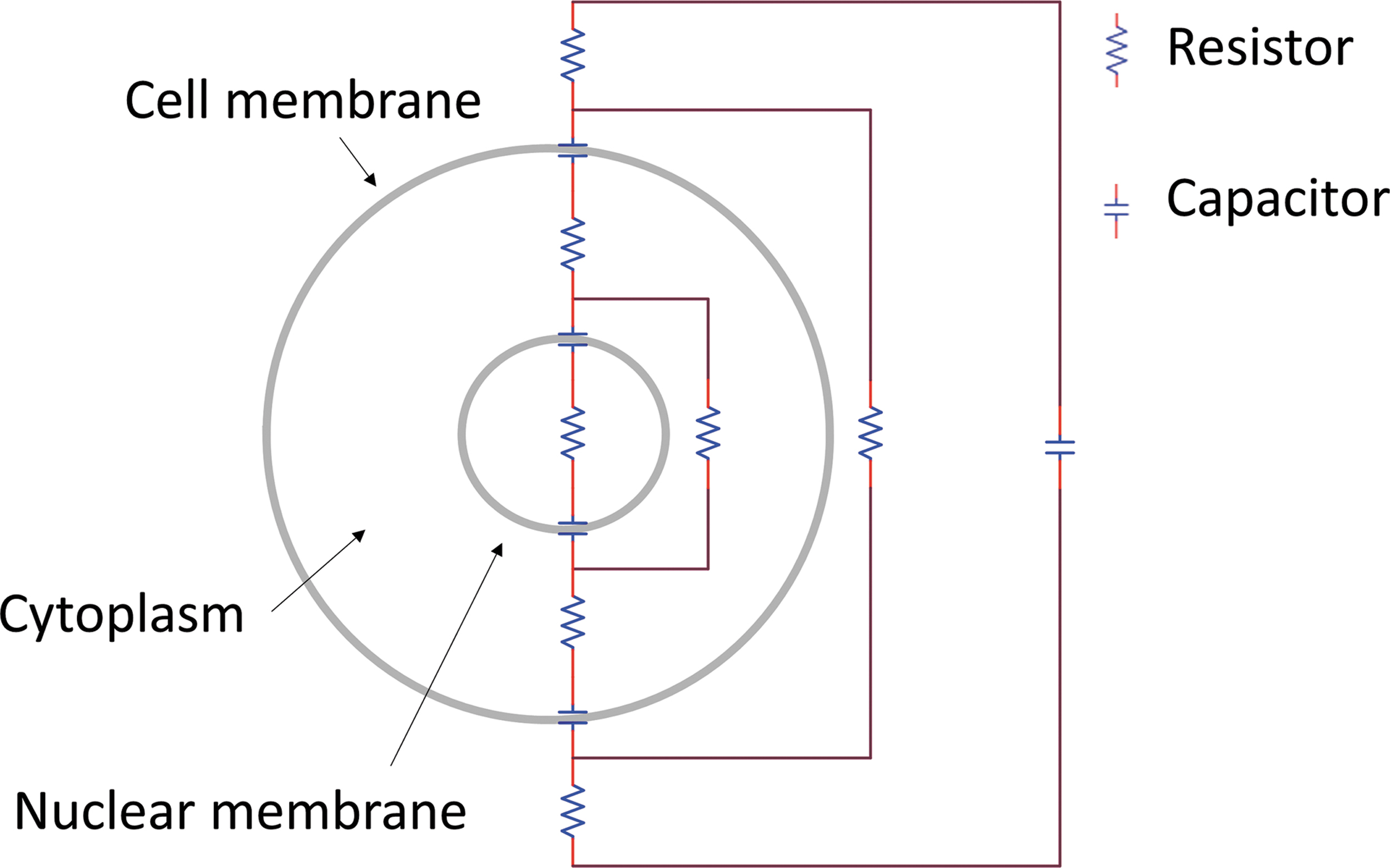
Biological cells can be represented as electrical circuits. The plasma membrane and intracellular membranes can be compared with circuit capacitors, whereas the cell cytoplasm acts as a resistor.
When considering a typical biological cell, the charging time constant τm of the cell membrane is in the order of a 100 ns. The effects observed on the biological cell subjected to pulsed electric field application are dependent on the type of pulse considered. These can be predicted using a passive linear model, in which structural changes of the cell due to the electric field application are not considered, and the plasma membrane is a stable element with fixed resistivity and permeability. When pulses of long duration are applied, compared with the charging time constant of the plasma membrane, the effects of the applied field are confined to the cell plasma membrane. In this case, a significant voltage across the plasma membrane is created, whereas that across intracellular membranes is negligible. 18
In the case of ultrashort pulses, where the pulse rise time is shorter than τm, the applied field can reach the interior of the cell and affect subcellular membrane potentials without permanently altering the outer membrane.9,19,20 Intracellular effects due to the application of ultrashort pulsed electric fields will be observed, including organelle membranes alterations, 18 calcium mobilization,21,22 protein effects, 21 and cell death regulation.23–25
Dependence on dielectric properties
When modeling the effects of pulsed electric fields on biological cells, the capacitive component of the cytoplasm and the cell culture medium are neglected. 26 However, this assumption is only valid when considering pulse durations long compared with the dielectric relaxation time of the cytoplasm (∼700 ps), defined as the ratio of its permittivity (ɛcp) and conductivity (σcp). 20 Experimental evidence 27 revealed that for pulse durations approaching 700 ps, the conductance of the plasma membrane cannot be assumed to be zero anymore, and the cytoplasm cannot be considered as a resistive element. The distribution of the electric field is therefore determined by the dielectric permittivity of the cell components and of the cell's environment rather than their resistivity.11,17,26 Therefore, when considering the case of subnanosecond pulses, the distribution of the electric field in the cell is determined by the dielectric properties of the cell culture media—the polarization of its molecules in response to an applied electric field—rather than its resistive characteristics.
Changes in protein conformation
Direct application of pulsed electric fields has been shown to affect protein structure and aggregation. 28 Specifically, pulses in the microsecond and nanosecond range can induce changes in secondary and tertiary protein structure and affect denaturation and aggregation.29–33
Application of pulses in the range of nanosecond and picosecond pulses results in the arising of intracellular effects, bypassing the plasma membrane. One of the debated questions in the field is whether ultrashort pulses can deliver enough energy, power or high-frequency components to disrupt molecular bonding and ultimately result in intracellular protein structures alterations.
Molecular dynamics (MD) simulations found that the application of intense nanosecond pulsed electric fields can affect protein structure and dipolar properties.34,35 Application of pulses as short as 10 ns can promote the transition from α-helix to β-sheet structure in amyloid aggregates.36,37 Additionally, electric pulses can induce reversible or irreversible protein denaturation, proportionally to the intensity of the applied field. 35 Simulations and experimental data elucidating the effects of picosecond pulsed electric fields on protein are not readily available. Results observed upon application of longer pulses and from picosecond pulses simulations suggest that the latter may affect protein aggregation and warrant an extended research effort.
Cell membrane permeabilization
Pulsed electric fields in the range of milli-, micro-, and nanoseconds have been widely studied for their ability to induce transient or permanent cell membrane permeabilization. These phenomena are commonly referred to as reversible and irreversible electroporation and have wide applications in the fields of cancer treatment and translational research. Irreversible electroporation results in irreparable damage to the cell membrane, and it has been applied in the clinical field for the direct ablation of desired tissues without damaging the nearby areas. 38 It has been adopted for the treatment of tumors39,40 and cardiac arrhythmias. 1 Reversible electroporation has found extensive application for the intracellular delivery of drugs, 41 leading to the development of the field of electrochemotherapy, where accessible tumors are treated with the intracellular delivery of chemotherapeutics.42,43 This type of electroporation is also utilized for the intracellular delivery of nucleic acids44,45 and contributes to the development of the field of gene therapy.46,47
The formation of pores upon application of electric pulses that permeabilize the cell membrane to solutes that would be impermeant under standard conditions is a widely accepted concept in the field. However, details concerning the mechanisms that drive poration have yet to be elucidated, due to the limitations of nanoscale observational instrumentation. MD simulations of lipid bilayers have been a key tool in providing insights of such processes at a molecular level. They stipulated that the formation of pores is initiated by field-driven water intrusion in the lipid bilayer,48,49 that pore initiation time is a function of the applied electric field, 50 and have provided tentative steps in the evolution of a pore, from its creation to its annihilation.48,51
MD simulations have also been applied to the study of membrane effects of subnanosecond pulses. Vernier et al. showed that with the application of electric fields of magnitude above 1.5 GV/m, pore formation can occur with the application of pulses as short as 1 ns and 50 ps. 22 Therefore, it is reasonable to expect membrane effects upon application of high-intensity, ultrashort pulsed electric fields on biological cells. Simulations also show that delivery of picosecond pulses with appropriate intensity might be used to selectively permeabilize internal organelles. 52
Picosecond pulsed electric fields have shown to induce a response when applied to excitable biological cells. Three hundred twenty picoseconds pulses can initiate action potentials in hippocampal neurons, whereas 500 ps pulses can induce calcium transients in NG108 rat neuroblastoma-glioma hybrid cells. 22 The mechanisms by which subnanosecond pulsed electric fields might cause cell permeabilization are not yet experimentally defined.
A study performed by Semenov et al. aimed at defining the impact of the applied fields on membrane proteins, specifically focusing on voltage-gated calcium channels. 21 The authors used GH3 and NH108 cells, which present voltage-gated calcium channels, as well as Chinese Hamster Ovary (CHO) cells, which do not. Upon application of picosecond pulsed direct current (DC) stimuli (190 kV/cm, 500 ps at 50% height), voltage-gated channels in GH3 and NH108 cells opened and elicited calcium transients of the duration of multiple seconds, an effect that was enhanced by the application of multiple stimuli. Furthermore, they observed that this calcium mobilization remained active up to 10 s post-treatment. No response was observed in CHO cells. From these results, the authors concluded that the application of picosecond pulsed electric fields on biological cells can result in permeabilization of the cells by activation of voltage-gated calcium channels, rather than through lipid phase electroporation. However, the mechanisms behind the opening of voltage-gated calcium channels in response to applied electric fields are not known.
Apoptosis
Apoptosis is one of the mechanisms of regulated cell death normally occurring in the organism to ensure development and tissue homeostasis as well as proper functioning of the immune system and response to external stimuli. 53 When pulses of long duration are applied to achieve irreversible electroporation, the insult generated on the cell membrane is great, and accidental cell death occurs. Mechanisms of regulated cell death, such as apoptosis, can be induced with the application of pulses of shorter duration. Particularly, nanosecond pulses have been shown to induce apoptosis54,55 and have been a useful tool in the treatment of tumors.56,57
Application of picosecond pulsed electric fields has also been shown to permeabilize the cell membrane in in vitro models, altering cell permeability and inducing cell death by apoptosis.11,17 In a single study from Schoenbach et al., 800 ps pulses stimuli were shown to induce 50% lethality in B16 cells, after delivery of 18,000 pulses at 150 kV/cm, or of 125 pulses at 950 kV/cm. 17 A different study demonstrated that the pulsing conditions of 200 ps, 25 kV/cm, lead to death of B16 cells after at least 1.8 million pulses. Additionally, application of pulses of the same length, with electric field between 20 and 40 kV, increased cell conductance in NG108 cells even with the application of a much lower number of pulses. 11
The mechanisms through which apoptosis is induced in cells subjected to picosecond electric fields were studied in HeLa cells. After exposure of the cells to 800 ps pulses at 250 kV/cm, inhibition of cell growth and release of mitochondrial contents into the cytoplasm were observed, suggesting that cell apoptosis might occur through the mitochondrial-mediated pathways.24,25 Further studies revealed that picosecond pulsed electric fields might induce their apoptotic effects via the endoplasmic reticulum stress response and caspase-dependent signaling pathways. 23 Additionally, it was demonstrated that cell growth inhibition and apoptosis can be induced in a dose-dependent manner. 24
Neurodegeneration: Alzheimer's Disease and HD
Neurodegenerative diseases are conditions that arise from the progressive degeneration and death of cells in the brain and peripheral nervous system. Genetic predisposition and environmental factors are contributing aspects to the development of neurodegenerative disease, and the risk of being affected increases significantly with age. Two neurodegenerative diseases of interest for possible therapeutic applications of picosecond pulsed electric fields are HD and Alzheimer's disease.
HD is a rare autosomal dominant disease, which exhibits genetic anticipation of affected progeny of affected individuals. It is characterized by the insurgence of motor (chorea), neuropsychiatric, and cognitive disturbances. 58 HD is caused by a mutation in exon 1 of the huntingtin (HTT) gene, located on chromosome 4. The altered gene presents an expansion in the cytosine–adenine–guanine (CAG) trinucleotide repeat (TNR). Repeats of the CAG sequence are observed in healthy individuals; however, when the number of repeats is above 36–40, disease phenotype is observed. The CAG-repeat codes for the glutamine amino acid, and the mutation results in the production of a mutant huntingtin (mHTT) protein, which presents variable and abnormally long polyglutamine repeats.
The comprehensive mechanisms that lead to the formation of such unstable regions are unknown; however, studies conducted on HD human fibroblasts and induced pluripotent stem cells suggest a potential epigenetic involvement in the disease. More specifically, downregulation of DNA repair genes was observed in HD cells, suggesting that DNA repair mechanisms play a role in the maintenance of TNR stability.59,60 Although the mHTT gene is ubiquitously expressed in the organism, medium spiny neurons of the striatum are selectively vulnerable to the effects of the mutated protein. The cause of such vulnerability is yet to be determined; however, dopamine D2 receptors have been implicated in HDs pathogenesis and may be a factor.58,61
The presence of the mHTT protein results in extensive pathogenic effects of the vulnerable cells. Such proteins have a propensity to form intracellular aggregates, which are commonly identified as polyglutamine aggregates. The protein's presence is associated with pathogenic effects, such as transcription factors aggregation and mitochondrial dysfunction, and affects mechanisms of cellular proteostasis and axonal transport.58,59 Such alteration in the normal physiology of the cell ultimately results in the death of neurons and progressive loss of gray matter in the brain of HD patients. 58
Alzheimer's disease is the leading cause of dementia, and it is a disease observed with greatest prevalence in patients older than 65 years. It is characterized by the progressive insurgence of problems centered on episodic memory, followed by topographical and multitasking difficulties, which ultimately progress to interfere with the patients' daily activities. In most cases, the development of the disease is sporadic, whereas in <0.5% of the cases, it is due to mutations in the genes amyloid precursor protein (APP), presenilin 1 and presenilin 2, leading to a rare familial form of the disease.
Two distinctive features of Alzheimer's disease are the formation of amyloid plaques and neurofibrillary tangles. 62 Amyloid plaques consist in the extracellular accumulation of abnormally folded Aβ protein, a downstream product of APP metabolism. Aβ is produced by the organisms throughout life; however, the function of neither APP nor Aβ is clear. 63 It is hypothesized that the accumulation of abnormal forms of Aβ is a primary pathological process in Alzheimer's disease and that it is due to an imbalance between the production and clearance of the peptide. Neurofibrillary tangles are primarily constituted by hyperphosphorylated tau helical filament pairs and accumulate intracellularly. Tangle formation is strictly associated with the loss of neurons and synapses, and therefore, the severity of the disease is correlated with neurofibrillary tangles pathology. 62
HD and Alzheimer's disease are interesting targets for the application of picosecond pulsed electric fields, due to their shared characteristic presence of intracellular and extracellular protein aggregates. The ability of ultrashort pulses to directly target proteins might be advantageous in the approach of such neurodegenerative diseases.
Applications of Picosecond Pulsed Electric Field to Neural and Neurodegenerative Research
Picosecond pulse generators
One of the driving challenges in picosecond bioelectrics is the availability of pulsed power supplies. Picosecond generators have a trinity of high speed, high voltage, high current, and in some cases, high repetition rate. This combination presents challenges in finding a suitable switching and circuit topology. As a result, there are only select solutions in the literature.
State-of-the-art solid-state switching is typically silicon carbide or gallium nitride metal–oxide–semiconductor field-effect transistors. These switches have seen growing use in bioelectrics 64 ; however, they have limited pulse rise times and fall times and are thus limited to pulses of nanosecond duration or longer. To implement picosecond pulses in a solid-state circuit, a designer can select from a few reported switching technologies. First is avalanche transistor switching. 65 Avalanching is a property of bipolar junction transistors in which an electron avalanche occurs. This describes a chain reaction in which a free electron being accelerated by the electric field can impact other electrons creating a multiplication of free electrons, which allows for the fast switching times with high currents. This technique has previously been used for Pockel's cells.66,67
The next type of switching technique utilizes optical or photoconductive switching. In this method, a p-n junction is excited by a laser source. The light energy generates free carriers and modulates the conductance through the switch.68–70 The final type of solid-state switching utilizes drift step recovery diodes. 71 In this switching technology, the step recovery diode stores charge in the forward bias. When the bias reverses, there is a rapid opening of the diode forming a pulse. Fast Ionization Device generators, which utilize a drift step recovery diode, can produce pulses with voltage amplitude up to 10 kV, an extremely fast rise time, and a repetition rate up to 100 kHz. 26
All the before-mentioned solid-state switching methods have limitations in their voltage and current capability. To achieve higher voltage and currents without adding switching elements to the circuit, spark gap switching has become a viable technique for picosecond pulse generation. The advantages of a spark gap are low cost, high current handling, and ease of use. Spark gap-based generators can be manufactured from off-the-shelf components, greatly optimizing production costs, and the voltage amplitude can reach several hundreds of kV. One disadvantage of these systems is their inability to generate high repetition frequencies. 26 Pressurized and circulating systems of gas or oil can be used to increase the repetition frequency.72,73 However, this comes at increased complexity and cost. Improvements to a standard spark gap system include triggering and tail cut switching. Spark gaps have the ability to be triggered by a trigatron 74 or laser.75,76 Tail cut switching is a second spark gap used to sharpen the pulse and decrease the pulse width.
Any of the above switching techniques can be utilized in a picosecond pulse-forming circuit topology. The first method is a pulse-forming network. In this technique, energy is first stored in either a discrete capacitor–inductor or transmission line such as a coaxial cable. Pulse-forming network charging can be done by a DC or resonance pulse transformer.77,78 Once charging is complete, the switch closes and the transmission line forms an L-C network, which induces a square pulse on the load. The pulse width is determined by the length of the transmission line. The downside of a simple transmission line topology is that the voltage experiences at the load is half of the input (Vin/2). To overcome this, an alternative pulse-forming line topology can be used, notably a Blumlein line.
Another circuit topology that can be used for picosecond pulse generation is the Marx generator. In this circuit topology, capacitors are charged in parallel and discharged in series. This creates a voltage multiplier effect. By using a peaking capacitor, the time constant of the resistor–capacitor discharging can be controlled. For picosecond pulses, it is difficult to synchronize all the switches for each stage of the Marx generator.
It should be noted that picosecond pulse generators are extremely sensitive to stray inductive or capacitive elements. This is because the L-C elements forming the pulse are on the same order as the stray components. Special attention needs to be paid to the geometry to minimize these effects. Furthermore, impedance matching the pulser to the load presents challenges at short pulse widths. This is because the frequency elements of the pulse are ultra-wideband. Extra precautions should be taken to impedance match the system and prevent backward reflections at the load. One technique would be a attenuator as seen in Ref. 79 While this technique improves the reflections, a larger power supply is necessary to overcome the attenuator.
Pulse application methods
Applying picosecond pulses to cells or tissue can take a variety of forms and depend on the application. In vitro, an electric field is normally applied between two parallel wires.79,80 In vivo, picosecond pulses can be applied using needle or tweezer-based electrodes. In needle electrodes, two or more sharpened electrodes are placed subcutaneously and the electric field is established between the electrodes. 81 On the contrary, tweezer-based electrodes are superficial. The tissue to be pulsed is clamped between conducting plates and the field is applied.82,83
When using pulsed electric fields of nanosecond or longer duration, penetrating or contact electrodes are required to be in close contact with the treated area. This means that the affected area limits the possible treatments to tissues close or on the surface of the body. A distinctive characteristic of picosecond pulsed electric fields is the possibility for delivering energy in a noncontact manner. This is because for a pulse to radiate independently through free space, the originating geometry must be on the same scale as the pulse spatial width. For ultra-wideband pulses, the pulse spatial width given by:

where pw is the pulse spatial width, tp is transient full width half max, c is the speed of light, and εr is the relative dielectric permittivity. This indicates that picosecond pulse antennas on the order of <1 m can be constructed potentially allowing for noninvasive treatment of deep lesions.17,79 In addition, the pulse spatial width describes the resolution of the pulse in tissue. When using pulses in the range of 100–200 ps, antennas can be used to focus the radiation efficiently on a target point, with resolution in the subcentimeter range.11,17
Antennas have been developed for the expressed purpose of delivering picosecond pulsed electric fields to biological targets. The first efforts utilized a prolate spheroid reflector, which uses constructive interference to focus the pulse inside of tissue. An example of such a system was developed by Xiao et al. 84 Here, they demonstrated that using such system, with a 100 ps rise time, a focusing spot size of 2–3 cm along the z-axis can be achieved. Another approach could use multiple antennas in an array around the target. Again, constructive interference focuses the pulse in the tissue.
In both these approaches, the electric field at the target is greatly reduced by the reflection at the interface of air and tissue. This is due to the large difference in permittivity between the two. The problem has been approached by Petrella et al. with the development of a dielectric antenna. This system allows for direct contact with the tissue, resulting in increased coupling of the radiation to the tissue, and allowing delivery of the proper field to target 1–2 cm in depth. 85
A final aspect that must be considered for the application of picosecond pulsed electric fields as a therapeutic approach for neurodegenerative disease is the necessity of delivery to the brain in a safe and efficient manner. This problem was also approached by Petrella et al., with the development of a dielectric rod antenna for stimulation of neural tissue. 85 The antenna comprises three main components: a hollow conical transverse electromagnetic waveguide loaded with a dielectric, a dielectric rod, and a dielectric cone as the wave-emitting section. This system allows direct contact to the tissue and delivery of pulses to targets that are 1–2 cm in depth. The ability of the antenna to deliver electric fields able to overcome the threshold for biological effects (20 kV/cm) and starting from a pulse length of 100 ps was confirmed through simulations and is shown in Figure 2.
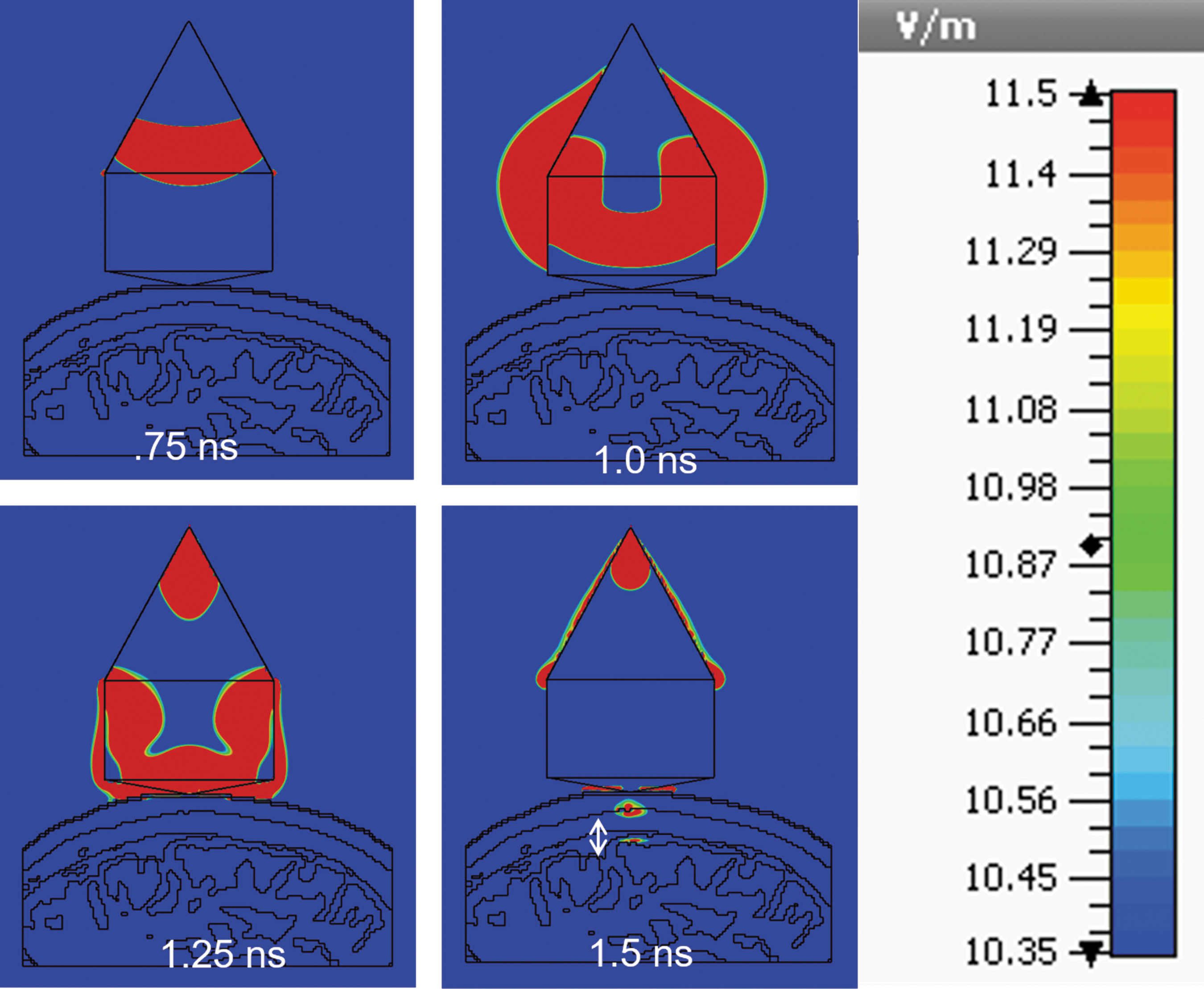
Simulation of delivery of a 100 ps pulse into the brain using a dielectric rod antenna. The snapshots describe the propagation of the wave through the antenna over 1.5 ns. The transient propagation of the electric field shows an excitation of the brain tissue at 11.5 V/m with a diameter of ∼11 mm.
It should be noted that current antenna data are all in silico. While in vitro experiments have been supportive of the approach, it is not yet clear whether an antenna system will translate to in vivo. However, the system holds promises for an application in the clinical field. The previously reported observations of direct effects of ultrashort pulses on cell proteins and their ability to easily penetrate the cell membrane without damaging it render their potential of this technology worth exploring. The ability to deliver pulses in the 100 ps range subcranially has the potential to innovate the way in which Alzheimer's disease and HD are treated.
Synergy of Picosecond Pulsed Electric Fields and Neurodegenerative Diseases
Disaggregation of proteins
The ability of electric fields to target and affect protein aggregates characteristic of neurodegenerative diseases has been confirmed through MD simulations36,37,86,87 and in vitro experiments.28,29 The applications of subnanosecond pulses in the context of neurodegenerative disease were explored by Schoenbach and Greene in a patent published in 2011. 88 The authors presented a method for targeting of protein amyloids, which consist of aggregated misfolded proteins or cut peptides. Such aggregates are aggravating of various conditions, comprising neurodegenerative diseases such as HD and Alzheimer's disease.
They showed that the application in vitro of one or multiple ultrashort pulses on samples containing cells and amyloid fibrils resulted in the disaggregation of the amyloids and preservation of the surrounding environment. More, specifically, no death, destruction, or serious injury of surrounding cells was observed. The change in molecular structure generated by the application of one or more ultrashort pulses was hypothesized to entail a break in hydrogen bonding between the amyloid fibrils' components. Finally, the authors also found that amyloid fibrils possess “weak points,” which are more easily broken with the application of electric fields. The method described by Schoenbach and Greene represents a novel and promising approach to the treatment of diseases aggravated by the presence of amyloid fibrils.
Optimization of pulsing parameters for the targeting of amyloids' “weak points” would possibly allow efficient breakdown of the aggregates with no or limited damage to surrounding healthy tissue. Once smaller fibrils are produced, it is easier for the body to process and eliminate them. Due to the short length of pulses applied, and the properties of ultrashort pulses described in the introduction, it is possible to envision an application of such methods to target intracellular protein aggregates. With this conception, various diseases, including Alzheimer's disease and HD, are optimal targets for the described therapeutic method, rendering research for the further development and application of this technology necessary.
Neural stem cell differentiation
For many years, it was believed that neurons are a terminally differentiated cell type, implying that loss of neuronal cells is a permanent event. However, it is now accepted that new neurons are generated throughout life and that neural stem cells are found in the adult brain. 89 In the context of neurodegenerative diseases, the neurogenic abilities of the brain and their potential stimulation through external factors are worth exploring.
The effects of picosecond pulsed electric fields on the differentiation of human neural stem cells were first reported by Petrella et al., who applied 1800 repetitions of 660-ps pulses to human induced pluripotent stem cells-derived neural stem cells in vitro. This resulted in the inhibition of cell proliferation over 24 h, and altered expression of neural differentiation-related markers was observed. Specifically, a significant increase in expression of the astrocyte marker gene (GFAP) was observed for application of both 20 and 40 kV/cm electric fields, whereas no increase in oligodendrocyte (GALC) and neuronal (DCX) markers was observed. The described results were found to be cell-type specific, as stimulation of mesenchymal stem cells under the same conditions did not result in a reaction. 80
The authors also introduced a novel method for the delivery of picosecond pulsed electric fields in vitro to cells grown in monolayer cultures. A coaxial cable transitioning into a pair of electrodes was anchored to the head of a 3D bioprinter, allowing for the noncontact delivery of pulses to the cells. This setup is shown in Figure 3. The stimulation of anchored cells was enabled by their close proximity with the electrodes, allowing the leakage electric field from a parallel transmission line to affect the target cells. This method allows for pulse delivery to a high number of cells with high yield, and in a coordinate-controlled manner, ideal for high-throughput studies and analyses. 70 How picosecond pulsed electric fields can induce the differentiation of neural stem cells to a specific cell line and whether it is possible to control this process are questions that have yet to be answered. The evidence reported here and the potential application in “neuroregeneration” and as a therapeutic approach for HD and Alzheimer's disease warrant further research.
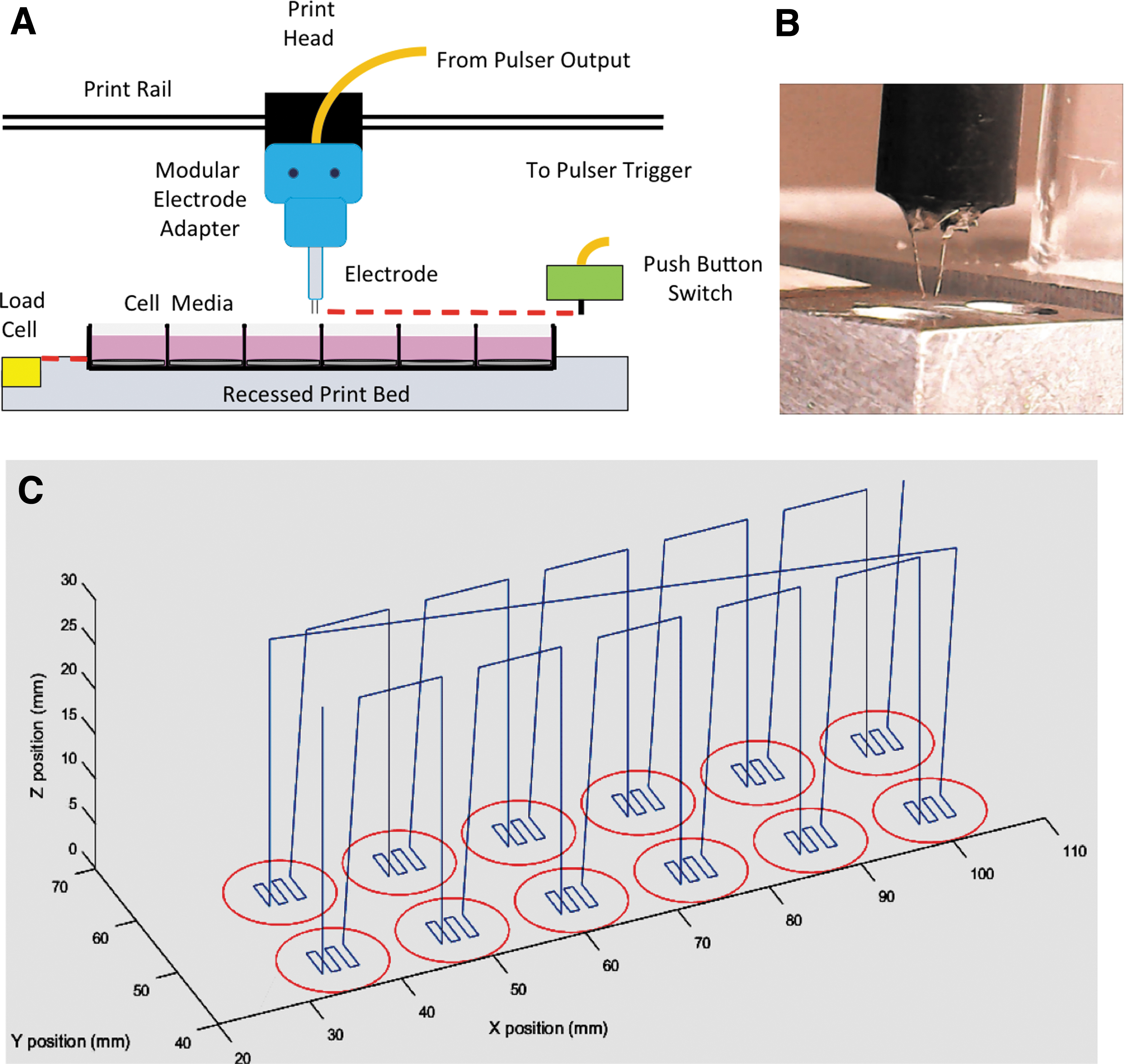
A coordinate-based picosecond pulsing system can be created combining a picosecond pulse electrode to a customized 3D printer. A cross-sectional view of the setup
Conclusions
Research and applications of picosecond pulsed electric fields are not as widely developed as for pulses in the milli-, micro-, and nanosecond range. Recent interest in the potential of ultrashort pulsed electric fields has shown that these possess unique properties, possibly resulting in yet another clinical application of pulsed electric fields. Picosecond pulses have been shown to penetrate the cell membrane and induce intracellular effects, directly affect plasma membrane proteins, and be able to induce cell permeabilization as well as cell death by apoptosis. Additionally, a unique feature of picosecond pulses is the possibility of noncontact delivery using focusing antennas and conceivably target deep tissues and organs inaccessible with traditional electrode designs.
One particular field of interest for the therapeutic application of picosecond pulsed electric fields is that of neurodegeneration, with focus on HD and Alzheimer's disease. These both are characterized by progressive degeneration and death of neurons and present the formation of intracellular and extracellular protein aggregates throughout the disease progression. The delivery of picosecond pulsed electric fields has been shown to induce the disaggregation of protein amyloids without inducing cell death or significant damage to surrounding healthy tissue. The pulses have also been shown to affect neural stem cell differentiation. Finally, such stimuli can be delivered in a noncontact manner, and simulations have shown the possibility of delivering ultrashort pulses able to induce biological effects subcranially.
Extensive research efforts for the advancement and improved understanding of all aspects of subnanosecond pulsed electric fields are necessary before a clinical therapeutic application can even be conceived. Particularly, further research in this field is necessary to gain a comprehensive understanding of potential side effects. However, based on the current knowledge in the field, picosecond pulsed electric fields have the potential to be applied as a safe and noninvasive therapeutic approach to neurodegenerative diseases targeting protein aggregates.
Footnotes
Authors' Contributions
M.Z. contributed to the article design and developed the majority of the article. R.P. contributed to the article design and developed the sections “Picosecond Pulse Generators” and “Pulse Application Methods.” P.A.M. contributed to the article design and revision. All the three authors have reviewed and approved the article before submission. The submitted article is not published, in press, nor submitted elsewhere.
Author Disclosure Statement
No competing financial interests exist.
Funding Information
No funding was received.