




Abstract
Objective:
The present study aimed to demonstrate the possible effects of increased 2-arachidonoylglycerol (2-AG) by applying the monoacylglycerol lipase inhibitor KML-29 on rats with ovarian ischemia–reperfusion (IR) model.
Methods:
Forty-eight female Wistar albino rats were divided into six groups. Group 1: Sham, Group 2: Ischemia, Group 3: IR, Group 4: IR + KML-29 (2 mg/kg), Group 5: IR + KML-29 (20 mg/kg), and Group 6: IR + vehicle (dimethyl sulfoxide). Three hours of ischemia followed by 3 h of reperfusion. Two different doses of KML-29 (2 and 10 mg/kg) were administered intraperitoneally in Groups 4 and 5, 30 min before reperfusion. Ovarian IR injury and ovarian reserve were evaluated histopathological and by using nuclear factor (NF)-κB, interleukin (IL)-1β, tumor necrosis factor (TNF)-α, transforming growth factor (TGF)-β1, superoxide dismutase, glutathione peroxidase pre-/postoperative blood antimullerian hormone, and inhibin B.
Results:
In the KML-1 and KML-2 groups, this damage was significantly reduced compared to the ischemia group. NF-κB, IL-1β, TNF-α, and TGF-β1 immunoreactivities increased statistically significantly in the ischemia group compared to the control group (p<0.001). Immunoreactivities of these proteins were significantly decreased in the KML-1 and KML-2 groups (p<0.001). It was observed that the number of these apoptotic cells decreased significantly in the KML-1 and KML-2 groups compared to the ischemia group (p<0.001). The postoperative inhibin level showed a significant decrease in the ischemia group compared to the sham group, while a significant increase was observed in the KML-1 and KML-2 groups compared to the ischemia group.
Conclusion:
It was seen that anti-inflammatory, antioxidant, and antiapoptotic activity was formed, and the ovarian reserve was preserved with 2-AG in ovarian IR damage. The protective effect of endocannabinoids on the ovaries may create a promising new treatment strategy for many pathologies that will affect the ovarian reserve.
Introduction
Ovarian torsion is a gynecological emergency seen in 3% of adolescents and young adults. In ovarian torsion, the ischemia process begins with the interruption of the flow in the arteria and vena ovarica. Adenosin triphosphate (ATP) production decreases and anaerobic glycolysis begins. When the ischemic period is prolonged, the damage becomes irreversible and cell death usually results in necrosis. Restoring the blood circulation of the ischemic tissue allows the cells to heal in the reversible phase. However, this creates another problem, “reperfusion damage.” Reoxygenation of ischemic tissues causes the production of reactive oxygen species (ROS), including superoxide anions, hydrogen peroxide (H2O2), and hydroxyl radicals. ROS damages the phospholipids and proteins of the cell membrane and further increases the mitochondrial permeability, which can lead to a reduction in ATP and cell death. 1
Following this process, the results of studies on how ovarian functions and ovarian reserve affect show variations.2,3 In ischemia–reperfusion (IR) injury studies in which anti-inflammatory and antioxidant agents were used, the histopathological improvement was observed in the ovary. However, the processes associated with this damage have not yet been revealed.
The growing demand for medical marijuana worldwide increases the urgent need for the scientific evaluation of cannabinoids as a treatment. With the isolation of Cannabis sativa, Δ9-tetrahydrocannabinol in 1964, a new era began in the scientific world. The endocannabinoid system (eCS) comprises two major ligands, anandamide (AEA) and 2-arachidonoylglycerol (2-AG), and two receptors, cannabinoid receptor 1 (CBR1) and cannabinoid receptor 2 (CBR2). Endocannabinoids control essential biological processes, including cell survival, death, and progenitor/stem cell proliferation and differentiation. 4 2-AG, which belongs to the monoacylglycerol family of endocannabinoids, acts as a full potent agonist on CB1 and CB2.
Some data suggest that this molecule may affect ovarian functions associated with obesity, menstrual irregularity, chronic oligo-anovulation, and infertility by modulating pathways involved in energy balance and metabolic control. Examining the literature, a strong neuroprotective effect of 2-AG is of attention. 5 Although this effect was associated with the apoptotic processes, the pathways in which 2-AG is effective remains unknown. Another unknown issue is the effects of endocannabinoids on ovarian IR injury and ovarian reserve in general. For this purpose, the present research aimed to show the effects of 2-AG on the ovary by using KML-29, an inhibitor of monoacylglycerol lipase (MAGL) enzyme that breaks 2-AG into glycerol and arachidonic acid. It was observed that KML-29 exerts anti-inflammatory effects against acute injury and inflammation in mice.
In the present study, the possible effects of 2-AG, which shows an endogenous increase with KML-29, on ovarian IR injury both histopathologically and tissue nuclear factor (NF)-κB, interleukin (IL)-1β, tumor necrosis factor (TNF)-α, transforming growth factor (TGF)-β1, superoxide dismutase (SOD), glutathione peroxidase pre-/postoperative blood levels were presented for the first time by the evaluation of antimullerian hormone (AMH) and inhibin B levels.
Materials and Methods
Animals and study design
The local Animal Ethics Committee approved the experimental procedures used in the present study (via KOBAY D.H.L. A.Ş.Registry No:476). All experiments were carried out according to the Guide for the Care and Use of Laboratory Animals, as confirmed by the National Institute of Health (USA). The female Wistar Albino rats (180–240 g, 8 weeks old) were randomized into six groups (n=8):
Group 1: control, the rats underwent laparotomy operation; Group 2: Ischemia (I), 3 h of ischemia was created; Group 3: IR, 3 h of ischemia and following 3 h of reperfusion was created; Groups 4 (KML-1): The rats received a signal dose of 2 mg/kg KML-29 and vehicle (dimethyl sulfoxide [DMSO]) intraperitoneally, 30 min before induction of IR; Group 5 (KML-2): The rats received a signal dose of 10 mg/kg KML-29 and vehicle (DMSO) intraperitoneally, 30 min before induction of IR; Group 6: The rats received vehicle (DMSO) intraperitoneally, 30 min before induction of IR.
Surgical procedure
As an anesthetic drug for surgical interventions, 50 mg/kg Ketamine (Ketalar; Parke Davis, Eczacıbaşı, Turkey) and 10 mg/kg Xylazine (Rompin, Bayer AG, Leverkusen, Germany) were used in combination. After anesthesia, blood samples were taken from all the rats for biochemical evaluation. Rats were kept in a supine position and 2% iodine alcohol was used for antisepsis of the lower abdominal region. Then, a 2.5 cm longitudinal incision was made in the lower abdominal region and the right ovary was visualized. For the induction of ischemia, vascular clamps were applied to the right ovarian vessels. At the end of the 3 h ischemia period, the vascular clamps were removed and 3 h reperfusion was continued. The vital signs of all rats remained stable throughout the surgical procedure. At the end of the reperfusion period, 1 mL blood samples were obtained from the rat's jugular vein for measurement of postreperfusion.
The rats were sacrificed by decapitation in accordance with the ethical committee principles and the bilateral ovarian tissue was harvested. The ovarian tissue was vertically divided into halves: one half of the ovary was put into a 10% neutral-buffered formalin solution for 24 h for histologic examination. The other half was cleaned of retroperitoneal white adipose tissue and rapidly stored in a −80°C freezer until required for biochemical analysis. All collected blood samples were immediately centrifuged for 10 min at 5000 rpm at +4°C. The serum obtained was transferred into Eppendorf tubes and stored at −80°C until assayed.
Drugs and vehicle
KML-29 (MAGL inhibitor; 1-piperidinecarboxylic acid, 4-[bis(1,3-benzodioxol-5-yl) hydroxymethyl]-, 2,2,2-trifl uoro-1-(trifluoromethyl)ethyl ester) was obtained from Sigma-Aldrich (St. Louis, MO).
DMSO was obtained from Merck, Germany [Assay (GC) ≥99.7].
Histopathological examination
Ovarian tissues taken from the subjects were fixed in 10% formaldehyde for histopathological evaluation. Tissue tracking and paraffin embedding procedures were applied. Then, 5-μm-thick sections were taken from the paraffin blocks. Sections were stained with hematoxylin and eosin. Images (Olympus BX53; Olympus, Tokyo, Japan) were analyzed under a microscope. 6 The criteria for ovarian injury were determined as interstitial edema, dilatation, hemorrhage, polymorphonuclear leukocyte infiltration (PNL), and follicular cell degeneration (granulosa cells). Histopathological scores were given in 10 different areas. Accordingly, this scoring was evaluated as 0: No damage, 1: Slightly damaged, 2: Moderately damaged, and 3: Heavily damaged. Also, primordial, primary, secondary, and tertiary follicle values were counted in 10 different areas. 7
Immunohistochemistry review
The avidin-biotin-peroxidase method was applied immunohistochemically to determine the immunoreactivity of ovary NF-κB (sc-514451; Santa Cruz Biotechnology, Dallas, TX), IL-1β (sc-52012; Santa Cruz Biotechnology), TNF-α (sc-52746; Santa Cruz Biotechnology), and TGF-β1 (sc-130348; Santa Cruz Biotechnology). Sections of 5 μm thickness taken from paraffin blocks were kept in the oven at 60°C overnight and then passed through xylene and decreasing alcohol series. Then, it was treated with citrate buffer for antigen recovery after washing with distilled water. H2O2 was applied after washing with phosphate-buffered saline. The subsequent procedures were carried out using the Large Volume Detection System kit. Then, the serum block was applied for 5 min. After the serum block, NF-κB, IL-1β, TNF-α, and TGF-β −1+4°C were applied overnight as primary antibodies.
After the application of the primary antibody, secondary streptavidin-horseradish peroxidase and 3,3′-diaminobenzidine (DAB) chromogen with biotin was applied for 1 min and counterstained with Gill hematoxylin. Finally, it was passed through the gradually increasing alcohol series and xylene and sealed with entellan.8,9 The scores were given for immunoreactivity measurements from 10 different areas from the images taken from the preparators. This scoring was done as follows; 0: no staining, 1: Mild staining, 2: Moderate staining, and 3: Intense staining. 10
Biochemical analysis
Serums and ovary tissues taken from the subjects were stored at −80°C. Rat AMH (Cat. No.: E-EL-R3022; Elabscience, Houston, TX) and Rat inhibin B (Cat. No.: EA0059Ra; Bioassay Technology Laboratory, Shanghai, China) kits were studied in serums adopting the enzyme-linked immunosorbent assay (ELISA) method. In the ovary samples, Rat-2 Arachidonoylglycerol (Cat. No.: E1322Ra; Bioassay Technology Laboratory), General AEA (Cat. No.: EA0024Ge; Bioassay Technology Laboratory), Rat superoxidase Distumatse (Cat. No.: EA0168Ra; Bioassay Technology Laboratory), Rat Glutathione (Cat. No.: EA0113Ra; Bioassay Technology Laboratory), and Rat Malondialdehyde (Cat. No.: EA0156Ra; Bioassay Technology Laboratory) were measured. The quantities were determined at 450 nm in an ELISA reader.11–13
Terminal deoxynucleotidyl transferase dUTP nick end labeling
We used the in situ apoptosis detection kit (ApopTagÒ Plus Peroxidase In Situ Apoptosis Detection Kit, S7101; Millipore, Burlington, MA) to detect apoptosis. Sections (5 μm) were cut from paraffin blocks of ovarian samples of the six groups. The sections were deparaffinized in xylene, rehydrated, and incubated with 20 μg/mL proteinase K (Cat. No.: 20S-001) for 10 min, and rinsed in distilled water. Endogenous peroxidase activity was inhibited with 3% H2O2. The sections were incubated with equilibration buffer for 10–15 sec and terminal deoxynucleotidyl transferase enzyme in a humidified atmosphere at 37°C for 60 min. They were put into prewarmed working strength stop/wash buffer at room temperature for 10 min subsequently and incubated with antistreptavidin–peroxidase for 45 min. The staining was done with DAB and nuclei were counterstained with Gill hematoxylin.
Statistical analyses
All statistical analyses were carried out by using the GraphPad Prism version 7.00 for Mac (GraphPad Software, La Jolla, CA). D'Agostino Pearson omnibus test was used to identify the normal distribution of the data. If the numerical data were nonparametric, the Kruskal–Wallis test was conducted, if it was parametric, a one-way analysis of variance test was carried out and Tukey's correction was used for the post hoc assessment. The data were expressed as the mean of normalized data±standard deviation of the mean. p Value <0.05 was considered statistically significant.
Results
Histopathological findings
Hematoxylin–eosin staining results are shown in Figure 1. The normal histological structure was seen in the Sham group. Hemorrhage and follicular degeneration damages were observed in the ischemia group. The ischemia group showed a statistically significant increase in histopathological damage compared to the control group. In the KML-1 and KML-2 groups, this damage was significantly reduced compared to the ischemia group. Also, in the follicle counts, it was observed that there was a decrease in the number of primordial, primary, secondary, and tertiary follicles in the ischemia group.

Ovary hematoxylin and eosin staining image, histopathological scoring images.
Immunohistochemical findings
Immunohistochemistry images are shown in Figure 2. NF-κB, IL-1β, TNF-α, and TGF-β1 immunoreactivities increased statistically significantly in the ischemia group compared to the control group (p<0.001). Immunoreactivities of these proteins were significantly decreased in the KML-1 group (p<0.001). The immunoreactivities of NF-κB, IL-1β, and TNF-α proteins were significantly decreased in the KML-2 group (p<0.001).
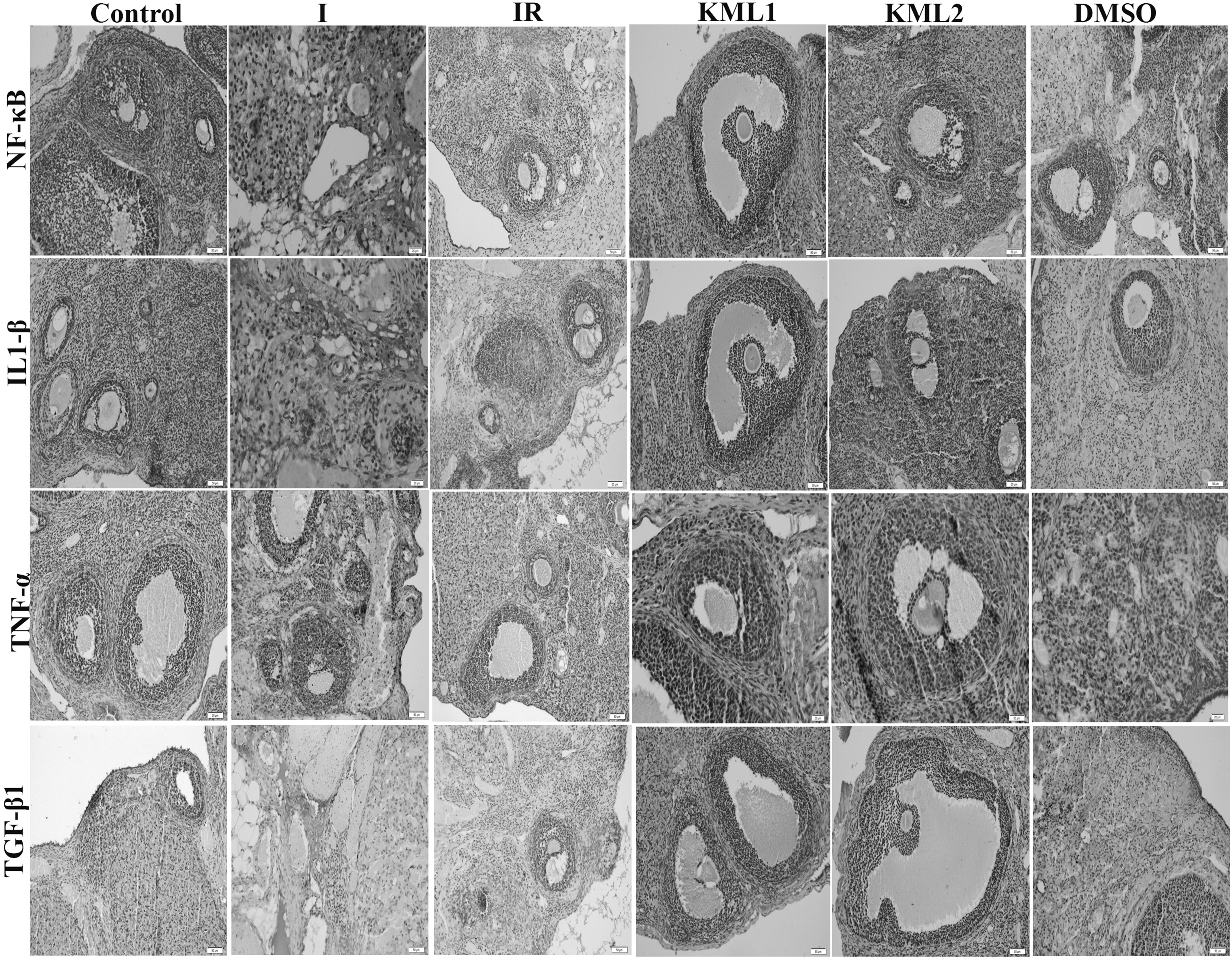
Ovary NF-κB, IL-1β, TNF-α, and TGF-β immunohistochemistry images (X200). IL, interleukin; NF, nuclear factor; TGF, transforming growth factor; TNF, tumor necrosis factor.
Terminal deoxynucleotidyl transferase dUTP nick end labeling
Terminal deoxynucleotidyl transferase dUTP nick end labeling (TUNEL) results are shown in Figure 3. The number of apoptotic cells increased significantly in the ischemia group compared to the sham group (p<0.001). It was observed that the number of these apoptotic cells decreased significantly in the KML-1 and KML-2 groups compared to the ischemia group (p<0.001).
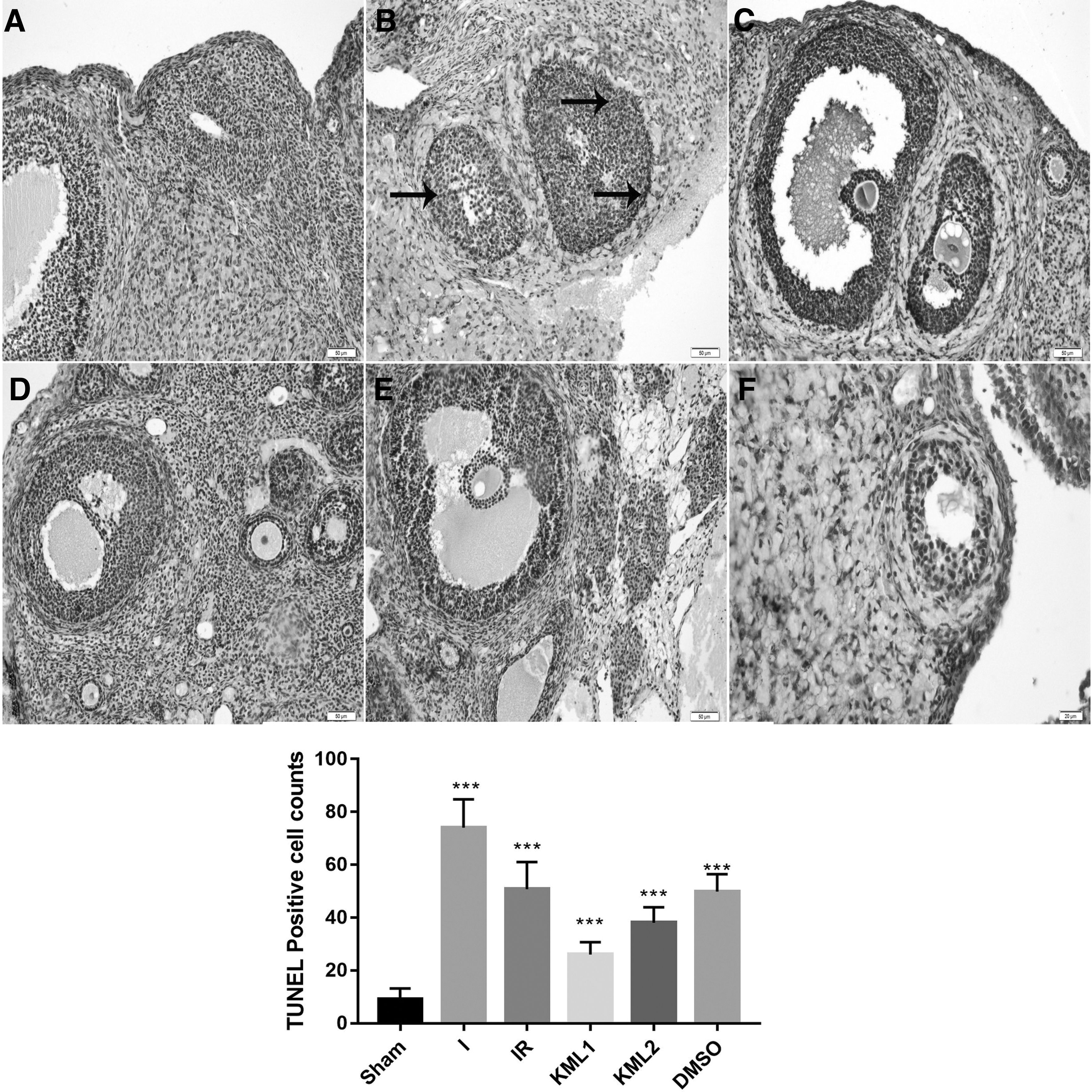
The ovary TUNEL results and graph.
Biochemical findings
The results of the biochemical analyses are shown in Table 1. Serum AMH and inhibin levels were similar between groups in the preoperative period. In the postoperative period, the AMH level decreased in the ischemia group compared to the sham group. A significant increase was observed in the KML-1 group compared to the ischemia group. The postoperative inhibin level showed a significant decrease in the ischemia group compared to the sham group, while a significant increase was observed in the KML-1 and KML-2 groups compared to the ischemia group.
Biochemical Findings
All data are expressed as the mean±SD (n=8). All data are expressed as the mean±SD.
p<0.05 Compared to the sham group.
p<0.001 Compared to the sham group.
p<0.0001 Compared to the sham group.
2-AG, 2-arachidonoylglycerol; AEA, anandamide; AMH, antimullerian hormone; DMSO, dimethyl sulfoxide; I, ischemia; IR, ischemia–reperfusion; MDA, malondialdehyde; SD, standard deviation; SOD, superoxide dismutase.
Arachidonoylglycerol, general AEA, SOD, and glutathione levels significantly decreased in the ischemia group compared to the sham group. Arachidonoylglycerol, SOD, and glutathione levels increased significantly in the KML-1 group compared to the ischemia group. In the KML-2 group, the SOD level showed a significant increase compared to the ischemia group. The malondialdehyde (MDA) level showed a significant increase in the ischemia group compared to the sham group, while the MDA level in the KML-1 and KML-2 groups showed a significant decrease compared to the ischemia group, Table 1.
Discussion
MAGL has been considered a promising therapeutic target for many diseases in recent years. 14 KML-29 is the most selective MAGL inhibitor identified by Chang et al. 15 However, due to the complex biology of eCS, its effectiveness remains unclear. In the present study, the effects of 2-AG, which increased by MAGL inhibition in ovarian tissue, on IR damage were presented for the first time. According to the data obtained in the present study, an improvement in ovarian IR damage was observed in the IR group, in which low dose KML-29 was administered histopathologically and biochemically. However, low-dose KML significantly increased AMH values, which are indicators of ovarian reserve, compared to the IR group.
Endocannabinoid levels increase with tissue damage. However, since the increased endocannabinoids are rapidly degraded by MAGL and fatty acid amide hydrolase enzymes in tissues, their effects cannot be fully revealed, so their potential effects are unknown. 16 Reviewing the studies in which MAGL inhibition increased 2-AG levels, it was seen that almost all of them focused on the central nervous system. The neuroprotective activity of 2-AG in the central nervous system is associated with the prevention of dopaminergic neuron loss and the reduction of proinflammatory cytokine and prostaglandin release. 17 Ovarian tissue is a special organ with its structure, in which follicles show active changes and participate in hormone production. The IR damage of this organ, inflammatory, and oxidative pathways are activated and cell damage occurs by apoptosis. 7 A suggested pathogenesis of tissue injury in the course of reperfusion is buildup of the stimulated neutrophils that produce ROS. 18
There are several enzymatic activities involved in the generation of ROS: the reduction of dioxygen in the mitochondria can lead to various intermediate forms of ROS; within peroxisomes, the reduction of amino acid oxidase triggers the oxidative deamination in the α-keto acids and copper and iron ions generate ROS that may be released into the blood flow by transferrin, albumin, and ceruloplasmin. 19 Lipid peroxidation in the cell is a common detrimental outcome of free radicals that end up decreasing the membrane potential and cell damage. MDA causes severe cell injury via stimulation of polymerization and crosslinking in components of the membrane. 20 Various antioxidant and anti-inflammatory agents acting on these pathways have organ protective effects. 7 Cannabinoid derivatives inhibit toll like receptor 4 and NF-κB on neurons.
NF-κB involves a family of pleiotropic transcription factors. It plays an essential role in regulating the expression of genes involved in various cell processes, including inflammation. NF-κB is held in the cytoplasm in an inactive state by specific inhibitors. Upon degradation of the inhibitor, NF-κB moves to the nucleus and activates transcription of specific genes. 20 NF-κB activation can be stimulated by different molecules such as cytokines and ROS. Upon its activation, NF-κB arouses the transcription of various proinflammatory mediators such as IL-6, TNF-α, and inducible nitric oxide synthase. 21 It has been reported that cannabinoids produces anti-inflammatory activity and exerts neuroprotective effects through activation of the signaling pathway.
In one of our previous studies, we demonstrated the protective effect of anti-inflammatory activity with CB2 agonistic property over the NF-κB pathway in ovarian IR injury. 22 Examining the literature, it was seen that the anti-inflammatory effects caused by KML-29 were shown on the joint brain tissue. This efficacy was evaluated by Philpott and McDougall with the osteoarthritis model leukocyte rolling and adhesion. 16 It was thought that the effects of cannabinoids on inflammatory processes occur through cytokine dysregulation and their interactions on immune system cells. 23 Therefore, in the present study, the inflammatory parameters revealed in the ovarian tissue was evaluated to show the efficacy of 2-AG, and it was found that NK-FB, IL-1b, TNF-α, and values were lower in the low-dose KML-29 group compared to those of the IR group. NF-κB executes the inflammatory process.
It was thought that the increase in 2-AG decreases IL and TNF-α levels with the activation of this pathway. Evaluating the oxidative mechanisms related to the damage, it was observed that SOD and glutathione values significantly increased in the IR group that received low-dose KML-29 compared to the IR group, and the MDA values did not change. Increased ROS production and impaired antioxidant capacity have been shown as the main factors contributing to ovarian dysfunction during ovarian IR injury. 24 In the present study, only SOD levels increased significantly in the group in which high doses of KML-29 was administered. As a result, MAGL inhibition, antioxidant mechanisms take action. Therefore, it was thought that the increased antioxidative parameters may have been effective in protective efficiency against IR damage.
Ovarian reserve is the key point of all ovarian treatments for clinicians. However, it was thought that reserve changes in ovarian torsion have not been studied in detail. Özler et al., in their study on evaluating the number of follicles in ovarian torsion, found that the average number of preantral and small antral follicles in the torsion and detorsion groups was significantly lower than those in the sham group. 2 On the contrary, in another study in which the torsion duration was extended, it was found that there was a nonsignificant decrease only in the number of primordial follicles. 3 In the present study, the numbers of primordial and primary follicles were found to be significantly lower in both the I and IR, that is, the detorsion group.
The important difference of the present study from similar studies in the literature is that an agent belonging to the eCS, which we think may affect the ovarian reserve, was applied and oxidative, inflammatory, biochemical, and apoptotic parameters were also evaluated in addition to the histological evaluation. The AMH is a member of the TGF superfamily and is secreted from the granulosa cells of primary growing follicles in the human ovaries until the early antral stage. 25 Serum AMH level is a marker for preantral follicles in the primary pool. 26 In the present study, in addition to histological findings, there was an increase in the AMH and inhibin B values. This result, as seen in the histopathological evaluation, can be associated with the increase in the number of primordial and primary follicles seen with KML-29 application.
Also, the effect of increased 2-AG on TGF-β should be considered. In the present study, it was seen that TGF-β, which increases especially with I, decreased in the IR group as a result of a low-dose-KML-29 application. We think that this effect, which will cause a decrease in fibrosis, may contribute to the preservation of the ovarian reserve. It has been reported in the literature that TGF-β changes depending on synthetic derivatives such as cannabinoids. 27 To the best of our knowledge, the effect of 2-AG on TGF-β was demonstrated for the first time. Another process that will affect the reserve can be associated with the active activation of antiapoptotic mechanisms that increase the number of surviving cells. The cellular protective effect caused by KML-29 in ovarian IR damage is seen using TUNEL.
Zhong et al. have reported that apoptosis significantly decreased in hippocampal neurons with a noncaspase-dependent pathway in the mice that they established a cerebral ischemia model and gave 2-AG. 5 The authors associated this antiapoptotic effect in neurons with the 2-AG-dependent modification of mitochondrial membrane depolarization due to ischemia. It was thought that the anti-apoptotic effect on the ovary may be related to the activation of the NF-κB pathway due to endocannabinoids. 22 Here, the interaction of the NF-κB pathway with antiapoptotic B-cell lymphoma family members was thought to be effective. In ovarian IR damage, the NF-κB pathway has also been reported to generate anti-inflammatory activity. 7 As a result, all these data showed that many mechanisms that increase the number of surviving cells work together in ovarian protective effect due to endocannabinoids.
Some studies have drawn attention to the dose-dependent efficacy of KML-29, especially in mechanical and cold allodynia related to analgesia. 22 However, chronic high-dose KML-29, despite the benefits of MAGL inhibition may lead to the desensitization and downregulation of CB1 receptors by developing physical dependence and cross-tolerance to the cannabinoid receptor. 28 No increased efficacy was found with high-dose-KML-29 in the ovarian IR injury. On the contrary, a lower dose was found to be more effective. Although KML-29 is a potent MAGL inhibitor, we do not know why the administration of higher doses of KML-29 produces such a result.
However, it is stated that endogenous cannabinoids are at different levels even within the same organ (cerebellum and hippocampus regions of the brain).29–31 Variable levels may have caused changes in enzymatic reactions depending on the metabolism differences occurring with the experimental model. The expected efficacy with fewer side effects due to low-dose use can be considered as a desirable situation. However, since the model created in the present study was based on the surgical approach, it was not possible to fully evaluate the side effects.
The strongest aspect of the present study is that the tissue levels of 2-AG and AEA values belonging to eCS, which can be affected by the use of KML-29, were shown together. Also, examining the markers of both oxidative, inflammatory, and apoptotic processes together strengthen the study. However, it can be thought that different results can be obtained with different IR durations. The fact that IR duration was not applied except the one for 3 h in the present study can be considered a limitation. Another limitation was that especially the central side effects that KML-29 may cause could not be evaluated.
eCS-focused treatment modalities offer promising new possibilities for many diseases. The cellular protective effect caused by MAGL inhibition also appears to be effective in the ovarian IR damage. At this point, the most important detail was the protection of the ovarian reserve. With the present study, it was revealed that new researches on cannabinoids in gynecological pathologies may be useful in revealing the different usage areas of these agents.
Footnotes
Author Disclosure Statement
All authors declare no conflicts of interest.
Funding Information
No funding was received for this article.