
Abstract
Abstract
Purpose:
To evaluate the effects of Triesence® (TRI), a new preservative-free triamcinolone approved by the U.S. Food and Drug Administration (FDA) for intraocular use, on human retina pigment epithelial (ARPE-19) and rat neurosensory (R28) cells in culture.
Methods:
ARPE-19 and R28 cell cultures were treated 24 h with 1,000, 500, 200, or 100 μg/mL of crystalline (cTRI) or 1,000, 500, or 200 μg/mL of solubilized (sTRI). TRI was solubilized by centrifuging the drug, discarding the supernatant containing the vehicle and then resuspending the drug pellet in an equivalent amount of Dimethyl sulfoxide to achieve the same concentration as the commercial preparation. Percentage of cell viability (CV) was evaluated by a trypan blue dye-exclusion assay. The mitochondrial membrane potential (ΔΨm) was analyzed with the JC-1 assay. The caspase-3/7 activity was measured by a fluorochrome assay.
Results:
In the ARPE-19 cultures, the cTRI caused a decrease in CV at 1,000 μg/mL (13.03±6.51; P<0.001), 500 μg/mL (28.87±9.3; P<0.001), 200 μg/mL (54.93±5.61; P<0.001), and 100 μg/mL (82.53±0.65; P<0.005) compared with the untreated controls (96.98±0.16). In R28 cultures, the cTRI treatment also reduced CV values significantly (P<0.001) for the 1,000 μg/mL (22.73±2.44), 500 μg/mL (34.63±1.91), 200 μg/mL (58.70±1.39), and 100 μg/m (75.33±2.47) compared with the untreated controls (86.08±3.54). Once the TRI was solubilized (sTRI), the CV and ΔΨm remained similar to the untreated controls for both ARPE-19 and R28 cells. The sTRI treatment with 1,000, 500, and 200 μg/mL increased in caspase-3/7 activity in ARPE-19 cells (P<0.01) and in R28 cells (P<0.05) compared with dimethyl sulfoxide equivalent controls.
Conclusion:
The crystalline form of TRI (cTRI) can cause a significant decrease in CV to cultured retinal cells. Once the TRI is solubilized (sTRI), at the same concentrations, the cells remain viable with no decrease in CV or ΔΨm. The sTRI can, however, increase caspase-3/7 activity, thus suggesting some degree of apoptosis.
Introduction
However, as an off-label application, its intraocular safety has long been a concern. 16 Kenalog (Brystol-Meyers-Squibb, Princeton, NJ), initially developed for intra-articular or intramuscular applications, is the most commonly used TA for intravitreal injection. However, it has not been developed for intraocular use; and intravitreal injections have been discouraged by its manufacturer. Its vehicle is described as an aqueous solution containing sodium chloride to maintain isotonic osmolarity, 0.99% benzyl alcohol (BA) as a preservative and bactericidal agent, 0.75% sodium carboxymethylcellulose for increasing viscosity and distribution of the suspension, and 0.04% polysorbate as a preservative and sterile water.
The most common adverse effects of ocular steroid therapy are glaucoma and cataract formation.17–19 In addition, given that the commonly used formulation of TA is not formulated for the eye, there is a known risk of pseudoendophthalmitis and a hypothetical potential for clinical retinal toxicity from the vehicle when it is injected intravitreally.20–22
The toxicity of TA to retinal cells has been shown in vitro23–29 and in morphologic rabbit studies. 30 Electroretinographic (ERG) data are inconclusive, as some papers show toxicity, 31 whereas others conclude that it is safe for intraocular use.32,33 The mechanism of toxicity is still to be determined: some authors attribute at least part of the retinal cells TA toxicity to its preservative BA.31,34,35 Other data suggest that the direct contact between TA crystals and the retinal cells could promote the cell death.29,36,37
Recently, a preservative-free TA developed and approved by the U.S. Food and Drug Administration (FDA) for intraocular use known as Triesence® (TRI; Alcon Labs, Forth Worth, TX) has become commercially available. TRI is a synthetic corticosteroid with anti-inflammatory action. The aqueous suspension provides 40 mg/mL of TA, with sodium chloride for isotonicity, 0.5% carboxymethylcellulose sodium, and 0.015% polysorbate 80. It also contains potassium chloride, calcium chloride (dihydrate), magnesium chloride (hexahydrate), sodium acetate (trihydrate), sodium citrate (dihydrate), and water for injection.
Despite the numerous evidence showing in vitro toxicity of TA, the in vitro toxicity of TRI has not been previously reported. New evidence indicates that different TA preparations have unequal particle sizes: Kenalog particles can reach up to 40 μm, whereas TRI particles reach less than 30 μm. 38 The difference in particle size may lead to different outcomes when retinal cells are exposed to different TA forms in vitro.
The purpose of the present study is to evaluate the effects of the 24 h exposure of TRI on human retinal pigment epithelial (ARPE-19) and rat neurosensory (R28) cells in culture. To determine the effects of TRI crystals on the cells, the TRI were solubilized (sTRI) and exposed to the retinal cells. We found that the sTRI form of TRI was less toxic than the crystallized form (cTRI), thereby suggesting that the crystals may be causing some sort of damage to the cells.
Methods
Cell culture
ARPE-19 cells were obtained from ATCC (Manassas, VA). Cells were grown in 1:1 mixture (vol/vol) of Dulbecco's modified Eagle's and Ham's nutrient mixture F-12 medium (Invitrogen-Gibco, Carlsbad, CA), nonessential amino acids 10 mM 1×, 0.37% sodium bicarbonate, 0.058% L-glutamine, 10% fetal bovine serum, and antibiotics (penicillin G 100 U/mL, streptomycin sulfate 0.1 mg/m, gentamicin 10 μg/mL, amphotericin B 2.5 μg/mL).
R28 cells, which are rat embryonic precursor neurosensory retinal cells, were derived from postnatal day 6 rat retina in the laboratory of one of the authors (G.M.S.). 39 R28 cells express genes characteristic of neurons 40 and functional neuronal properties. 41 R28 cells were cultured in Dulbecco's modified Eagle's medium, high glucose (Invitrogen-Gibco) with 10% fetal bovine serum, 1× minimum essential medium, 10 mM 1× nonessential amino acids, 0.37% sodium bicarbonate, and 10 μg/mL gentamicin.
Before being exposed to the drug, the cells were plated onto 6-well tissue culture plates and incubated at 37°C in 5% CO2 to reach 80%–90% confluence, and then they were transferred to a serum-free environment for 24 h to reach a sessile nonproliferative state, similar to what is observed in the retina.
Exposure to TRI
ARPE-19 and R28 cultures were treated for 24 h with 100, 200, 500, and 1,000 μg/mL concentration of crystalline commercially available TRI (cTRI); and cell viability (CV) analyses were performed. To determine the role of the TRI crystals on the toxicity observed, ARPE-19 and R28 cultures were treated with 200, 500, and 1,000 μg/mL of TRI dissolved on dimethyl sulfoxide (DMSO) (sTRI) for 24 h. Briefly, commercially available TRI (Alcon Laboratories) was centrifuged at 5,000 rpm for 1 min; and the supernatant containing the vehicle was discarded. The drug pellet was resuspended in an equivalent amount of DMSO to achieve the same TRI concentrations of that in the commercial TRI suspension. For the equivalent doses of 1,000 μg/mL, 500 μg/mL and 200 μg/mL, 250 μL, 125 μL and 50 μL of DMSO were, respectively, added per mL of culture media, and they represent the same amount of DMSO used to solubilize the drug at the concentration tested. CV along with caspase-3/7 activities and JC-1 mitochondrial membrane potential (ΔΨm) were assessed for the sTRI at the following concentrations: 200, 500, and 1,000 μg/mL.
CV assay
The CV assay was performed as previously described. 26 Briefly, cells were harvested from 6 well-plates by treatment with 0.2% trypsin-EDTA and incubated at 37°C for 5 min. Before trypsinization, the original medium in each well was transferred to a sterile 15 mL centrifuge tube (Corning, Inc., Corning, NY) to include the floating cells in CV count. The 15 mL tube containing the cell suspension and the medium with the floating cells was centrifuged at 1,000 revolutions per minute (rpm) for 5 min. The supernatant was then discarded, and the cell pellet was resuspended in 1 mL of culture medium. Automated CV analysis was performed (ViCell™ analyzer; Beckman Coulter, Inc., Fullerton, CA). The analyzer performs an automated trypan blue dye-exclusion assay and gives the percentage viability of cells.
Caspase-3/7 activity
Caspase-3/7 activities were detected with the help of detection kits (Carboxyfluorescein FLICA Apoptosis Detection Kits; Immunochemistry Technologies LLC, Bloomington, MN). The FLICA reagent has an optimal excitation range from 488 to 492 nm and an emission range from 515 to 535 nm. Apoptosis was quantified as the level of fluorescence emitted from FLICA probes bound to caspases. Nonapoptotic cells appeared unstained, whereas cells undergoing apoptosis fluoresced brightly. The caspase activity was measured as average signal intensity of the fluorescence of the pixels in a designated spot—mean signal intensity (msi).
At the designated time period, the wells were rinsed with fresh culture media, replaced with 300 μL/well of 1× FLICA solution in culture media, and incubated at 37°C for 1 h under 5% CO2. Cells were washed with PBS. The caspase-3/7 activity was measured as average signal intensity of the fluorescence of the pixels in a designated spot—msi. The following controls were included: untreated ARPE-19 and R28 cells without FLICA to exclude autofluorescence from cells; untreated ARPE-19 and R28 cells with FLICA for comparison of caspase activity of treated cells; and tissue culture plate wells without cells with buffer alone to represent the background levels. Fluorescence intensity was measured by Fluorescence Image Scanning Unit (excitation λ=488 nm, emission λ=520 nm; FMBIO III; Hitachi).
Mitochondrial membrane potential (ΔΨm) measurements
Detection of ΔΨm values was performed using the JC-1 mitochondrial membrane potential detection kit (Biotium, Hayward, CA). JC-1 (5,5′,6,6′-tetrachloro-1,1′,3,3′-tetraethyl-benzimidazolyl-carbocyanine-iodide) is a cationic dye that accumulates as aggregates in the mitochondrial membranes of healthy cells which result in a red fluorescence (590 nm). The damaged cells, which have diminished ΔΨm values, show a green fluorescence (529 nm). The ratio of red (live cells) and green (dead cells) fluorescence is measured in each sample. The fluorescent signal was measured with the scanning unit (FMBio III; Hitachi) set to detect green (510–525 nm) and red (590 nm) emissions. Ratios of red to green fluorescence were calculated, and the data were analyzed by unpaired Student's t-test.
Statistical analysis
Data were subjected to statistical analysis by ANOVA (Prism, ver. 3.0; GraphPad Software, Inc., San Diego, CA) and Unpaired Student's t-test (two-tailed; GraphPad Software, Inc.). Newman-Keuls multiple comparison test was done to compare the data within each experiment. P<0.05 was considered statistically significant. Error bars in the CV graphs represent SEM with experiments performed in duplicate and repeated thrice. Error bars in the JC-1 and Caspase-3/7 graphs represent SEM with experiments performed in triplicate and repeated thrice.
Results
CV in the cTRI-treated cultures
ARPE-19 cells
cTRI caused a significant decrease in CV at all concentrations tested (Fig. 1). The mean viability of ARPE-19 cells after 24 h of exposure to crystalline (nonsolubilized) TRI was: 13.03%±6.51%, 28.87%±9.3%, and 54.93%±5.61% for 1,000, 500, or 200 μg/mL, respectively (P<0.001), and 82.53%±0.65% for 100 μg/mL (P<0.05 when compared with untreated controls (96.98±0.16%).

Percentage of cell viability of ARPE-19 (mean±SEM) exposed to crystalline Triesence (cTRI), n=3. There was a significant decrease in cell viability in all concentrations tested after 24 h. * = Statistically significant.
R28 cells
Cells exposed to cTRI showed a significant decrease in CV compared with untreated control cells (22.73%±2.44%, 34.63%±1.91%, 58.70%±1.39%, and 75.33%±2.47% for 1,000, 500, 200, or 100 μg/mL, respectively, versus 86.08%±3.54% for untreated controls, P<0.001, Fig. 2).
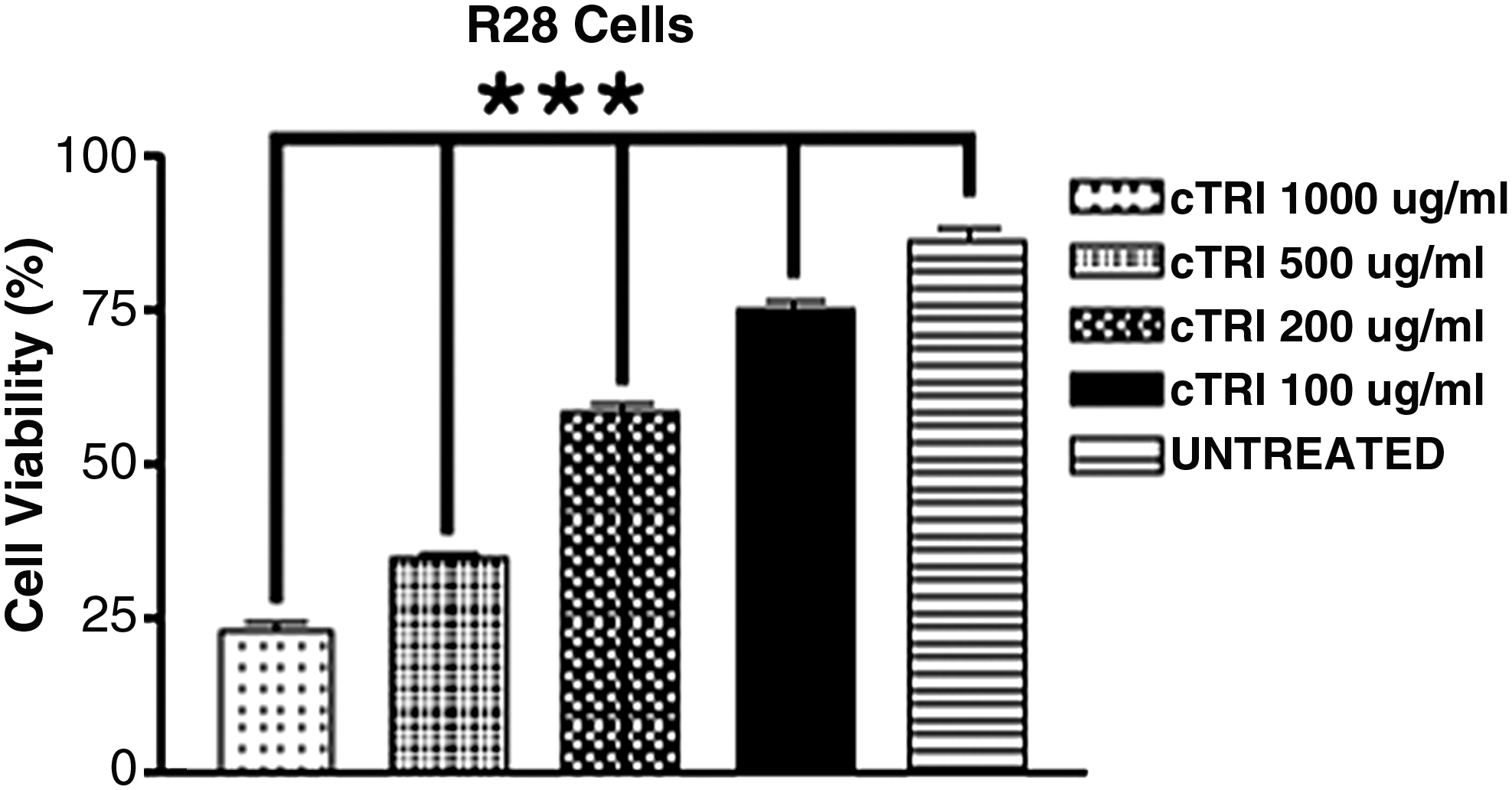
Percentage of cell viability of R28 cells (mean±SEM) exposed to cTRI, n=3. There was a significant decrease in cell viability in all concentrations tested after 24 h. * = Statistically significant.
CV assay—sTRI
ARPE-19 cells
sTRI did not significantly reduce the CV of ARPE-19 cells at any concentration tested. The mean viability of ARPE-19 cells after 24 h of exposure to sTRI was sTRI 1,000 (78.63%±27.14%), 500 (89.60%±9.27%), and 200 (95.57%±0.64%) μg/mL, respectively, versus 98.27%±0.35%, 97.73%±0.71%, or 97.07%±1.70% for cells treated with DMSO equivalent doses or 96.98±0.16 for untreated controls (Fig. 3).

Percentage of cell viability of ARPE-19 (mean±SEM) exposed to TRI solubilized in dimethyl sulfoxide (DMSO) (sTRI), n=3. There was no significant decrease in cell viability in all cell lines and concentrations tested.
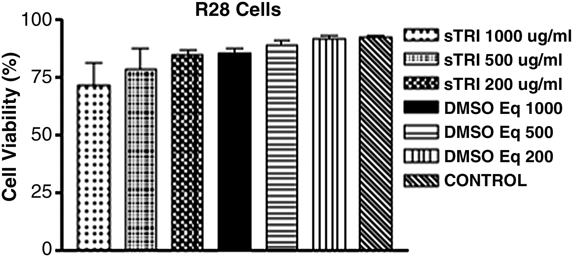
R28 cells
Similar results were obtained when R28 cells exposed to sTRI were compared with cells exposed to DMSO equivalent doses or untreated controls (Fig. 4). There was no statistical difference in CV between cells exposed to sTRI (71.93%±16.83%, 78.60%±16.05%, or 84.90%±4.10% for 1,000, 500, or 200 μg/mL, respectively) and cells exposed to equivalent doses of DMSO (85.40%±3.63%, 88.70%±3.70%, or 91.97%±2.31% for doses equivalent to 1,000, 500, or 200 μg/mL, respectively) or untreated controls (92.60%±0.97%).
Percentage of cell viability of R28 cells (mean±SEM) exposed to TRI solubilized in DMSO (sTRI), n=3. There was no significant decrease in cell viability in all cell lines and concentrations tested.
Caspase-3/7 activity in the sTRI-treated cultures
ARPE-19 cells
Cells treated for 24 h with 1,000, 500, and 200 μg/mL sTRI had higher caspase-3/7 activity compared with corresponding DMSO-treated controls (16,432.400±596.26 msi vs. 11,862.05±557.46, P<0.001; 16,299.89±205.21 msi vs. 11,836.19±114.19 msi, P<0.001; and 13,002.63±152.83 msi vs. 10,709.12±326.65 msi, P<0.01 for 1,000, 500, and 200 μg/mL, respectively). Caspase-3/7 activity of cells treated with sTRI was significantly higher than untreated controls (10,345.70±171.82 msi). However, the 1,000 and 500 μg/mL DMSO controls had a significantly higher caspase-3/7 activity than untreated controls (P<0.05 and P<0.01, respectively, Fig. 5).
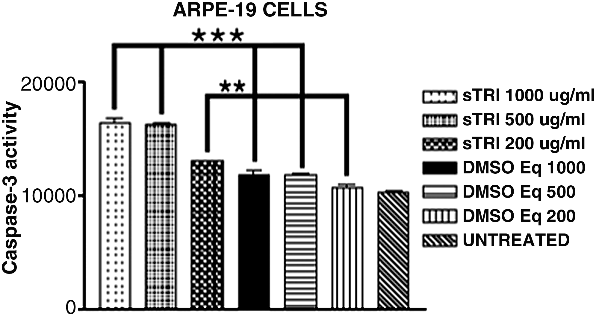
Caspase-3/7 activity in ARPE-19 cells exposed to sTRI (mean±SEM), triplicates, n=3. Cells treated for 24 h with 1,000, 500, and 200 μg/mL sTRI had higher caspase-3/7 activity compared with corresponding DMSO-treated controls (DMSO 250, 125, and 50 μL per mL, P<0.01). The 1,000 and 500 μg/mL DMSO controls had a significantly higher caspase-3/7 activity when compared with untreated controls (P<0.05 and P<0.01, respectively). * = Statistically significant.
R28 cells
Cells treated for 24 h with 1,000, 500, and 200 μg/mL sTRI had increased caspase-3/7 activity compared with corresponding DMSO-treated controls (14,958.29±779.81 msi vs. 1,713.04±1,632.04 msi, P<0.01; 14,541.38±3,337.16 msi vs. 3,465.48±2,445.89 msi, P<0.01; 14,322.28±2,755.96 msi vs. 6,997.07±56.56 msi, P<0.05 for 1,000, 500, and 200 μg/mL, respectively). However, just the highest dose tested had significantly higher caspase-3/7 activity compared with untreated controls (8,298.51±900.79 msi, P<0.001). Despite the lower concentration of DMSO controls when compared with untreated controls, DMSO controls were not statistically different than the untreated controls (Fig. 6).

Caspase-3/7 activity in R28 cells exposed to sTRI (mean±SEM), triplicates, n=3. Cells treated for 24 h with 1,000, 500, and 200 μg/mL sTRI had higher caspase-3/7 activity compared with corresponding DMSO-treated controls ((DMSO 250, 125, and 50 μL per mL, P<0.05). The DMSO controls showed a lower caspase activity when compared with untreated controls, but that difference was not statistically significant. * = Statistically significant.
Mitochondrial membrane potential assay (ΔΨm) in the sTRI-treated cultures
ARPE-19 cells
There was no statistically significant difference between cells treated with sTRI (2.022±0.07, 2.043±0.332, or 2.308±0.570 for 1,000, 500, and 200 μg/mL, respectively), equivalent doses of DMSO (2.363±0.432, 2.550±0.377, or 2.282±0.656 for equivalent doses of 1,000, 500, or 200 μg/mL), or untreated controls (1.567±0.334) (Fig. 7).
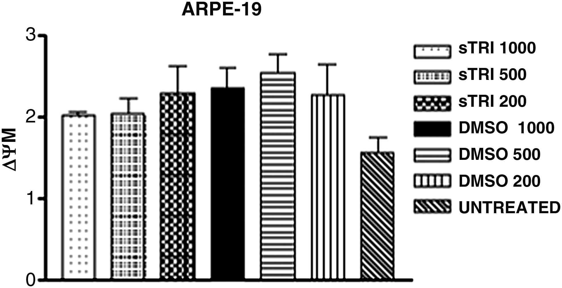
Evaluation of the mitochondrial membrane potential (ΔΨm) of ARPE-19 cells exposed to sTRI (mean±SEM), triplicates, n=3. There was no statistical difference between cells treated with sTRI or its equivalent DMSO controls.
R28 cells
Similar results were observed when R28 cells were exposed to sTRI for 24 h. There were no statistical differences in JC-1 potential between cells exposed to sTRI (0.882±0.029, 0.838±0.035, or 0.819±0.065 for 1,000, 500, or 200 μg/mL) (DMSO equivalent doses of 1,000, 500, or 200 μg/mL were 0.945±0.292, 0.886±0.278, or 0.891±0.227, respectively) or to untreated controls (0.714±0.326) (Fig. 8).

Evaluation of the mitochondrial membrane potential (ΔΨm) of R28 cells exposed to sTRI (mean±SEM), triplicates, n=3. There was no statistical difference between cells treated with sTRI or its equivalent DMSO controls.
Discussion
The toxicity of TA in vitro has been shown;23,26,27,29,36 but the source of the problem has been controversial. Some authors attribute the toxicity to components of the solution, such as the dispersion agent polysorbate 80 or its preservative, BA. 28 Polysorbate 80 has been associated with hypersensitivity with intravenous formulations of multiple medications.42–44 BA is an aromatic alcohol that has been incorporated into many parenteral preparations as a preservative.
However, it has not been clear as to whether the BA is the cause of the TA toxicity. A minority of studies has reported that the vehicle from TA is not toxic;23,24 but many others have demonstrated cell damage from BA. For example, Chang et al. found necrosis after in vitro exposure of retinal cells to the BA vehicle. 28 Morrison et al. found that at a 3.3-fold higher concentration than that injected into a human eye, BA injected into rabbit eyes caused retinal whitening and intraretinal hemorrhages, which eventually lead to atrophy as histologically confirmed. 45 Moreover, at high concentrations of BA, the ERG responses of superfused bovine retinas showed decreased b waves. 46 This led some authors to suggest that TA be cleared from most of its vehicles before injection. 45
TRI is a new preservative-free triamcinolone developed for intraocular use. Its composition does not contain the preservative BA and has 2.5 times lower concentration of polysorbate compared with the commercially available TA. However, the present study shows loss of CV after treatment with cTRI in 2 different cell lines. These findings are similar to our previous article that showed diminished CV in ARPE-19 and R28 cells after exposure to the commercially available TA (Kenalog; Bristol-Meyers Squibb, Princeton, NJ). 23 The fact that cTRI is toxic to retinal cells in vitro indicates that factors other than BA, such as direct contact of steroid crystals on the retinal cells, may be involved in TA toxicity in vitro. Trivaris, another preservative-free triamcinolone compound, has already been used in humans in clinical trials. However, it is an aqueous gel suspension, and that would make it hard to homogenize the drug into culture media, requiring a different methodology to expose the retinal cells to the drug, which was considered to be beyond the scope of this article.
In the present study, TRI was solubilized in DMSO to determine the role of the TRI crystals (found in the commercially available solution) on retinal cells. To obtain sTRI, we centrifuged the drug, removed the supernatant, and added the same amount of DMSO to dilute the drug. Previous articles have shown that the final concentration achieved by the centrifugation method is identical to the original commercial concentration. 47 We chose the centrifugation technique, because the sedimentation process reduced the expected dose by 25%, and the filter technique showed a reduction between 45% and 75% of the original concentration, depending on the pore size. 47 In both cell lines, we observed a marked increase in CV with the sTRI preparation compared with the cTRI. This suggested that lack of crystals in the solubilized TA preparation correlated with higher CV compared with the cultures treated with the identical but crystal-containing preparations. We also observed caspase upregulation when ARPE-19 cells were treated with DMSO equivalent doses of 1,000 or 500 μg/mL, but not when r28 cells were exposed to the same concentration of the solvent, which is in agreement with previous ERG studies in rats that showed a dose-dependent decrease in retinal function for DMSO concentrations of 1.77×10⁻⁴ mM or more which was retinal layer specific. 48 We have also found lower caspase activity in DMSO controls than in untreated controls in r28 cells, but this difference was not statistically significant. The fact that the solvent used just caused an increase in caspase-3/7 upregulation in ARPE-19 cells may warrant future investigations.
According to our findings, the mechanical effects of the crystals may play a role in the cytotoxicity. TA particles can range in size from 2 up to 80um depending on the purification technique. To differentiate the mechanical effect from a chemical effect of the TA particles, Szurman et al. 29 exposed confluent cultured ganglion cells to either TA particles or glass pearls approximately the same size as the TA particles and showed a strong and rapid cytotoxicity for TA, but not for glass pearls. Despite their data suggesting that the mechanical effect of even large epicellular particles seems to be insignificant, the shape of the crystals could not be ruled out as a causative factor for the cell death. 29 Some studies have demonstrated that TA is not toxic if there is no direct contact of the TA crystals with the cell surface.29,36 The TA crystals seem to exert a strong, local, concentration-dependent chemical toxicity if directly adhering to the apical surface of retinal cells. 2 If the TA concentration falls below the solubility equilibrium of 36 μg/mL then no toxicity is found; but with the appearance of minute cell-adhering crystals, rapid progressive toxicity has been observed. Some authors have suggested that the epiretinal TA crystals act through a localized, chemical toxic effect rather than the mechanical weight effect.2,29,36
Szurman et al. used anterior capsules removed from patients who had undergone phacoemulsification as a basement membrane sheet over a layer of ganglion cells in culture and observed that areas covered by an overlying basement membrane during TA exposure were completely devoid of dead cells; and the interface between vital and dead cells was clearly depicted following the edge of the basement membrane. Similarly, in areas where the ganglion cells were covered by porcine vitreous, no dead cells were found. The inner limiting membrane and the vitreous were considered to be protective factors. 29 These findings reinforce the data from the present study, as different methodology suggests that the TA crystals play an important role in retinal cell toxicity in vitro.
In addition to a mechanical protective effect, it has been proposed that the internal limiting membrane would act as a filter, so that the real concentration of the drug on the retinal pigment epithelium would be much lower than in the vitreous. Additionally, retinal and choriocapillaris circulation would wash away high levels of drug concentration. 33 Those protective effects just mentioned could explain the dissociation between tissue culture experiments showing toxicity of TA and most in vivo studies that show no toxicity.
Injection of subretinal TA in rabbit eyes showed normal ERG but significant retinal thinning in the area of TA injection, providing evidence that high concentration of TA in the subretinal space can be toxic to the retinal pigment epithelium and the neurosensory retina. 49 In clinical practice, this may not be a major concern, as the drug is injected intravitreally. However, inadvertent subretinal injection of TA or TRI, as well as its use to stain the vitreous or preretinal membranes during vitrectomies for macular holes or rhegmatogenous retinal detachments, may lead to high concentration of crystals in the subretinal space around the macular region, which could be potentially toxic.
Although the levels of CV were not diminished by the sTRI preparation, more subtle effects could be occurring with the sTRI solutions. Therefore, we examined changes in both mitochondrial membrane potential (ΔΨm) and also caspase-3/7 activity in cultures exposed to sTRI. We found that in both cell lines, the ΔΨm was not affected by sTRI but the caspase-3/7 activity increases at the 1,000 μg/mL, 500 μg/mL, and 200 μg/mL concentrations, suggestive of cell apoptosis. Other reports have already shown in retinal cell cultures that TA (Kenalog) exposure increased caspase-3/7 activity.24,26 It may be that although the sTRI is solubilized in DMSO, when the higher concentrations are placed in the tissue culture media, the TA drops out of solution and forms minute amounts of crystals that can come into contact with the retinal cells. Alternatively, the use of DMSO as a solvent for the TA crystals alters the solubility equilibrium of 36 μg/mL, thus increasing the concentration of soluble drugs.
Caspase upregulation without a decrease in CV has already been reported. Zhang et al. observed apoptosis in human bronchial epithelial cells after chrysotile fiber exposure without a decrease in CV. 50 Patil et al. exposed R28 cells to 1,000, 400, 200, and 100 μM of benzo(e)pyrene and noted a decrease in CV after 1,000 or 400 μM 24 h exposure. However, caspase-3/7 upregulation was observed in all concentrations tested. 51 Sharma et al. exposed ARPE-19 cells to benzo(e)pyrene and noted a decrease in CV after 1,000, 400, and 200 μM, but not at 100 μM exposure, whereas caspase upregulation was noted in all concentrations tested. 52 These results suggest that exposure of retinal cells to a sub-lethal dose may trigger the apoptotic cascade without causing a decrease in CV after 24 h.
In conclusion, preservative-free triamcinolone compounds, such as TRI, present a decrease in CV in vitro, so the preservative agent BA cannot be considered the only causative factor involved in TA toxicity in vitro. Our data show that exposure to cTRI in doses comparable to those used in clinical practice caused a decrease in CV to human ARPE-19 and rat neurosensory R28 cells in vitro. Once solubilized (sTRI), the CV was not affected, although some increased caspase-3/7 activity was noted, thus suggesting that the TRI may have precipitated to yield some small crystals to come into contact with the cells. These data may be clinically relevant in specific situations, such as when TRI is located at the subretinal space, or when the potentially protective mechanisms against steroid crystals toxicity are removed, such as in a vitrectomized eye with or without internal limiting membrane peeling. More studies should be performed to analyze the safety of intraocular steroids in those particular situations.
Footnotes
Acknowledgments
This work was supported by Pan-American Association of Ophthalmology Foundation (David & Julianna Pyott Pan-American - Retinal Research Fellowship), Discovery Eye Foundation, Iris and B. Gerald Cantor Foundation, Gilbert Foundation, Lincy Foundation, Ko Family Foundation, and Research to Prevent Blindness Foundation.
Disclosure Statement
All authors have no financial disclosure. The authors have no proprietary interests in this study.