




Abstract
Abstract
In contrast to adult stem cells, induced pluripotent stem cells (iPSCs) can be grown robustly in vitro and differentiated into virtually any tissue, thus providing an attractive alternative for biomedical applications. Although iPSC technology is already being used in human biomedicine, its potential in animal production has not been investigated. Herein, we investigated the potential application of iPSCs in dairy production by generating bovine iPSCs and establishing their ability to generate mammary epithelial tissue. iPSCs were derived by retrovirus-mediated expression of murine Oct4, Sox2, Klf4, and c-Myc in mammary epithelium and dermal fibroblasts. The resulting reprogrammed cells stained positive for alkaline phosphatase and showed renewed expression of pluripotency genes, including Lin28, Rex1, Oct4, Sox2, and Nanog. In addition, injection of epithelial- or fibroblast-derived reprogrammed cells into nonobese diabetic (NOD/NOD) mice resulted in the formation of teratomas containing differentiated derivatives of the three germ layers, including cartilage, membranous ossification, stratified squamous epithelial tissue, hair follicles, neural pinwheels, and different types of glandular tissue. Finally, mammary epithelium-derived iPSCs could be induced to differentiate back to a mammary phenotype characterized by epithelial cells expressing cytokeratin 14 (CK14), CK18, and smooth muscle actin (SMA) as a result of treatment with 10 nM progesterone. This study reports for the first time the generation of iPSCs from bovine epithelial cells and demonstrates the potential of using iPSCs technology for generating bovine mammary tissue in vitro.
Introduction
T
Induced pluripotent stem cells (iPSCs) are generated by inducing the expression of pluripotency-related genes such as Oct4, Sox2, Klf4, and c-Myc in differentiated somatic cells (Takahashi and Yamanaka, 2006). Since the original report in mice (Takahashi et al., 2007), iPSCs have been generated from humans and several other species including monkey (Liu et al., 2008), rat (Li et al., 2009; Liao et al., 2009), pig (Esteban et al., 2009; Ezashi et al., 2009; West et al., 2010; Wu et al., 2009), sheep (Sartori et al., 2012), goat (Song et al., 2013), cow (Cao et al., 2012; Han et al., 2011; Malaver-Ortega et al., 2012; Wang et al., 2013), and horse (Donadeu, 2014; Nagy et al., 2011; Sharma et al., 2014). From a biomedical perspective, iPSCs offer several advantages over embryonic stem cells (ESCs) because they can be generated without using embryos from virtually any individual, i.e., in a patient- and disease-specific manner, for subsequent therapeutic or research applications. Considerable efforts are now being placed on developing improved methods to efficiently generate human iPSCs that can be safely used for clinical transplantation (Inoue et al., 2014).
Cattle have significant commercial value and may provide an attractive large animal model for biomedical and biotechnology research. iPSC technology may enhance the ability to generate transgenic cattle for the purposes of producing therapeutic proteins in milk, improving disease resistance and other valuable traits, and generating large animal models of human disease. Bovine iPSCs have been generated in several studies from fetal or adult fibroblasts (Cao et al., 2012; Hu et al., 2012; Lei et al., 2013; Staszkiewicz et al., 2013; Sumer et al., 2011; Xiao et al., 2011). In livestock species, in particular in ungulates, low efficiency and partial reprogramming are reported in the derivation of iPSCs. Bovine and porcine iPSCs that have been obtained thus far are dependent on continued exogenous vector expression (Cao et al., 2012; Sumer et al., 2011; Telugu et al., 2010; West et al., 2010).
Even if new approaches are tested to select more fully reprogrammed cells, a reporter system that monitors pluripotency status is needed (Lei et al., 2013). Furthermore, in other species, epithelial cells have been shown to be more amenable for reprogramming fibroblasts, providing distinct advantages toward eventual biomedical applications (Aasen et al., 2008; Aoi et al., 2008; Sharma et al., 2014); however, this has yet to be tested in cattle. Finally, the ability to use bovine iPSCs to generate specific tissue types that may be of interest to the livestock industry has not been widely explored (Cao et al., 2012), and the use of different cell types, including fibroblasts, could help in this respect. Thus, the aim of this study was to determine whether adult bovine epithelial cells could be efficiently reprogrammed into iPSCs and whether these could be differentiated into cells with a mammary epithelial phenotype.
Materials and Methods
Tissue collection and cell culture
Mammary epithelial cells (MECs) were obtained at an abattoir from a 5-year-old Piedmontese cow. Cells were expanded until passage 7 (35 days of culture), before retrovirus infection, in EpiCult-B medium (Stem Cell Technologies, Vancouver, Canada) supplemented with 1,000,000 IU/mL penicillin/streptomicin (P/S; Invitrogen, Life Technologies, Paisley, UK), 1 mM
Cell reprogramming
Bovine cells were reprogrammed using an adaptation of the original Yamanaka protocol (Takahashi and Yamanaka, 2006). The preparation of Moloney murine leukemia virus (MMLV) constructs coding for murine oct4, sox2, klf4, and c-Myc, as well as a control green fluorescent protein (GFP)-coding virus (Fig. S1), have been described (Breton et al., 2013). MECs and BSFs were subjected to two rounds of transduction, after which cells were placed in iPSC medium. Putative biPSC colonies were observed between 15 and 20 days posttransduction and were manually picked up and replaced for further expansion. At each passage, cells were collected after Accutase dissociation (Invitrogen).
Alkaline phosphatase analysis
Each expanded clone was subjected to alkaline phosphatase analysis. Cells were fixed in acetone/methanol (1:1 vol/vol) for 30 sec. Subsequently, after a brief wash in phosphate-buffered saline (PBS), cells were analyzed using a Leukocyte Alkaline Phosphatase Kit (Sigma-Aldrich,) according to the manufacturer's instructions.
PCR
Total RNA was extracted using the RNeasy Blood and Tissue Kit (Qiagen, Crawley, UK) and incubated with DNase (DNA-Free Kit, Ambion, Life Technologies, Paisley, UK) according to the manufacturers' instructions. Two micrograms of RNA were reverse-transcribed using the SuperScript III Reverse Transcriptase Kit (Invitrogen). Genomic DNA was extracted using the DNeasy Blood and Tissue Kit (Qiagen). Genomic DNA and cDNA were analyzed by PCR using GoTaq Green Master Mix (Promega, Fitchburg WI, USA) and bovine-specific primers (Table 1). PCR reactions included an initial denaturation step at 94°C for 5 min followed by 30 cycles of denaturation at 94°C for 1 min, annealing at 58°C for 45 sec, and extension at 72°C for 75 sec, followed by final extension at 72°C for 10 min. PCR products were run in a 1% agarose gel at 100 V for 1 h.
Karyotype analysis
Cells were cultured for 75 days in iPSC medium until 60% confluence and then subjected to 120 min of colcemid (0.05 μg/mL) treatment, followed by centrifugation steps and hypotonic (KCl 75 mM) and fixative methanol/glacial acetic acid (3:1) treatments according to standard cytogenetic methods, as previously described (Iannuzzi and Di Bernardino, 2008). According to the same authors, metaphase spreads were also treated with a solution at 5% of barium hydroxide [Ba(OH)2] at 50°C for 30 min to detect the arrangement of the constitutive heterochromatin (C banding). C banding is also the best banding technique to identify sex chromosomes very easily because their C-banding patterns differ completely from those of the autosomes (Iannuzzi and Di Meo, 1995).
Both preparations were stained with Acridine Orange (0.01% wt/vol) and mounted in phosphate buffer before the observation. The slides were observed at 100× magnification with a Nikon Eclipse E600 fluorescence microscope equipped with fluorescein isothiocyanate (FITC)-specific filter. Digital images were captured in gray scale and analyzed for conventional karyotypes.
Teratoma formation assay
Female nonobese diabetic (NOD/NOD) mice were bred and housed at the animal facility of the Department of Veterinary Sciences of the University of Turin according to the procedures and guidelines approved by the Italian Ministry of Health. Three million cells were injected into dorsal subcutaneous tissue of 4- to 6-week-old mice. After 6 weeks, tumors were collected and fixed in 4% paraformaldehyde, embedded in paraffin, sectioned at 4 μm, and processed for Hematoxylin & Eosin staining.
Mammary differentiation assay
Reprogrammed clones were left in iPSC medium or transferred to Differentiation Medium [iPSC medium supplemented with 0.1% bovine serum albumin (BSA; Sigma, St. Louis, MO, USA], 10 ng/mL epidermal growth factor (EGF; Immunotools, Friesoythe, Germany), 10 ng/mL cholera toxin (Sigma-Aldrich), 1 μg/mL insulin (Sigma-Aldrich), 0–100 nM progesterone, 4 ng/mL LIF, and 0.5 μg/mL hydrocortisone (Sigma-Aldrich)]. Negative controls were cultured in EpiCult-B medium containing 10% FBS supplemented with 0.1% BSA, 4 ng/mL LIF, 10 ng/mL EGF, 10 ng/mL cholera toxin, 1 μg/mL insulin, and 0.5 μg/mL hydrocortisone. Primary MECs were cultured in EpiCult-B medium with 10% FBS.
Histochemistry and Immunohistochemistry
Cells were cultured on glass slides (BD Biosciences, Oxford, UK), fixed in 4% ice-cold paraformaldehyde for 20 min, and washed three times with PBS. Cells used in mammary differentiation assays were fixed with acetone/methanol (1:1 vol/vol). Fixed cells were incubated in blocking solution with 10% goat serum (Sigma-Aldrich) and PBS Tween 0.2% for 60 min at room temperature and then incubated with anti-Oct4 (C-10, Millipore) or anti-Lin28 (PR3G10, Millipore) diluted 1:100 in blocking solution with 0.1% Triton X100 overnight at 4°C. After extensive washing with PBS, cells were incubated at room temperature for 1 h with goat anti-mouse immunoglobulin M (IgM) Alexa Fluor 488 (Invitrogen) diluted 1:500 in blocking solution. After three washes with PBS, the slides were mounted in Vectashield plus 4′,6-diamidino-2-phenylindole (DAPI; Sigma-Aldrich). For teratoma analysis, tissue sections were dewaxed and processed either for immunochemistry or immunofluorescence, as previously described (Martignani et al., 2010).
Briefly, after dewaxing, sections were incubated at room temperature for 1 h in TBS-Tween (0.1 M Tris, 0.07 M NaCl, 0.05% Tween 20, pH 7.6) supplemented with 10% goat serum (Sigma-Aldrich) and then for 1 h at room temperature with one or two primary antibodies as required. Then sections were washed three times with TBS-Tween. The secondary antibodies were applied, and the sections were incubated for another hour. Sections were counterstained with DAPI (Sigma-Aldrich). Primary antibodies used were against cytokeratin 14 (CK14; 1:500, polyclonal AF-64, Covance, Princeton, NJ, USA), CK18 (1:200, clone KS-B17.2, Sigma-Aldrich), p63 (1:200, clone 4A4, Thermo Fisher Scientific, Fremont, CA, USA), milk proteins (1:500, Nordic Immunology, Tilburg, The Netherlands), and epithelial cell adhesion molecule (EpCAM; 1:100, E144, AbCAM, Cambridge, UK). Alexa Fluor 488–labeled goat anti-rabbit IgG and Alexa Fluor 594–labeled goat anti-mouse IgG (Invitrogen) were used as secondary antibodies. In each case, control cells were used in which the primary antibody was replaced with a suitable isotype (normal mouse IgG or normal rabbit IgG from Santa Cruz Biotechnology Inc., Santa Cruz, CA, USA) at the same concentration.
Results
Twelve days after transduction, distinct luminescent colonies arose from MEC cultures (Fig. 1A). Similar colonies in BSF cultures were visible after day 15 posttransduction. The numbers of colonies at day 15 for MECs and fibroblasts, evaluated for 50,000 cells, were 0.11% and 0.09%, respectively. A total of 20 MEC- and 10 BSF-derived colonies were picked and expanded in iPSC medium. The resulting clones were tested for alkaline phosphatase activity (Fig. 1B). Four MEC-derived reprogrammed clones (bMRC1, 2, 3, 4) and one clone derived from BSFs (bFRC1) that were positive for alkaline phosphatase assay were selected for further characterization.

Phase-contrast photomicrograph of alkaline phosphatase–specific activity in reprogrammed clones: (
PCR analyses
We confirmed genome integration of the four reprogramming factors by PCR analysis (Fig. S3) (Supplementary Data are available at www.liebertpub.com/cell/). We also found that all four exogenous factors continued to be expressed at variable levels in the putative iPSC analyzed clones (Fig. S2). Moreover, RT-PCR analysis showed that the reprogrammed clones expressed the endogenous counterparts of the reprogramming factor genes as well as the genes of pluripotency, OCT4, SOX2, NANOG, LIN28, and REX1, as well as c-MYC and KLF4 (Fig. 2).
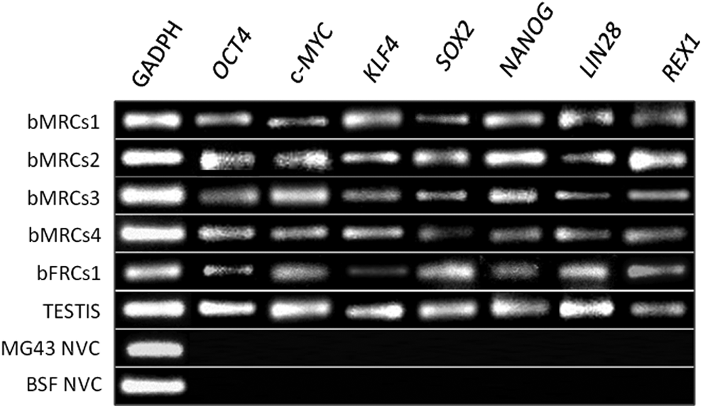
Results of PCR analyses of reprogrammed clones, positive control (bovine testis), nontransduced parental bovine mammary epithelial cell (line MG43), and bovine skin fibroblasts (BSF).
Immunofluorescence assay
We analyzed the expression of the two core pluripotency markers, OCT4 and LIN28, in bMRC4 (Fig. 3) and bFRC1 clones by immunohistochemistry. We reported OCT4 located in cell nuclei and OCT4 and LIN28 in both cytoplasm and nucleoli in both cell types.

Immunoflourescent staining of the mammary epithelial-derived clone, bMRC4 (passage 4) showing specific staining for OCT4 (
Karyotype analysis
The chromosomal arrangement of the investigated cells was evaluated by conventional karyotype and by the position of constitutive heterochromatin (HC) (Figure 4). The first preparation revealed a normal chromosome number for the bovine species (2n=60, XX). No aneuploidy conditions were found for the investigated cells (Fig. 4a, b). The HC was analyzed by using the chromosome banding analysis (CBA) technique, which revealed very strong fluorescence in the centromeric regions of all the autosomes, whereas the remaining part of the chromosomes showed dull fluorescence. All autosomes showed an acrocentric position of the centromeres, thus evidencing a normal arrangement. As expected for cattle chromosomes, no C-positive regions were present on the X chromosomes (Fig. 4c).

Conventional fluorescent staining for metaphase chromosomes: (
Teratoma formation assay
Subcutaneous injection of bRMC4 and bFRC1 into NOD/NOD mice gave rise to tumors that were collected 6–8 weeks later. Tumors were 1×1 cm in size, well demarcated, encapsulated within subcutaneous adipose tissue, and infiltrative. Histopathological analysis revealed a predominance of epithelial and mesenchymal cells, with evidence of cartilage, membranous ossification, epidermis containing stratified squamous epithelial tissue, hair follicles, neural pinwheels, and different types of glandular tissue (Fig. 5). In addition, immunofluorescence analysis (Fig. 6) revealed the presence of CK14, p63, and SMA in the basal layer, and CK18 and MUC1 in the luminal cells in such glandular structures. Milk proteins were also found in ductal lumen and alveoli of bRMC4-derived tissues, resembling a mammary phenotype (Fig. 7).
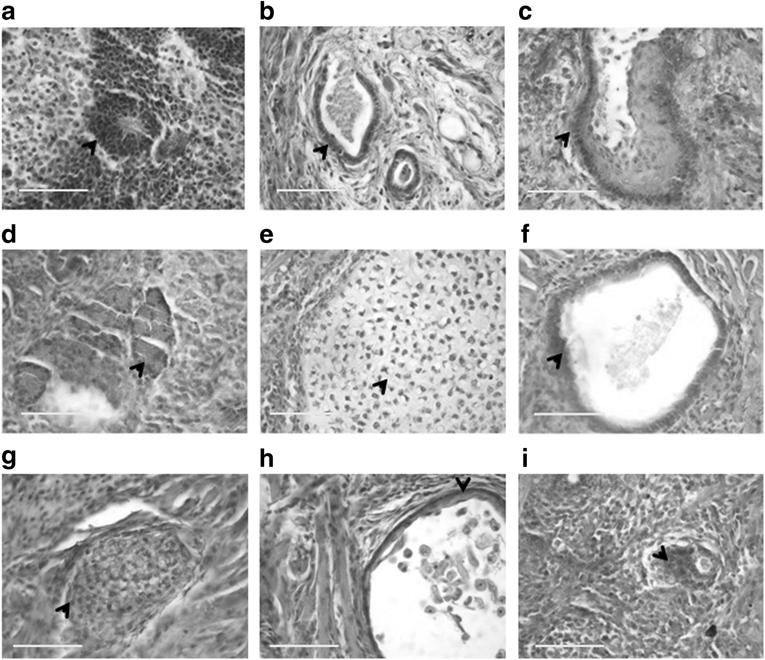
Hematoxilin & Eosin–stained sections of teratomas derived from bMRC4 (
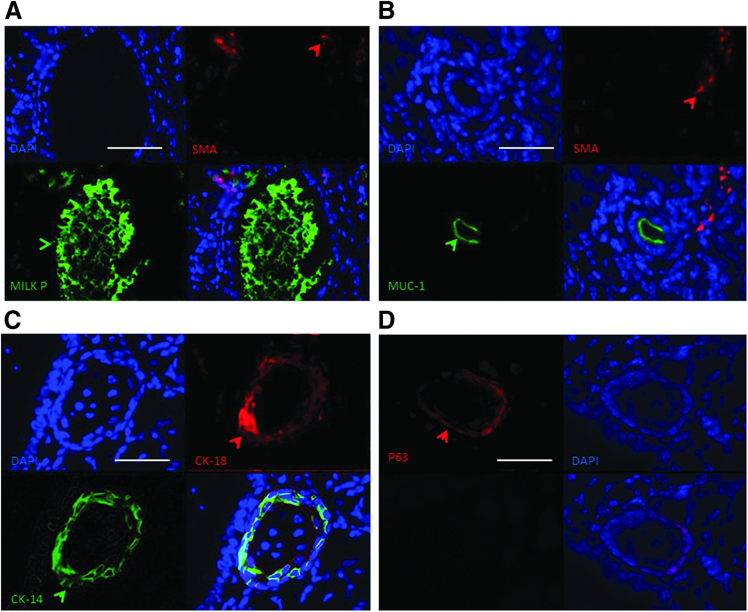
bMRC4-derived teratomas showing mammary epithelial outgrowths with alveoli and ducts positively stained for: (

Expression of mammary markers in bMRC4 clone after 2 weeks in differentiation medium. (
Partial mammary differentiation potential of bovine MEC-derived iPSCs
To verify if the hormonal environment could have an effect in addressing a mammary phenotype, we organized a series of experiments to evaluate whether progesterone could induce the expression of surface markers typical of differentiated MECs. Among these markers, attention has focused on K14, a myoepithelial marker of differentiated cells, and K18, a luminal marker of MECs. After 20 passages in iPSC medium (100 days), bMRC4 cells were induced to differentiate into mammary epithelium. A first goal was to evaluate the possibility of growing the cells in different concentrations of LIF. It was not possible to rule out this factor because the cells rapidly lose the ability to adhere to the solid support of the plate. We tested different concentrations of LIF, and we observed that cells were able to maintain good viability with a concentration of 4 ng/mL. Furthermore, we tested different concentrations of progesterone up to 100 nM. After 2 weeks (three passages) in differentiation medium with at least 10 nM, cells were detected that were positive for CK14, CK18, and SMA (Fig. 6A, B). These cells were bigger than typical iPSCs and displayed no luminescence. They showed a heterogeneous epithelial-like morphology, including small polygonal cells, small spindle-shaped cells, and larger cells (Fig. 6C).
Discussion
iPSC technology offers the possibility of generating a variety of cell types from a defined genotype for tissue regeneration or in vitro modeling of developmental and disease processes. Such potential has already been exploited using human iPSCs, with remarkable results (Daley and Solbakk, 2011). However, iPSC technology has lagged significantly behind in domestic species, and the potential of iPSCs to improve livestock productivity has not been explored. From a practical point of view, given the limitations intrinsically associated with low numbers of adult stem cells from native tissues and maintenance in undifferentiated in vitro, the prospect of generating in vitro a permanent source of cell progenitors that can be differentiated into MECs is extremely attractive for use in the production of transgenic proteins in cow milk. This is particularly true because the potential to use adult mammary stem cells for transgenes has already been demonstrated (Martignani et al., 2010). In addition to providing an abundant progenitor cell source, bovine iPSCs could be readily amenable to genetic manipulation for mammary transgenesis purposes.
Although the generation of bovine iPSCs has already been reported, studies to date have used a limited number of cell types for reprogramming, namely fibroblasts (Takahashi and Yamanaka, 2006) or undefined testicular cells (Wang et al., 2013). Studies in human (Aasen et al., 2008), mouse (Aoi et al., 2008), and horse (Sharma et al., 2014) showed that epithelial cells can reprogram with a relative higher efficiency than fibroblasts. With this in mind, we reprogrammed bovine MECs and dermal fibroblasts simultaneously. Our results agree with those in other species and are consistent with the notion that, unlike fibroblasts, epithelial cells can do so with higher efficiency, which may due to the circumvention of mesenchymal-to-epithelial transition (MET) during reprogramming.
Bovine iPSCs generated in this study displayed reactivated expression of endogenous pluripotency markers previously reported for iPSCs of bovine (Sumer et al., 2011) and other species (Sharma et al., 2014; Takahashi et al., 2007). Those include Rex1 and Lin28, which were robustly associated with bona fide iPSCs in previous studies (Buganim et al., 2012; Chan et al., 2009). More compelling proof of pluripotency was provided by the demonstration that upon injection into NOD/NOD mice both bovine mammary epithelial- and fibroblast-derived reprogrammed cells could generate well-differentiated teratomas containing derivatives of the three germ layers.
The retroviral transduction system is generally considered one of the most efficient methods for obtaining iPSCs. It selects exclusively cycling cells inside a differentiated cell population and induces the process in progenitors (Takahashi et al., 2007). Reprogramming is thought to happen through a “stochastic route” in which the cells have to overcome well-defined “roadblocks” to carry on iPSC generation. For example, it has been reported that mouse mesodermal cells like fibroblasts need to pass through the MET to be reprogrammed. Blocking MET impairs the reprogramming of fibroblasts, whereas preventing epithelial-to-mesenchymal transition (EMT) in epithelial cells cultured with serum can produce iPSCs without Klf4 and c-Myc (Li et al., 2010). These authors proposed that the reprogramming process is more efficient for epithelial cells than for mesenchymal cells, also in relation to the number of reprogramming factors involved.
An important finding of the present study was that biPSCs could be readily induced to differentiate back toward a mammary cell phenotype. We started from the analysis of the concentration of LIF in culture (10 ng/mL as the starting level; data not shown) and observed that this factor is essential for the maintenance of cell viability at least at the concentration of 4 ng/mL. After that, cells were unable to remain adherent to the solid support and began the degenerative processes. To test if a differentiation phase may be induced by steroid treatment, we investigated if mammary markers may be expressed as reported for both luminal (CK18) and basal (CK14, SMA) cells both in human and bovine species (Martignani et al., 2009; Rauner and Barash, 2012; Stingl et al., 1998). After 2 weeks of culture in the presence of 10 nM progesterone, we observed the expression of these markers. We were not able to detect them in the presence of only hydrocortisone, a typical differentiation factor for the mammary tissue that was present in controls (Kabotyanski et al., 2009).
This observation is of interest because progesterone is one of the putative factors that drive progenitors toward a more mature and differentiated phenotype (Ayyanan et al., 2011; Hilton et al., 2014). Important studies have reported, at least in mice, a dynamic role for progesterone in activating adult stem cells within the mammary stem cell niche during the reproductive cycle (Ayyanan et al., 2011; Joshi et al., 2010) and that mouse mammary stem cells are highly responsive to steroid hormone signaling, despite lacking estrogen and progesterone receptors. In fact, ovariectomy markedly diminished the mammary stem cell number and outgrowth potential in vivo, whereas stem cell activity increased in mice treated with estrogen plus progesterone (Asselin-Labat et al., 2010). Further studies are being carried out to assess the actual mammary enrichment of precursors through hormonal conditioning and their ability to generate functional mammary alveoli in vivo, as just reported for adult mammary stem cell cells (Martignani et al., 2010).
In summary, we report the generation of biPSCs from bovine epithelial cells and fibroblasts. Interestingly, we were able to show a partial redirection toward a mammary phenotype in vitro after a treatment with progesterone. Further studies are necessary for a complete cell differentiation, but these reprogrammed clones are promising as an interesting model in the understanding of cell differentiation mechanisms in bovine mammary gland development.
Footnotes
Acknowledgments
This work was supported by PRIN 2010–2011 and University of Torino grant 2012 (M.B.) and by Institute Strategic Grant funding from the BBSRC (F.X.D.). R.S. was supported by a University of Edinburgh Staff Scholarship. We thank for Mrs. Cristina Cecere for technical help.
Author Disclosure Statement
The authors declare that no conflicting financial interests exist.
References
Supplementary Material
Please find the following supplemental material available below.
For Open Access articles published under a Creative Commons License, all supplemental material carries the same license as the article it is associated with.
For non-Open Access articles published, all supplemental material carries a non-exclusive license, and permission requests for re-use of supplemental material or any part of supplemental material shall be sent directly to the copyright owner as specified in the copyright notice associated with the article.
