
Abstract
Abstract
The recent discovery of ovarian stem cells in postnatal mammalian ovaries, also referred to as putative stem cells (PSCs), and their roles in mammalian fertility has challenged the long-existing theory that women are endowed with a certain number of germ cells. The rare amount of PSCs is the major limitation for utilizing them through different applications. Therefore, this study was conducted in six phases to find a way to increase the number of Fragilis- and mouse vasa homolog (MVH)-positive sorted cells from 14-day-old NMRI strain mice. Results showed that there is a population of Fragilis- and MVH-positive cells with pluripotent stem cell characteristics, which can be isolated and expanded for months in vitro. PSCs increase their proliferation capacity under the influence of some mitogenic agents, and our results showed that different doses of stem cell factor (SCF) induce PSC proliferation with the maximum increase observed at 50 ng/mL. SCF was also able to increase the number of Fragilis- and MVH-positive cells after sorting by magnetic-activated cell sorting and enhance colony formation efficiency in sorted cells. Differentiation capacity assay indicated that there is a basic level of spontaneous differentiation toward oocyte-like cells during 3 days of culture. However, relative gene expression was significantly higher in the follicle-stimulating hormone-treated groups, especially in the Fragilis- sorted PSCs. We suggest that higher number of PSCs provides us either a greater source of energy that can be injected into energy-impaired oocytes in women with a history of repeat IVF failure or a good source for research.
Introduction
R
It has long been accepted that production of ovarian oocytes is ceased before birth (Beard, 1900; Zuckerman, 1951), but recent investigations have indicated that OSCs are not only present in juvenile and adult ovaries but also actively participate in oogenesis (Johnson et al., 2004, 2005; Kerr et al., 2006; Zou et al., 2009). Due to the low number of OSCs in juvenile and adult ovaries, isolation and proliferation of OSCs in vitro are a laborious procedure. Therefore, many attempts have been made to investigate a novel method for isolating OSCs with maximum purity and to proliferate them without losing their potential. The presence of BrdU–MVH (mouse vasa homolog), double-positive cells in the ovarian surface epithelium (OSE), showed that OSCs have in situ proliferation potential in the ovary after birth. Also, injection of green fluorescent protein (GFP)-conjugated OSCs into oocyte-free ovaries indicated that OSCs have the potential to differentiate into oocytes and recover fertility in sterile mice (White et al., 2012; Zou et al., 2009).
Different isolation methods have been used for the isolation of OSCs from ovarian tissues (Pacchiarotti et al., 2010; Parte et al., 2011; Parvari et al., 2015; Virant-Klun et al., 2008, 2011, 2013; White et al., 2012; Woods and Tilly, 2013; Yazdekhasti et al., 2016; Zou et al., 2009). Tilly and his group introduced a detailed isolation protocol by fluorescent-activated cell sorting (FACS) and magnetic-activated cell sorting (MACS) using MVH and Fragilis marker.
They showed that purified germline stem cells have a gene expression profile that is consistent with primitive germ cells and they also have the ability to produce oocytes in vitro and fertilization-competent eggs in vivo. Moreover, they indicated that these cells can be expanded for months and can spontaneously generate 35–50 μm oocytes (Woods and Tilly, 2013). Zou et al. (2009) also purified female germline stem cells (FGSCs) by MACS using the two markers MVH and Fragilis, and observed that Fragilis-based magnetic bead sorting method had better results compared with MVH. Izadyar et al. (2010) used a transgenic mouse model in which GFP was expressed under a germ cell-specific Oct-4 promoter for isolation and derivation of germline stem cell from postnatal mouse ovary (Pacchiarotti et al., 2010).
Effects of different supplements on the proliferation of OSCs have been evaluated in vitro. Parte et al. (2013) cultured ovarian tissue fragments and observed that follicle-stimulating hormone (FSH) and basic fibroblast growth factor (bFGF) induce proliferation and migration of OSCs from inserts and retain their potential to differentiate into oocyte-like structures in extended cultures spontaneously (Bhartiya et al., 2016). They also observed that pregnant mare serum gonadotropin (PMSG) increases FSH receptor (FSHR) gene expression and PCNA immunostaining in OSE and oocytes of primordial follicles. In a study, Hu et al. (2012) evaluated effects of GSK3 inhibitor BIO, leukemia inhibitory factor (LIF), and some other growth factors (epidermal growth factor [EGF], bFGF, glial-derived neurotrophic factor [GDNF], and PDGF) on proliferation and colony formation of OSCs. They indicated that BIO promotes proliferation of FGSCs through activation of β-catenin and upregulation of E-cadherin, and LIF has significant positive effects on the number of colonies (Hu et al., 2012).
Park et al. (2013) also showed that bone morphologic protein 4 (BMP4) increases the number of OSCs and in vitro-derived oocytes in a dose-dependent manner. Bui et al. (2014), through the isolation and culture of PSCs, showed that 40 ng/mL stem cell factor (SCF) has a high potential for inducing proliferation in PSCs in adult pig ovary. They indicated that SCF not only increased the proliferation of PSCs but also the proportion of c-Kit-positive PSCs.
Some groups of researchers believe that OSCs may originate from bone marrow and peripheral blood (Bukovsky et al., 2005; Johnson et al., 2004, 2005). They observed the expression of germline markers in bone marrow and also demonstrated that bone marrow transplantation restores oocyte production in wild-type mice (Johnson et al., 2005). Another group also claimed that very small embryonic-like stem cells (VSELs) could be a subpopulation of primordial germ cells (PGCs) or originated from them (De Felici, 2010). With the hypotheses that OSCs may originate from bone marrow and SCF is one of the most important factors in hematopoiesis, migration, and survival of PGCs (De Felici and Pesce, 1994; Pesce et al., 1993), we designed this study to evaluate the effect of SCF on different potentials of sorted PSCs through Fragilis and MVH markers in vitro in mice.
Materials and Methods
Ethics statement
The treatment of the mouse used in this research followed guidelines approved by Tehran University of Medical Sciences' Ethics Committee.
Animals
The 14-day-old NMRI strain mice used in this study were purchased from the animal housing of pharmacology faculty at Tehran University of medical sciences and maintained on 12-hour day/12-hour night cycles with free access to food and water. In each experiment, 20 ovaries were collected and minced after removal of adherent tissue.
Study overview
The schematic summary of this study has been shown in Figure 1. This study was conducted in six phases such that the findings of each step were used in designing the next phase. In the initial phase of this study, MACS efficiency based on the number of sorted cells and growth curve was evaluated and compared between the two markers Fragilis and MVH. In addition, cell morphology and gene expression profile of freshly isolated PSCs were evaluated at the initial phase. In the second phase, PSCs were sorted by Fragilis marker, and the effects of different doses of SCF on PSC proliferation were assessed using the BrdU cell proliferation assay.
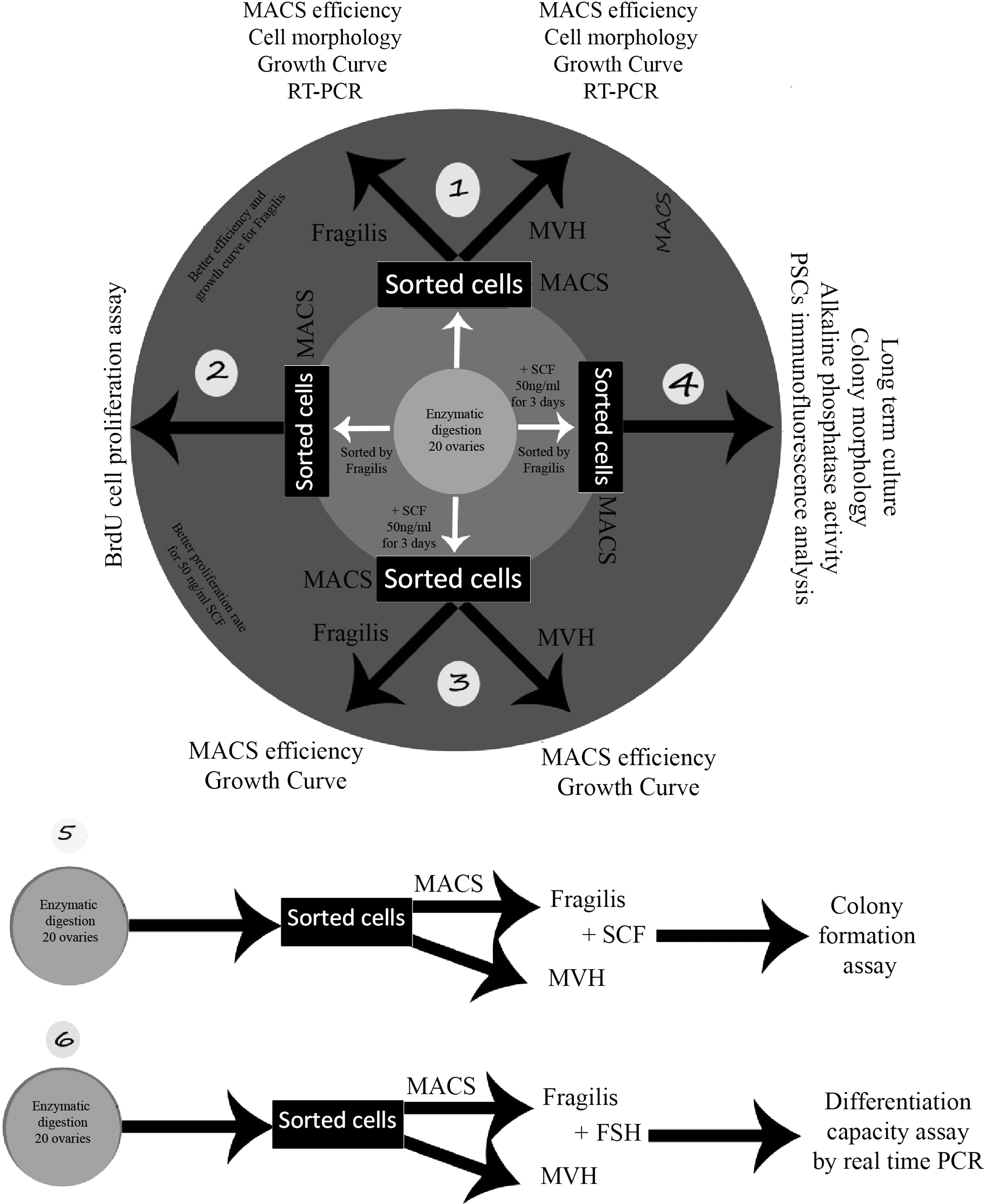
An overview of the study. This study has been designed in six phases, and findings of each phase have been used for the next phase. Evaluation of MACS efficiency in the first phase, proliferation effect of different doses of SCF in the second phase, growth curve and MACS efficiency under the influence of SCF in the third phase, long-term culture of PSCs in the fourth phase, the effect of SCF on PSC colony formation in the fifth phase and differentiation capacity of PSCs in the sixth phase. MACS, magnetic-activated cell sorting; PSCs, putative stem cells; SCF, stem cell factor.
In the third phase, the effects of 50 ng/mL SCF (concentration selected based on previous findings) on the number and growth curve of sorted cells from both Fragilis and MVH were evaluated. In the fourth phase, cells were incubated with 50 ng/mL SCF for 3 days and were then sorted by Fragilis. Sorted cells were utilized for long-term culture and other assessments. In the fifth phase, the effects of different doses of SCF on colony formation of sorted PSCs were assessed. In the last phase of the study, the differentiation capacity of sorted PSCs under the influence of 0.5 IU FSH was examined.
Isolation of PSCs from mouse ovary
The ovaries (20 for each experiment) were aseptically collected from 14-day-old NMRI strain mice, and the attached fat pad, bursa, and oviduct were carefully removed from each ovary. The ovaries were then dissected and minced into slurry in an enzymatic solution consisting of 800 U/mL collagenase IV (Cat. No. LS004188; Worthington Biochemical Corporation) dissolved in Hank's Balanced Salt Solution (HBSS, Cat. No. 14175-095; Gibco) and 1 mg/mL DNase I (D5025; Sigma, St. Louis, MO), and kept in a prewarmed (37°C) orbital shaker for 15 minutes at 250 r.p.m. After 15 minutes, the tube was removed from the orbital shaker and the pieces of ovary were manually dispersed by gentle pipetting using a 5 mL glass serological pipette. The tube was incubated one more time on the orbital shaker at 37°C for an additional 15 minutes at 250 r.p.m.
The ovarian pieces were again manually dispersed by gentle pipetting until no visible pieces of ovary were present. The cell suspension was filtered through a 40 μm nylon mesh cell strainer, and the filtrate was collected in a 15 mL tube. Ten milliliters of warm HBSS was added to the tube containing the strained cell suspension, and the tube was centrifuged at 300 g for 5 minutes at room temperature. Supernatant was removed and the cell pellet was resuspended in a primary culture medium (PCM) consisting of 10 mL α-MEM (Invitrogen, Grand Island, NY) supplemented with 10% fetal bovine serum (F2442; Sigma), 1% GlutaMAX (35050-061; Invitrogen), 1% nonessential amino acids (M7145; Sigma), 0.1 mM β-mercaptoethanol (M3148; Sigma), 100 mg/mL penicillin/streptomycin (p4333; Sigma), and 103 U/mL LIF (L5283; Sigma). The cells were then placed into 0.1% gelatin-coated 60 mm tissue culture plates (SPL) and incubated overnight at 37°C in an atmosphere of 5% CO2 in the air. This preplating step was performed to reduce the number of fibroblasts. The following morning, the floating cells were harvested and centrifuged at 300 g for 5 minutes at room temperature. The cell pellet was resuspended in the above-mentioned culture medium plus 50 ng/mL SCF, human recombinant (Cat. No. 300-07; PeproTech UK), and placed into a 35 mm culture plate for 3 days. The obtained cells were directly used for long-term culture.
Purification of PSCs by MACS
To compare the efficiency of the purification method for the two antibodies that we used, PSCs were isolated based on their expression of MVH and Fragilis by magnetic bead sorting. After trypsinization and centrifugation of ovarian cells at 300 g for 5 minutes, at room temperature, the ovarian cells were incubated in the MACS buffer solution for 20 minutes on ice. After rinsing, the ovarian cells were incubated with MVH (Rabbit anti-DDX4 antibody, Cat. No. ab13840; Abcam) and Fragilis (anti-Fragilis antibody, Cat. No. ab15592; Abcam) antibodies for 20 minutes on ice. The ovarian cells were then incubated with Goat anti-rabbit IgG antibody-conjugated microbeads (Cat. No. 130-048-602; Miltenyi Biotec). After one additional wash, the cell suspension was loaded onto MACS Cell Separation columns (MS Columns, 130-042-201; Miltenyi Biotech) and was separated according to the manufacturer's specifications (Miltenyi Biotec). The magnetic beads spontaneously detached from the cells and were removed by magnetic bead separator during the cell passage process.
Culture and growth of PSCs
All cells sorted by Fragilis and MVH methods were counted and then centrifuged and resuspended in a complete OSC culture medium consisting of the above-mentioned medium plus 1 mM sodium pyruvate (Cat. No. 11360-070; Invitrogen), 1 × concentrated N-2 supplement (Cat. No. AR009; R&D Systems), 10 ng/mL EGF, human recombinant (Cat. No. PHG0311; Invitrogen), 1 ng/mL bFGF, human recombinant (Cat. No. 13256-029; Invitrogen), and 40 ng/mL GDNF, human recombinant (Cat. No. 212-GD-010; R&D Systems). Then, cells were placed in one well of a four-well mitomycin C-treated mouse embryonic fibroblast (MEF) feeder layer (seeded at a concentration of 5 × 10³ cells/well) plate after vigorous pipetting at 37°C in 5% CO2 in the air. To make the cells settle down, the culture medium was maintained for 1 week while a drop of fresh complete OSC culture medium was added every day. After the first 7 days, the culture medium was refreshed every 2–3 days and digital images were taken. At 70%–80% confluence, PSCs were subcultured using trypsin (0.25% trypsin-EDTA; Gibco) in combination with mechanical pipetting every 5–8 days at a 1:1–3 dilution.
Growth curves
To construct a growth curve for both MVH- and Fragilis-sorted PSCs, the number of PSCs at 24, 48, 72, and 96 hours were counted. A day after preplating culture, freshly isolated PSCs were aliquoted into four wells of 48-well plates (without MEF feeder) in a complete OSC culture medium at an initial concentration of 1 × 10³ cells/ml.
Factorial experiment
For the factorial design experiment, three independent experiments were performed. 2.5 × 10³ freshly isolated PSCs by Fragilis method were cultured in 96-well culture plates without feeder layer in the PCM supplemented with five different doses of SCF (0, 10, 30, 50, and 70 ng/mL) for 3 days, and PSC proliferation rates were assessed by the BrdU cell proliferation assay. After 3 days of incubation with 50 ng/mL SCF following preplating of culture, isolated PSCs by Fragilis and MVH were aliquoted into 5 wells of 48-well plates (without MEF feeder) in a complete OSC culture medium at an initial concentration of 1 × 10³ cells/mL, to evaluate the proliferative effect of the optimized dose of SCF (50 ng/mL). The cells were counted at 24, 48, 72, 96, and 120 hours and the values were used to draw a growth curve.
To examine the effects of treatment with and without 50 ng/mL SCF on the number of cells sorted by Fragilis and MVH method, isolated cells from the digestion step were cultured (one night after preplating culture) in PCM plus 50 ng/mL SCF, and placed into 35 mm culture plate for 3 days before MACS. After 3 days, the number of cells before and after performing MACS was counted.
Colony formation assay
To determine the efficiency of colony formation of PSCs in the presence of different doses of SCF, freshly sorted PSCs were cultured on mitomycin-treated MEF feeder layer in 24-well culture dishes at an initial density of 1 × 10³ cells/mL in the presence of 10, 30, 50, and 70 ng/mL SCF in a complete OSC medium and the number of colonies was counted after 2 weeks.
Characterization of PSCs
RT-PCR
Total cellular RNA was extracted using Trizol reagent (Invitrogen) according to the manufacturer's instructions, and examined for purity and concentration using a photometer (NanoDrop ND). Incubation with DNase I (Sigma) for 30 minutes at 37°C removed possible DNA contamination. One microgram RNA and primers (Bioneer, CycleScript RT PreMix) were utilized for cDNA synthesis (complementary cDNA synthesis kit; Bioneer, South Korea).
Polymerase chain reaction was performed using the GeneAmp PCR system 9600 (PerkinElmer Life and Analytical Sciences, Wellesley, MA). Specific primer pairs of pluripotency-related markers, including, Oct-4, Nanog, c-Kit, germ line-related markers such as Fragilis, Dazl, and Mvh, and oocyte-related markers such as Zp3 (Table 1) were used for the polymerase chain reaction (PCR). Cycling parameters were as follows: 5 minutes of initial denaturation at 94°C, then 33 cycles at 94°C for 30 seconds, corresponding to the annealing temperature of primer pairs for 30 seconds, and 72°C for 1 minute. Ovarian cells from adult ovaries were used as positive controls and MEF cells were used as negative controls. Polymerase chain reaction products were loaded on 2% agarose gel, stained with ethidium bromide, and visualized by UVItec Cambridge-Gel Documentation Systems (CB4 1QB-UK). Expression of β-actin as a housekeeping gene was evaluated as an internal control.
Immunofluorescence analysis
Attached positive Fragilis-sorted PSCs at passage three were fixed in 4% paraformaldehyde (Merck, Darmstadt, Germany) for 15 minutes and rinsed twice with cold phosphate-buffered saline (PBS; pH 7.4), followed by permeabilization with 0.2% TritonX-100 (Sigma) for 15 minutes. Nonspecific binding sites were blocked with 1% bovine serum albumin (Sigma) at 37°C for 30 minutes. Primary antibodies against Oct-4 (Mouse monoclonal Oct-4 antibody, ab59545; Abcam), c-Kit (Rabbit polyclonal c-Kit antibody, ab5506; Abcam), Fragilis (Rabbit anti-Fragilis antibody, Cat. No. ab15592; Abcam), and MVH (Rabbit anti-DDX4 antibody, Cat. No. ab13840; Abcam) were diluted in a blocking buffer and incubated overnight at 4°C. Following three washes, appropriate FITC-conjugated secondary antibody (F1262 Sigma for Fragilis, MVH, and c-Kit, ab7064 Abcam for Oct-4) was added for 1 hour at room temperature in the dark. Nuclear counterstaining was carried out using either 4,6-diamidino-2-phenylindole (DAPI; Sigma) or Hoechst 33342 (62249; Gibco) for 10 minutes. For the negative controls, the primary antibodies were omitted. Cells were analyzed under a fluorescent microscope (Nikon, Germany).
Alkaline phosphatase staining
The alkaline phosphatase activity was assayed by Fast Red TR/Naphthol AS-MX Tablets (F4648; Sigma) according to the manufacturer's instructions. Briefly, the alkaline dye solution was prepared by dissolving Tris tablet in 1 mL of distilled or deionized water, and then one Fast Red TR/Naphthol AS-MX tablet was dropped into the Tris buffer. Cultured cells on the plates were fixed with 4% paraformaldehyde for 1–2 minutes after which they were washed twice with PBS. After washing, the cells were incubated with the provided solution for 15 minutes. The cells were then observed under an invert microscope.
BrdU cell proliferation assay
The proliferation of PSCs was assayed by the BrdU Cell Proliferation ELISA Kit (ab126572; Abcam) according to the manufacturer's instructions. Briefly, freshly isolated PSCs were cultured on 96-well culture plate at a density of 2.5 × 10³ cells/mL for 3 days in PCM. After the third day, 100 μL 2 × test reagent (0, 10, 30, 50, and 70 ng/mL SCF) was added to the appropriate wells. Twenty microliters of diluted 1 × BrdU was added to the appropriate wells and the plate was incubated for 2–24 hours. It was important that BrdU is incorporated into the proliferating cells and was thus added at least 2 hours before the end of the test reagent incubation period. On the next day, the media in the cell wells were aspirated and the provided fixation solution was added to the cells. The plate was washed thrice with 1 × Wash Buffer. One hundred microliters per well of anti-BrdU monoclonal detector antibody was added and the cells were incubated for 1 hour at room temperature. Cells were washed for the second time and 100 μL/well 1 × Peroxidase Goat Anti-Mouse IgG Conjugate was added, and the plate was incubated for 30 minutes at room temperature. The final wash was performed and 100 μL/well chemiluminescent substrate was added to the cells and incubated for 5–10 minutes. The plate was immediately read by a luminometer (0.2–1 second integration time).
Differentiation capacity assay
To compare differentiation capacity of the two groups of sorted cells (isolated by Fragilis and MVH method) into oocyte-like cells and their response to a differentiation inducer (FSH), freshly sorted cells were seeded on 35 mm culture dishes at an initial concentration of 4 × 10³ cells/mL in a complete OSC culture medium (without feeder layer) and were treated with 0.5 IU FSH (F2293; Sigma) for 3 days. After 3 days, the cells were trypsinized and stored at −80°C and levels of transcripts for oocyte-related markers (Nobox, Scp3, and Gdf9) were analyzed by real-time PCR. Total RNA was extracted as mentioned before and the comparative threshold cycle (Ct) values were calculated by CFX 96 Real-Time PCR system (Bio-Rad Laboratories, Hercules, CA) using SYBR Green chemistry (Bio-Rad). Using the ΔΔCt method, relative expression of targets was calculated by normalizing Ct values of targets against 18 seconds.
Statistical analysis
Statistical analysis was performed using two-way ANOVA to examine the effects of treatment with and without 50 ng/mL SCF on the number of cells sorted by Fragilis- and MVH-based MACS method. One-way repeated measurement ANOVA and subsequent post hoc test (Tukey) were performed for other evaluations using Prism 5 software. Each experiment was repeated at least thrice. Results are presented as mean ± standard deviation, and p-value of ≤0.05 was considered statistically significant.
Results
Purification and long-term culture of PSCs
Cells obtained from enzymatic digestion (Fig. 2a) were cultured overnight to reduce fibroblast contamination. To recover cells due to enzymatic digestion, floating cells were harvested and cultured for three more days. After 3 days, the cells were trypsinized and prepared for MACS using MVH and Fragilis markers. Isolated cells both from Fragilis and MVH method were similar in morphology and size. Most of the sorted cells were small and spherical with high nuclear to cytoplasm ratio and a bubble-like structure. Two distinct populations of PSCs were found in flushed positive cells both for Fragilis and MVH, small cells with 5–6 μm and larger ones with 9–10 μm in diameter (Fig. 2b). Sorted Fragilis- and MVH-positive cells found spindle-like structure after attachment to dish bottom (Fig. 2e, h). In each experiment, an average number of 0.8–1.5 × 10⁵ cells was obtained from enzymatic digestion of 20 ovaries, and after MACS, the mean number of cells was 3–6 × 10³ (4%) and 1.6–3 × 10³ (2%) (including contaminated false-positive cells) for Fragilis and MVH, respectively (Fig. 3a). Supplementation with 50 ng/mL SCF for 3 days before MACS showed that SCF could induce proliferation in both Fragilis- and MVH-positive cells; SCF increased the number and percentage of interested cells after MACS (Fig. 3a, b).

Cell and colony morphology of PSCs.

The efficiency of MACS sorting method for Fragilis and MVH marker and effect of SCF on growth and colony formation of PSCs.
From our PSC sorting results, we found that Fragilis had a better efficacy compared with MVH (Fig. 3a, b); so we decided to continue with the Fragilis method for the factorial experiment, long culture period, and immunofluorescence, and RT-PCR analysis. Fragilis-sorted PSCs were cultured on an inactivated MEF feeder in a well of a 24-well culture plate at an initial density of 4 × 10³ cells/well (Fig. 2c). Cell culture was maintained for 2 months without any significant change in cell morphology. During the first week of culture, one drop of culture medium was added each day instead of complete removal of culture medium. After 3–4 days, some cell clusters were observed (Fig. 2d) and real cell colonies appeared after 2 weeks (Fig. 2f–h). PSCs formed round, compact, flat colonies with most of them having a clear boundary; however, some appeared as monolayer colonies without a clear border. The colonies maintained their embryonic stem cell characteristics and were positive for the alkaline phosphatase activity (Fig. 2i). In some colonies, dispersing to single cells by trypsinization was difficult; therefore, we could not count the exact number of cells in those colonies. At a confluency of 70%–80%, the colonies and the surrounding PSCs were treated with 0.05% trypsin-EDTA for 2 minutes to disassociate the PSCs, while leaving most of the colonies intact.
MACS efficiency
Comparison of the number of sorted cells from both the Fragilis and MVH method and their growth curves (Fig. 3a, c) showed that Fragilis significantly improved the efficiency of MACS. Percentage of Fragilis- and MVH-positive PSCs was 4% (4920 ± 32 cells) and 2% (2340 ± 24 cells), respectively (Fig. 3a), and Fragilis-positive sorted PSCs had a sharper log phase compared with those sorted by MVH (Fig. 3c). From these results of the initial phase, the Fragilis method proved the efficiency and was thus employed for the next step.
Effect of SCF on the number, growth curve, and colony formation of PSCs
BrdU cell proliferation assay results showed that 50 and 70 ng/mL SCF had a higher impact on the proliferation of Fragilis-positive sorted PSCs. Even though there was no significant difference between 50 and 70 ng/mL, the effect of 50 ng/mL SCF was slightly higher (Fig. 3d). Incubation with 50 ng/mL SCF for 3 days before MACS significantly increased the number and growth curve of both sorted PSCs (Fig. 3a, b, e). In our experiment, the number and percentage of PSCs under the influence of 50 ng/mL SCF increased from 4% and 2% to 9% (11,210 ± 133 cells) and 5% (6160 ± 24 cells) for Fragilis and MVH, respectively (Fig. 3a). The growth curve of PSCs indicated that Fragilis-sorted cells under the influence of SCF showed a higher log phase compared with those sorted by MVH. Moreover, PSCs sorted by MVH had a sharper log phase when they were supplemented with 50 ng/mL SCF, higher than untreated Fragilis-sorted PSCs (Fig. 3e).
In the fifth phase of the study, we compared the effects of different doses of SCF on colony formation efficiency of Fragilis- and MVH-positive PSCs. Our results show that all doses of SCF significantly increased the number of colonies after 2 weeks of culture compared with the control group that did not receive SCF. However, colony formation efficiency was higher in the Fragilis-positive PSCs compared with the MVH-positive PSCs in the 50 ng/mL SCF group (Fig. 3f).
Characterizations of PSCs
To determine and confirm that the sorted cells from both Fragilis and MVH methods are female germ stem cells, RT-PCR and immunofluorescence analysis were performed (Figs. 4 and 5). RT-PCR results showed that both groups of sorted cells expressed pluripotency-related markers (Oct-4, Nanog, and c-Kit) and classic primitive germline-related markers (Fragilis, MVH, and Dazl), but did not show oocyte-related marker (ZP3) (Fig. 5). It can be deduced from these results that, these cells display characteristics of female germ stem cells, but not oocytes. Moreover, immunofluorescence analysis of proteins coded by the four genes (Fragilis, MVH, c-Kit, and OCT-4) confirmed pluripotency and germline characterizations of the sorted cells at passage 3 (Fig. 4). Nevertheless, the colonies were positive for the embryonic stem cell marker, alkaline phosphatase (Fig. 2i).
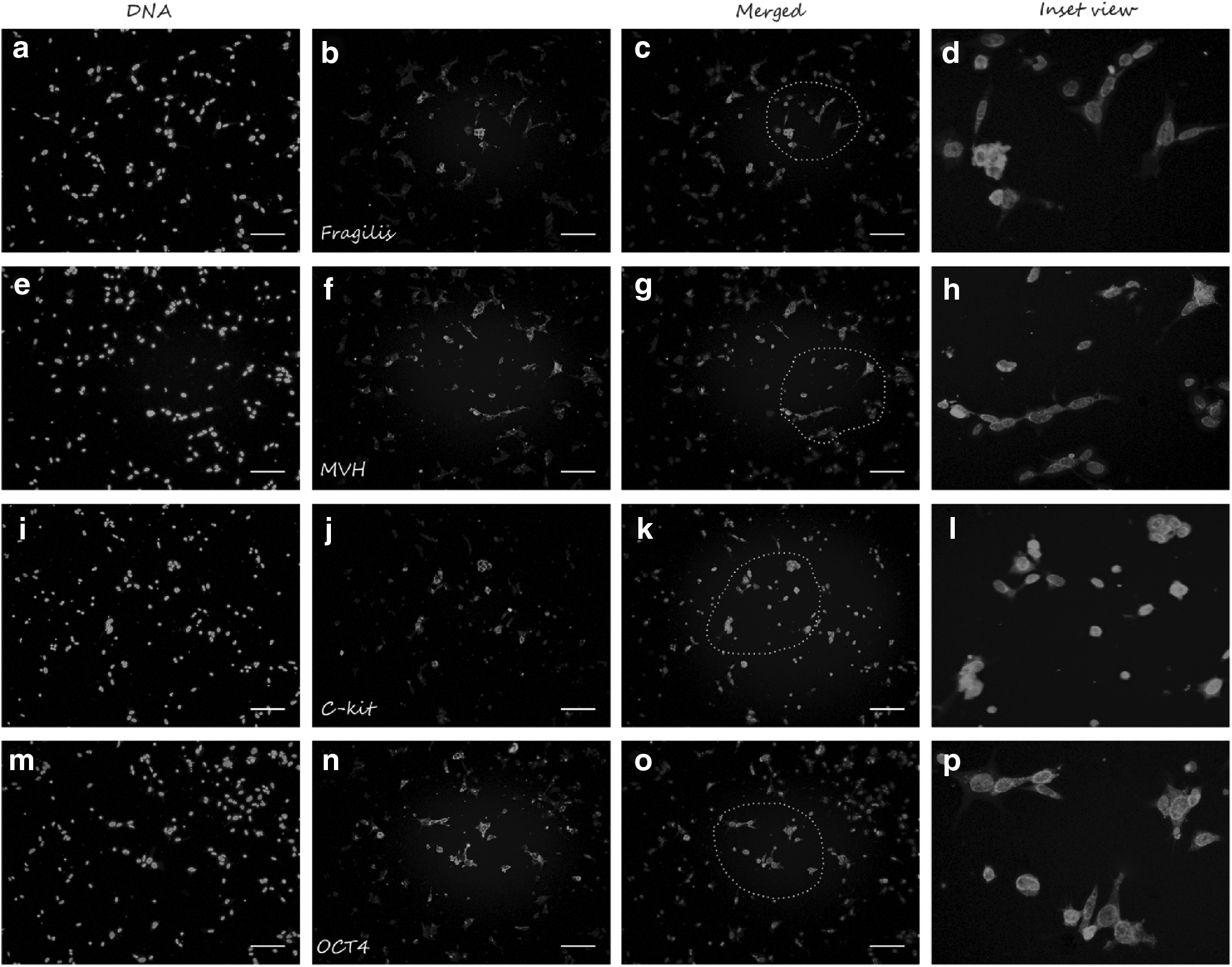
Immunofluorescence analysis of PSCs at passage 3. Sorted PSCs at passage 3 were positive for gemline markers, including Fragilis
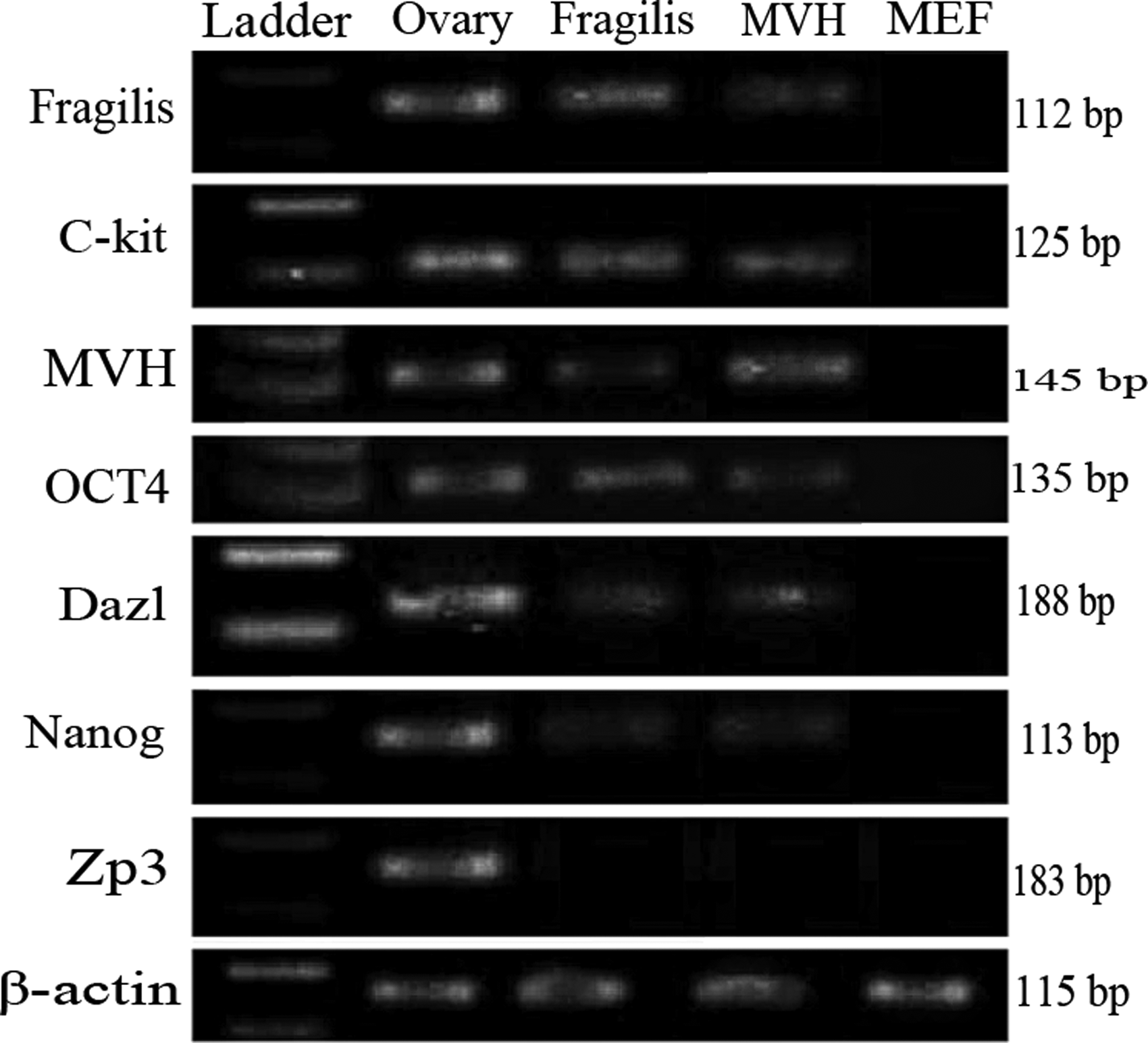
Reverse transcriptional PCR analysis for expression profile of sorted PSCs by Fragilis and MVH marker. RT-PCR analysis of both Fragilis- and MVH-positive PSCs show that PSCs express pluripotency-related markers (Oct-4, Nanog, C-kit, and Dazl) and classic primitive germline-related markers (Fragilis and MVH), but they did not express the oocyte-related marker (ZP3). ZP3 gene was not expressed in both sorted cells, and this shows that both MACS-positive fractions were free of oocyte contamination. MEF cells were considered negative control, which does not express pluripotency-, germline-, and oocyte-related markers, and β-actin as a housekeeping gene and internal control was also studied. RT-PCR.
Differentiation capacity assay
These results show that cells sorted either by Fragilis or MVH methods have different capacities based on their proliferation rate and MACS efficiency. In the final phase of the study, we evaluated differentiation potential of Fragilis- and MVH-sorted PSCs in the culture medium supplemented with 0.5 IU FSH. Our results indicate that there is a basic degree of spontaneous differentiation in all the groups during 3 days of culture, but relative gene expression was significantly higher in the treated groups, especially in the Fragilis-sorted PSCs (Fig. 6). However, relative gene expression was significantly higher in all groups except Gdf9 between the Fragilis- and MVH-sorted cells.

Real-time (quantitative) PCR analysis of oocyte-related markers after treatment with 0.5 IU/mL FSH. Relative gene expression of oocyte-related markers (Nobox, Zp3, and Gdf9) was significantly higher in all groups between Fragilis and MVH except for Gdf9. However, transcripts levels for all three genes were considerably more in all groups under the influence of 0.5 IU FSH. Values are mean ± SD; n = 3. *p < 0.05, **p < 0.01, ****p < 0.0001. FSH, follicle-stimulating hormone.
Discussion
This study has shown that postnatal mouse ovary possesses a population of Fragilis- and MVH-positive cells with stem cell characteristics. However, due to their unknown nature, origin, and function, it has not been vividly established that they are “ovarian stem cells” and have mostly been referred to as “putative stem cells” in literature (Bui et al., 2014; Virant-Klun et al., 2008). The presence of PSCs in postnatal mammalian ovary has been proven, but their role in routine ovarian function and folliculogenesis remains elusive. It has been reported previously that two distinct populations of stem cells exist in adult organs, active and dormant stem cells. It has been proposed that PSCs are a representative of dormant stem cells with no contribution in oogenesis (Bhartiya et al., 2013; Park and Tilly, 2015; Yuan et al., 2013).
Some other population of stem cells known as “Very Small Embryonic-like Stem Cells” has been found in many adult tissues such as the lung, heart, and brain, as well as in the umbilical cord blood and peripheral blood in the adult human bone marrow. They have been found to share some common characteristics with pluripotent stem cells such as the expression of pluripotency-related markers (Oct-4, Nanog, and SSEA-1), and also with PGCs such as the expression of c-Kit gene, as SCF receptor, Nanog, and Sox2 (Parte et al., 2011; Ratajczak et al., 2008; Virant-Klun et al., 2008; Wojakowski et al., 2011). Expression of the c-Kit gene in PGCs is consistent with our results suggesting that PSCs might be a representative of VSELs in adult ovary.
The stem cell niche is a specialized microenvironment that supports stem cells' capacity to self-renew and differentiate (Morrison and Spradling, 2008), and the factors secreted by the cells of each niche play a critical role in their communications. However, the structure and cell composition of PSC's niche are largely unknown. Also, the question why PSCs do not actively contribute to tissue repair like other VSELs in different adult tissue is still elusive.
Culture of OSE scraping cells has shown that these cells coexpress SCF and c-kit (Parrott et al., 2000) suggesting that in PSC's niche SCF is an important autocrine factor in normal folliculogenesis, Moreover, other studies have indicated that SCF is a crucial factor for proliferation and migration of PGCs (De Felici and Pesce, 1994; Pesce et al., 1993). In one study, Bui et al. (2014) demonstrated that SCF affects proliferation of pig PSCs and lead to increased proportion of c-kit-positive PSCs. They showed that SCF significantly induces PSC proliferation in a dose-dependent manner; 40 ng/mL SCF had maximum impact on PSC proliferation (Bui et al., 2014). Some other investigations have indicated that SCF, acting through c-Kit, is a primary growth and survival factor for murine PGCs (De Felici and Dolci, 1991; Dolci et al., 1993; Matsui et al., 1991) and oocytes (Morita et al., 1999; Packer et al., 1994). It has also been reported that SCF has an antiapoptotic effect on PGCs and fetal ovarian germ cell pool (Morita et al., 1999).
In view of these findings on the function of SCF and its receptor (c-Kit), we evaluated its effect on PSC proliferation with more details. Our results show that using Fragilis marker for sorting PSCs from digested ovarian cells is more efficient compared with MVH marker. Zou et al. (2011) reported that sorting by Fragilis marker almost increased purification efficiency twofold. They suggested that MVH is an RNA binding protein that is mainly expressed in the cytoplasm, and it is possible that only some MVH molecules bind to the membrane at any given time. Fragilis, on the other hand, is a cell surface protein that gives it an advantage over MVH regarding the number of binding sites for antibodies during PSC sorting (Zou et al., 2011). Using preplating culture for decrease number of fibroblasts was an option that we used it in this study. In fact, this method was an alternative way to increase the number of interested cells and percentage of positive cells sorted by MACS.
We found that cells obtained by Fragilis marker had higher efficiency and showed a sharper log phase in their growth compared with those sorted by MVH (Fig. 3a–c). One possible explanation for this result is that MVH is routinely expressed in the cytoplasm (Zou et al., 2011), which might have increased the number of false positive cells sorted by MVH compared with those sorted by Fragilis. This was observed in the proliferation capacity of the sorted cells, which was lower in the MVH-sorted cells compared with that of Fragilis. The self-renewal potential was also higher in the Fragilis-sorted cells. From these findings, it can be deduced that cells sorted by Fragilis share more common characteristics with stem cells based on their proliferation ability.
Our results show that both Fragilis- and MVH-sorted cells have two distinct populations of PSCs according to their sizes. Some studies have reported findings similar to our results regarding the size of sorted PSCs (Lu et al., 2016; Pacchiarotti et al., 2010; Virant-Klun et al., 2008, 2013; Zou et al., 2009, 2011). White et al. (2012) investigated on the size of MVH-positive sorted cells using FACS and MACS, and they found that the cells have an average size of 5–8 μm. Bui et al. (2014) also reported that PSCs sorted by SSEA-4 marker have a similar size of 5–7 μm. On the other hand, Bhartiya et al. (2016) suggested that OSCs constitute two distinct populations, including spherical VSELs (which express nuclear OCT-4 and other pluripotent and PGC-specific markers) and slightly bigger ovarian germ stem cells (OGSCs with cytoplasmic OCT-4 (which are equivalent to spermatogonial stem cells in the testes).
They claimed that OGSCs be destroyed, like tumor cells under chemotherapy, but VSELs stay alive along with a compromised somatic niche (Bhartiya et al., 2016). It has also been proposed that an aged ovary possesses stem cells (Niikura et al., 2009) and a compromised somatic niche unable to support stem cell differentiation and leads to menopause (Bukovsky, 2011; Massasa et al., 2010; Tilly and Telfer, 2009). Ovarian tissue transplantation studies indicated that OSCs still retain the differentiation potential following transplantation of grafting of aged ovarian tissue of Oct-4-GFP transgenic mice onto wild-type young mouse ovary, whereas exposure of young ovarian tissue to an aged environment resulted in reduced number of immature follicles. Impairment of the somatic microenvironment has been proposed as the main reason rather than depletion/aging of the stem cells (Bhartiya et al., 2012b; Niikura et al., 2009). A similar observation in male mice also confirmed this fact that transplantation was only able to support colonization and not differentiation under spermatogonial stem cell transplantation in irradiated testis (Zhang et al., 2007).
BrdU proliferation assay showed that 50 ng/mL SCF has a profound effect on the proliferation of PSCs and these results are almost consistent with the results obtained by Bui et al. (2014) who reported that SCF could enhance PSC proliferation in a dose-dependent manner with the maximum increase observed at 40 ng/mL. RT-PCR and immunofluorescence results confirmed that PSCs possess the machinery necessary to respond to SCF directly (c-Kit gene expression and translation). Therefore, our results were not unanticipated. In this study, MACS efficiency and PSC growth curve showed that 50 ng/mL SCF has a higher proliferation effect on Fragilis-sorted cells than those sorted by MVH (Fig. 3a–c), demonstrating that a higher proportion of the cells sorted by Fragilis express the c-Kit gene. This result confirms the fact that sorting by Fragilis has a better efficiency compared with MVH (Zou et al., 2011).
In the last phase, we evaluated the differentiation capacity of PSCs sorted by both Fragilis and MVH method. Our results show that Fragilis-sorted PSCs in a culture medium supplemented by 0.5 IU FSH have a higher capacity to differentiate into oocyte-like cells, which was confirmed by real-time PCR results (Fig. 6). These results are consistent with some other reported results. For instance, Parte et al. (2013) demonstrated that FSH and bFGF have a prominent proliferative effect on OSCs migrated from ovarian tissue inserts, and induce primary follicle growth initiation.
They also reported that gonadotropin (PMSG) treatment in adult mice leads to an increased pluripotent stem cell activity in the ovaries and is associated with increased meiosis, the appearance of several cohorts of primary follicle, and their assembly in proximity to OSE (Bhartiya et al., 2012a). They declared that PSCs have the necessary machinery (FSH receptor) that supports the activity of FSH, and PSCs respond to FSH by undergoing self-renewal, clonal expansion, and initiation of neo-oogenesis by FSH-R3 (Patel et al., 2013; Sriraman et al., 2015). Both groups of sorted PSCs were able to differentiate into oocyte-like structures in vitro spontaneously (Bhartiya et al., 2016; Bukovsky et al., 2004, 2008; Parte et al., 2011; White et al., 2012), and real-time PCR results confirmed this fact that there are some degrees of spontaneous differentiation among both groups of sorted PSCs, however, this differentiation increased under the influence of 0.5 IU FSH (Fig. 6).
Conclusions
We showed that PSCs naturally comprise 2%–4% ovarian cells after several step purifications; however, the interaction between c-Kit and SCF can increase their populations in vitro. SCF is able to enhance colony formation efficiency and the pattern of growth of PSCs sorted by Fragilis. PSCs are a candidate for future clinical treatment because of their putative VESL nature. VESLs are important stem cells for regenerative medicine applications with maximum pluripotency potential and without teratoma formation ability. PSCs are an excellent source for research, for instance, in transgenic animal generation (Pan, 2014; Zhang et al., 2011; Zhou et al., 2014), in vitro differentiation into oocytes (Park et al., 2013; Sriraman et al., 2015), and infertility treatment in the clinic (Terraciano et al., 2014). To enhance the oogenic activity of PSCs in adult human ovaries in vivo to maintain or rebuild the oocyte reserve, it is possible to isolate a small ovarian biopsy for ex vivo expansion under the influence of mitogenic factors such as SCF and then return to the ovaries to generate new oocytes and follicles (Terraciano et al., 2014; White et al., 2012).
Another possible clinical application of PSCs is in the form of cellular energy production. There is a belief that a decline in oocyte quality is due to impaired energetic capacity in oocytes in advanced maternal age (Bentov et al., 2011; Schon et al., 2000); therefore, generation of fresh energy by injection of a small portion of cytoplasm of sorted PSCs from the underlying infertile woman into her oocytes can compensate impaired energetic capacity in oocytes (Woods et al., 2012). This application also bypasses possible ethical considerations regarding the transfer of donor egg ooplasm of a young woman to eggs of women with a history of repeat IVF failure, which was raised in the late 1990s (Barritt et al., 2001; Cohen et al., 1997). Hereby, higher number of PSCs provides us a greater source of energy that can be injected into energy-impaired oocytes.
Footnotes
Acknowledgment
This article is part of the thesis of a PhD degree that was supported by Grant 26085.30.04.93 from Tehran University of Medical Sciences.
Author Disclosure Statement
The authors declare no conflicting financial interests exist.