




Abstract
To improve the isolation efficiency of parthenogenetic embryonic stem cells (pESCs) in mice, it is necessary to optimize the method to increase in vitro developmental competence of mice parthenogenetic blastocysts. Therefore, this study aims to investigate an optimal method for the production of mouse parthenogenetic blastocysts and isolation of pESC colonies by comparing the effects of two methods: (1) the treatment of histone deacetylase inhibitor PXD101 before, during, or after parthenogenetic activation; (2) parthenogenetic embryo aggregation; and (3) their combination treatment. The results suggest that application of PXD101 treatment and embryo aggregation could both improve the development of mouse parthenogenetic blastocysts (50 nM PXD101 treated 4 hours during activation and further 4 hours after activation: 40.0% vs. 20.0%; p < 0.05; two-cell embryo aggregation: 38.3% vs. 20.0%; p < 0.05) and also enhance the isolation rate of pESC colonies (PXD101: 33.3% vs. 11.8%; p < 0.05; two-cell embryo aggregation: 36.4% vs. 11.8%; p < 0.05). The combination of their treatments had the higher rate of parthenogenetic blastocyst development (41.7%) and significantly higher rate of pESC colony isolation from parthenogenetic blastocysts (45.0%); therefore, we concluded that the combination of these two methods (50 nM PXD101 treated for 8 hours and then aggregated at two-cell stage with 0.25% pronase for 10 minutes in our self-made concave) is considered the optimal way for the in vitro development of parthenogenetic blastocysts and subsequent pESC colony isolation in mice, opening new opportunities for application of this combination method to improve the parthenogenetic embryo development in other species.
Introduction
It has been suggested that the epigenetic modification, especially the histone acetylation, plays a critical role in the epigenetic reprogramming and embryo development (Qiu et al., 2016, 2018). Abnormal epigenetic modifications, such as histone deacetylation, occurring during early embryo development will result in abnormal embryos and low efficiency of blastocyst development (Qiu et al., 2016, 2018; Santos et al., 2003; Wee et al., 2006; Xu et al., 2012).
A large number of studies have suggested that application of the histone deacetylase inhibitors (HDACis) is an effective method to increase the level of histone acetylation and improve the in vitro development of somatic cell nuclear transfer (SCNT) embryos in mice (Costa-Borges et al., 2010; Ono et al., 2010; Qiu et al., 2017a, 2017c), sheep (Wen et al., 2014; Yao et al., 2012), cattle (Luo et al., 2013; Zhang et al., 2014), and pigs (Cong et al., 2013; Luo et al., 2015).
However, the study about the effect of HDACis on the in vitro development of parthenogenetic embryo is very limited. Mammalian parthenogenetic embryos are not viable and die (McGrath and Solter, 1984; Surani et al., 1984) attributed to a lack of biparental imprinting and abnormal epigenetic modifications (Sturm et al., 1994; Surani et al., 1984; Walsh et al., 1994). It has been suggested that the normal epigenetic regulations play a crucial function in normal embryo development during preimplantation (Marcho et al., 2015). Successful mammalian development requires the differentiation of single-cell zygotes into diverse cell types, even though they contain the same genetic material.
The reprogramming haploid parental epigenomes to reach a totipotent state play a vital role in the successful preimplantation development. This process requires extensive erasure of epigenetic marks shortly after fertilization. During the few short days after formation of the zygote, epigenetic programs (e.g., histone acetylation) are established and are essential for the first lineage differentiation. It has been suggested that epigenetic modification, especially the histone acetylation and chromatin organization, plays a critical role in this establishment of a totipotent embryo (Marcho et al., 2015). However, abnormal epigenetic modifications, such as DNA methylation and histone deacetylation, occurring during early development of parthenogenetic embryos, result in their abnormal development or even death (McGrath and Solter, 1984; Surani et al., 1984; Xiao et al., 2008; Yang et al., 2014).
Therefore, we hypothesize that the HDACis, by increasing the level of histone acetylation and gene transcription and expression, could prevent such epigenetic errors or improve epigenetic reprogramming, and further improve the developmental potential of parthenogenetic embryos.
The development of parthenogenetic embryo has been improved in porcine (Xiao et al., 2008) and ovine (Yang et al., 2014) by using a HDACi—trichostatin A (TSA), where 50 nM TSA treated for 24 hours and 300 nM TSA treated for 10 hours could improve the development of parthenogenetic embryos in porcine and ovine, respectively, suggesting the positive role of HDACis on the development of mammalian parthenogenetic embryos.
PXD101 (belinostat, N-hydroxy-3- [phenylsulfamoyl phenyl] acryl amide) is a novel and low molecular weight HDACi that inhibits class I and II histone deacetylases and induces the acetylation of histones H3 and H4 (Plumb et al., 2003). PXD101 has been reported to improve significantly the in vitro developmental competence of SCNT embryos in porcine (Jin et al., 2015). Also, our recent study (Qiu et al., 2017a, 2017c) investigated the effect of PXD101 on mouse SCNT embryo and concluded that the treatment of 50 nM PXD101 for 10 hours after activation could most effectively improve the in vitro developmental capacity of mouse SCNT embryos; however, the study about its effect on the development of mouse parthenogenetic embryo is deficient; therefore, this study, for the first time, investigated the effect of this new HDACi—PXD101—on the in vitro development of parthenogenetic embryos in mice.
In addition to application of the HDACis, studies have indicated that embryo aggregation is also an effective method to improve the in vitro embryo development, including the IVF (in vitro fertilization) embryos (Lee et al., 2007) and the SCNT blastocysts (Terashita et al., 2011) in porcine. And in mice, our previous study (Qiu et al., 2017c) indicated that the embryos aggregating at four-cell stage improved the development of SCNT embryos. Saadeldin et al. (2015) suggested that blastomere aggregation is an efficient alternative for the development of porcine parthenogenetic embryos. Our recent study (Qiu et al., 2017b) found that aggregation of the parthenogenetic diploid embryo and the male embryo improved the blastocyst development and parthenogenetic embryonic stem cell (pESC) colony isolation in mice.
However, there is limited study about the effect of the aggregation of the parthenogenetic embryos each other, especially the aggregation stage on the development of in vitro mice parthenogenetic embryos and also limited study about the comparison of these two methods on mice parthenogenetic embryo, therefore we carried out this study for the method optimization.
This study investigated the effect of PXD101 and the effect of embryo aggregation stage, and their combination effect on the development of mice parthenogenetic embryos and subsequent isolation of pESC colonies, aiming to do the optimization of mouse parthenogenetic embryo production and pESC colony isolation.
Materials and Methods
Unless otherwise indicated, all reagents were purchased from Sigma.
Animals
Kunming female mice were used as oocyte donors. All animals were maintained in accordance with the Animal Experiment Hand Book at the Center for Developmental Biology and experiments were carried out in accordance with the U.K. Animals (Scientific Procedures) Act, 1986, and associated guidelines.
Collection of oocytes
Mature oocytes were collected from the oviducts of females 8–12 week of age, which were induced to superovulate with 5 IU of pregnant mare serum gonadotropin, followed by 5 IU of human chorionic gonadotropin (hCG) 48 hours later. Oocytes were collected from oviducts 16 hours after hCG injection, placed in Hepes-buffered CZB medium (H-CZB), and treated with 0.1% hyaluronidase until the cumulus cells dispersed. The oocytes were transferred to fresh droplets of H-CZB and denuded of almost all cumulus cells by gentle pipetting. The denuded oocytes were then washed twice in H-CZB and then kept in K-modified Simplex Optimization Medium (KSOM) containing nonessential amino acid and essential amino acid supplemented with 1 mg/mL bull serum albumin, covered with paraffin oil, and cultured at 37°C in an atmosphere of 5% CO2 in air with 100% humidity until use.
Preparation of PXD101
The preparation and treatment of PXD101 were described previously (Jin, et al., 2015). Its final concentrations (5 nM or 50 nM or 100 nM or 500 nM) were prepared by dilution of the stock solutions in the culture or activation media, depending on the experimental procedure.
Production of parthenogenetic embryo
Based on our previous study (Qiu et al., 2017b), 10 mM SrCl2 in Ca2+-free CZB medium in the presence of 5 μg/mL cytochalasin B for 4 hours was used for the basic parthenogenetic activation.
Treatments of PXD101 for parthenogenetic embryo
The oocytes were activated using 10 mM SrCl2 and 5 μg/mL cytochalasin B supplemented with PXD101 (5 nM or 50 nM or 100 nM or 500 nM) for different treatment time. Depending on the experimental procedure, different protocols for the PXD101 treatment are detailed in following section. After three washes in KSOM, activated embryos were cultured in the same medium for development. Usually, one droplet (10–15 μL) contained about 20 embryos and was cultured in KSOM medium until the end of the experiment. The cleavage of embryos was observed on the second day after activation, and the development of blastocysts was observed at 108–132 hours after activation.
Aggregation of parthenogenetic embryos
The parthenogenetic activated two- or four-cell embryos were treated with 0.25% or 0.5% concentration of the pronase for 10 or 20 minutes in the ordinary M16 droplet or M16 droplet culture system containing 0.06% phytohemagglutinin (PHA) or self-made concave hole culture system. The rate of bare embryo, rate of aggregated embryo, rate of development of aggregated embryo, and the rate of parthenogenetic blastocysts were observed and analyzed (the evaluation was referred to our previous study Qiu et al., 2017b).
Isolation of pESC colonies
The pESCs from parthenogenetic blastocysts of different protocols were isolated using the immunoserometry method as previously described for ESC isolation (Liu et al., 2015; Wang and Liu, 2007). The pESC colonies were formed after the inner cell masses (ICMs) were passaged for three generations. The process of pESC isolation was as shown in Figure 1A–F. The number and rate of pESC colonies from different protocols were counted and analyzed. And the stem cell was identified with alkaline phosphatase (AKP) staining.
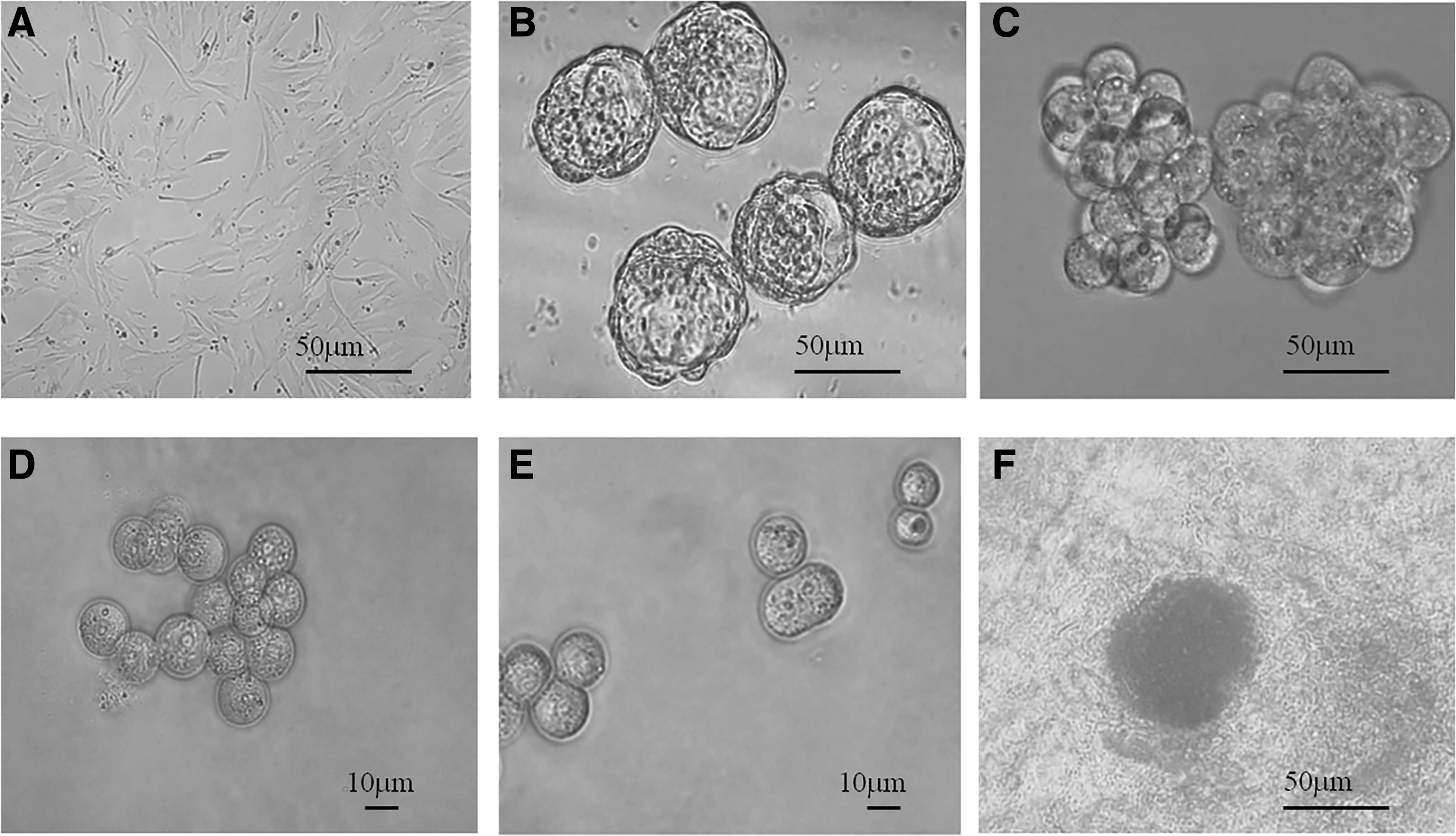
The isolation process of the mice pESC colony by immunoserometry method.
Experimental design
Investigation of the optimal concentration and timing of the PXD101 treatment
To determine the optimal concentration and timing of the PXD101 treatment, which most effectively improve the development, the parthenogenetic activated embryos were treated with 28 combinations of 4 different concentrations (5 nM or 50 nM or 100 nM or 500 nM) and 7 timing of the PXD101 treatments (below), and the number and rate of two-cell embryos and blastocysts of various groups were compared.
5 nM or 50 nM or 100 nM or 500 nM TSA:
Before (2 hours): before activation, the oocytes were put first in H-CZB medium supplemented with PXD101 (5/50/100/500 nM) for 2 hours, and then were activated by 10 mM SrCl2 in Ca2+-free CZB medium in the presence of 5 μg/mL cytochalasin B for activation of 4 hours; During (4 hours): the oocytes were activated by 10 mM SrCl2 in Ca2+-free CZB medium in the presence of 5 μg/mL cytochalasin B supplemented with PXD101 (5/50/100/500 nM) for 4 hours; After (4 hours): the oocytes were first activated by 10 mM SrCl2 in Ca2+-free CZB medium in the presence of 5 μg/mL cytochalasin B for 4 hours, and then put in H-CZB medium supplemented with PXD101 (5/50/100/500 nM) for 4 hours; Before (2 hours) and during (4 hours): the oocytes were put first in H-CZB medium supplemented with PXD101 (5/50/100/500 nM) for 2 hours, and then were activated by 10 mM SrCl2 in Ca2+-free CZB medium in the presence of 5 μg/mL cytochalasin B supplemented with PXD101 (5/50/100/500 nM) for 4 hours; Before (2 hours) and after (4 hours): the oocytes were put first in H-CZB medium supplemented with PXD101 (5/50/100/500 nM) for 2 hours and then were activated by 10 mM SrCl2 in Ca2+-free CZB medium in the presence of 5 μg/mL cytochalasin B for activation of 4 hours, and then put in H-CZB medium supplemented with PXD101 (5/50/100/500 nM) for 4 hours; During (4 hours) and after (4 hours): the oocytes were activated by 10 mM SrCl2 in Ca2+-free CZB medium in the presence of 5 μg/mL cytochalasin B supplemented with PXD101 (5/50/100/500 nM) for 4 hours, and then put in H-CZB medium supplemented with PXD101 (5/50/100/500 nM) for 4 hours; Before (2 hours), during (4 hours), and after (4 hours): the oocytes were put first in H-CZB medium supplemented with PXD101 (5/50/100/500 nM) for 2 hours and then were activated by 10 mM SrCl2 in Ca2+-free CZB medium in the presence of 5 μg/mL cytochalasin B supplemented with PXD101 (5/50/100/500 nM) for 4 hours, and then put in H-CZB medium supplemented with PXD101 (5/50/100/500 nM) for 4 hours.
Investigation of the optimal aggregation condition
The aggregation stage (two-cell stage and four-cell stage), the pronase concentration (0.25% and 0.5%), the treatment time (10 minutes and 20 minutes), and the aggregation system (the ordinary M16 droplet; M16 droplet culture system containing 0.06% PHA; and self-made concave hole culture system) were optimized.
Statistical analysis
Each experiment was repeated three times. Data were analyzed using the ANOVA test, with the treatment as the factor (Tables 2 and 3) or the concentration and timing as the two factors (Table 1). Statistical analyses were performed using SPSS 16.0 software. p values <0.05 were regarded as statistically significant.
Effect of PXD101 Treatment Concentration and Timing on the In Vitro Development of Mouse Parthenogenetic Embryo
PXD101 treatment time (hours) before, during, or after parthenogenetic activation.
Different lowercase superscripts in the same column indicate difference at p < 0.05.
M, mean; SD, standard deviation.
The Effect of Different Pronase Treatment and Different Aggregation Systems on the Aggregation and Development Potential of Mouse Parthenogenetic Two-Cell Embryo
Different lowercase superscripts in the same column indicate difference at p < 0.05.
PHA, phytohemagglutinin.
The Effect of Different Pronase Treatment and Different Aggregation Systems on the Aggregation and Development Potential of Mouse Parthenogenetic Four-Cell Embryo
Different lowercase superscripts in the same column indicate difference at p < 0.05.
Results
Effect of PXD101 concentration and timing on the in vitro development of mouse parthenogenetic embryo
Compared to the PXD101-untreated group, the treatments without “after (4 hours)” (including “before [2 hours]; during [4 hours]; before [2 hours], and during [4 hours]”) had no significant effect on the improvement of the parthenogenetic blastocyst rate for all the four concentrations (Table 1; p > 0.05), and as long as the treatments with “after (4 hours)” (including after [4 hours]; during [4 hours] and after [4 hours]; before [2 hours] and during [4 hours]; and after [4 hours]) all had a significant improved parthenogenetic blastocyst rate (Table 1; p < 0.05), indicating that the “after (4 hours)” treatment (PXD101 [5/50/100/500 nM] treated for 4 hours after activation) is very important for the improvement.
And among all the significant improved groups, 50 nM PXD101 treated “during (4 hours) and after (4 hours)” (the oocytes were activated using 10 mM SrCl2 in Ca2+-free CZB medium in the presence of 5 μg/mL cytochalasin B supplemented with 50 nM PXD101 for 4 hours, and then put in H-CZB medium supplemented with 50 nM PXD101 for 4 hours) had the highest rate of parthenogenetic blastocysts (Table 1; p < 0.05).
Effect of aggregation condition on the in vitro development of mouse parthenogenetic embryo
There was no effect of pronase concentration and treatment time on the rate of bare embryo when aggregated at two- or four-cell stage (p > 0.05; Tables 2 and 3). However, for whatever aggregated at two- or four-cell stage, there was both an effect of different aggregation systems on the aggregation and rate of parthenogenetic blastocysts (p < 0.05; Tables 2 and 3). For each type of pronase treatment, the parthenogenetic embryos aggregated in our self-made concave hole had significantly higher development rate and blastocyst rate (p < 0.05; Tables 2 and 3).
Aggregation of the embryos treated by 0.25% pronase for 10 minutes in our self-made concave hole had the highest aggregation rate, development potential, and blastocyst rate when aggregated at two-cell stage (47.5%, 42.5%, and 37.5%; Table 2), which was similar with that aggregated at four-cell stage (44.4%, 38.8%, and 33.3%; Table 3). Among all the groups, aggregation at two-cell stage treated by 0.25% pronase for 10 minutes in our self-made concave hole had the highest rate of parthenogenetic blastocysts (37.5%; Table 2).
Effects of PXD101 and embryo aggregation on the development of parthenogenetic embryos and on the isolation of pESC colonies
There is an effect of the combination method (PXD101 treatment and embryo aggregation) on the development of mouse parthenogenetic embryos and on the isolation of pESC colonies. Compared to the control group (untreated parthenogenetic embryo), treatment with 50 nM PXD101 for 8 hours (during [4 hours] + after [4 hours] activation) and two-cell parthenogenetic embryos aggregated with 0.25% pronase for 10 minutes in our self-made concave hole could both significantly improve the blastocyst formation and the subsequent isolation of pESC colonies (p < 0.05; Table 4), but there was no significant difference between both (p > 0.05; Table 4).
Effect of Two-Cell Embryo Aggregation, PXD101 Treatment, and Their Combinations on the Development of Mouse Parthenogenetic Embryo
50 nM PXD101 treated “during (4 hours) + after (4 hours)” first and then aggregated at two-cell stage with 0.25% pronase for 10 minutes in our self-made concave hole.
Different lowercase superscripts in the same column indicate difference at p < 0.05.
pESCs, parthenogenetic embryonic stem cells.
However, the treatment of their combination (50 nM PXD101 treated during [4 hours] + after [4 hours] activation first, and then aggregated at two-cell stage with 0.25% pronase for 10 minutes in our self-made concave hole) has a higher rate of blastocyst formation although not significant, but significantly higher rate of the subsequent isolation of pESC colonies from parthenogenetic blastocysts than that by their separate treatment (p < 0.05; Table 4).
The pESC colonies have a smooth surface and obvious boundary with the surrounding mouse embryonic fibroblast (MEF) cells and exhibit morphology typical of undifferentiated embryonic stem cells, and they were identified with AKP-positive staining (Fig. 1).
Discussion
At present, the efficiency of the parthenogenetic embryo development and the pESC isolation in mice is still very low (Cibelli et al., 2002; Ju et al., 2008). Therefore, to optimize the method to increase the in vitro developmental competence of mice parthenogenetic embryos, this study compared the effects of embryo aggregation and application of histone deacetylase inhibitor (PXD101) on the development potential of mice parthenogenetic embryos and on subsequent isolation of pESC colonies. The results suggested that both methods have a similar effect on the improvement of parthenogenetic embryos development; however, the combination of their treatments (50 nM PXD101 treated for 8 hours during and after activation and then aggregation of the two-cell embryos with 0.25% pronase for 10 minutes in our self-made concave hole) had the best development rate and subsequent pESC isolation rate.
Studies suggested that there are so far two main methods to improve the in vitro development of blastocysts: embryo aggregation and application of HDACis (Qiu et al., 2016, 2018), while most of the recent studies about the effect of embryo aggregation focused on IVF and SCNT embryos (Lee et al., 2013; Saadeldin et al., 2015; Siriboon et al., 2014; Terashita et al., 2011). Our previous study in mice (Qiu et al., 2017b) investigated the effect of the parthenogenetic embryo and the male embryo aggregation on the blastocyst development in mice. However, there is limited study about the effect of the aggregation of the parthenogenetic embryos on each other, especially the aggregation stage on the development of in vitro mice parthenogenetic embryo and limited study about the comparison of these two methods on mice parthenogenetic embryos; therefore, we carried out this study for the method optimization.
In this study, we concluded that the embryo aggregation improved in vitro development potential of mice parthenogenetic balstocysts (Table 4), which is consistent with the previous studies in pigs (Lee et al., 2013; Saadeldin et al., 2015; Siriboon et al., 2014; Terashita et al., 2011). It is suggested that the intercellular communication and interactions between blastomeres are very important for embryo development, and the embryo aggregation could enhance this intercellular communication (Siriboon et al., 2014). And we generated an increased rate of parthenogenetic blastocysts (38.3%) by two-cell embryo aggregation with 0.25% pronase treated for 10 minutes in our self-made concave (Table 4), which is similar with the result (40.0%) by Terashita et al. (2011) in pigs, but lower than that by Siriboon et al. (2014) (63.0%) where a different system (the well-of-the-well system) was used in pigs.
Using the same method of embryo aggregation, the improved rate of parthenogenetic blastocysts in this study (38.3%) was lower than our previous study (46.7%) (Qiu et al., 2017b) carried out for SCNT embryos where the male embryos were aggregated.
The rate of parthenogenetic blastocysts was 40.0% by 50 nM PXD101 treated for 8 hours (during [4 hours] + after [4 hours] activation) (Table 4), which is also similar with our previous study (40.2%) for the mice SCNT embryos (Qiu et al., 2017a) treated similarly by 50 nM PXD101, but treated for 10 hours (during [6 hours] + after [4 hours] activation). The effect PXD101 treatment on the in vitro developmental competence of SCNT embryos has been reported in porcine by Jin et al. (2015). However, its effect on mice parthenogenetic embryos has been much more limited and, therefore, fewer results are available.
This study suggested for the first time that compared to the PXD101-untreated group, the treatments without “after (4 hours)” had no significant effect on the improvement of the parthenogenetic blastocysts for all the four concentrations (Table 1; p > 0.05), and as long as the treatments with “after (4 hours)” (including after [4 hours]; during [4 hours] and after [4 hours]; before [2 hours] and during [4 hours] and after [4 hours]) all had a significant improved parthenogenetic blastocyst rate (Table 1; p < 0.05), indicating that the “after (4 hours)” treatment is very important for the improvement of mice parthenogenetic embryo development.
And the mechanism of the improvement is probably by its acting on the epigenetic reprogramming of embryos by regulating the acetylation of lysine residues in the positively charged histone tails of nucleosomes (Kishigami et al., 2006, 2007), which is beneficial for chromatin remodeling and gene transcription.
In this study, it was found that embryo aggregation and PXD101 treatment both significantly improved the subsequent isolation of pESC colonies in comparison to the untreated group (Table 4). Nowadays, pESCs are attracting more public attention due to their application in regenerative medicine (Hwang et al., 2005). However, there is a limit for its application due to its low establishing rates. Our study suggests that a new technique combined PXD101 treatment with embryo aggregation can be applied to improve the isolation rate of pESC colonies.
Our study concluded that the embryo aggregation at two-cell stage and application of PXD101 (50 nM treated for 8 hours) could both improve in vitro development potential of mice parthenogenetic embryo and subsequent isolation of pESC colonies, while their combination had the best effect; therefore, the combination of these two methods is considered the optimal treatment for in vitro development of parthenogenetic embryo in mice, opening new opportunities for the application of this combination method to improve the parthenogenetic embryo development in other species. Future study should be focused on the molecular mechanism underlying the improvement by this combination method.
Conclusion
The combination of PXD101 treatment and embryo aggregation (50 nM PXD101 treated for 8 hours and then aggregated at two-cell stage with 0.25% pronase for 10 minutes in our self-made concave hole) is considered the optimal way to improve the in vitro development of parthenogenetic embryo and subsequent isolation of pESC colonies in mice.
Footnotes
Author Disclosure Statement
The authors declare they have no conflicting financial interests.
Funding Information
This study was supported by the Innovation Team Building Program in Chongqing universities (Grant No. CXTDG201602004), the Based and Advanced Research Project of Chongqing (Grant No. cstc2017jcyjAX0477), and the National Natural Science Foundation of China (Grant No. 31572488).