




Abstract
By dissecting and comparing the transcriptional trajectories and epigenomic traits of reprogramming and transforming cells at the single-cell resolution, Huyghe et al discovered Bcl11b and Atoh8, two key transcription factors controlling cell plasticity during pluripotent reprogramming and oncogenic transformation.
During development, embryos progressively differentiate into functionally and phenotypically distinct cells to form multicellular organisms. This irreversible developmental process generates unique cellular identities with cell type-specific gene expression programs throughout the human lifespan. Stable fixed cellular plasticity and identification can be largely amended by pluripotent reprogramming (Takahashi and Yamanaka, 2006), direct reprogramming (Wang et al, 2021), and oncogenic transformation (Puisieux et al, 2018).
The high similarly between pluripotent reprogramming and oncogenic transformation is evident by their likeness in common biological processes [e.g., glycolytic metabolism (Warburg, 1956; Warburg et al, 1927)], transcription factors [e.g., c-Myc (Land et al, 1986, Land et al, 1983; Takahashi and Yamanaka, 2006), and p53 (Lee et al, 2015, Lee et al, 2012)], epigenetic modifiers [e.g., MLL1/WDR5 complex (Ang et al (2011)], etc. However, the molecular mechanisms involved in loss of original cell identity and reshaping through cellular plasticity during the early stages of reprogramming and transformation remain nebulous.
To understand the coordinated changes of cellular plasticity and identity that are critical for both reprogramming and transformation, a new study led by Huyghe et al (2022) dissected the comparative single-cell trajectories of pluripotent reprogramming (through Oct4, Sox2, Klf4, and c-Myc [OSKM]) (Takahashi and Yamanaka, 2006) and oncogenic transformation (through K-rasG12D and c-Myc) (Ischenko et al, 2013; Land et al, 1986, Land et al, 1983) (Fig. 1). The authors utilized powerful reprotransformable mice with conditionally doxycycline-induced OSKM expression or tamoxifen-induced K-rasG12D expression, combined with c-Myc overexpression, in the same population of somatic cells.
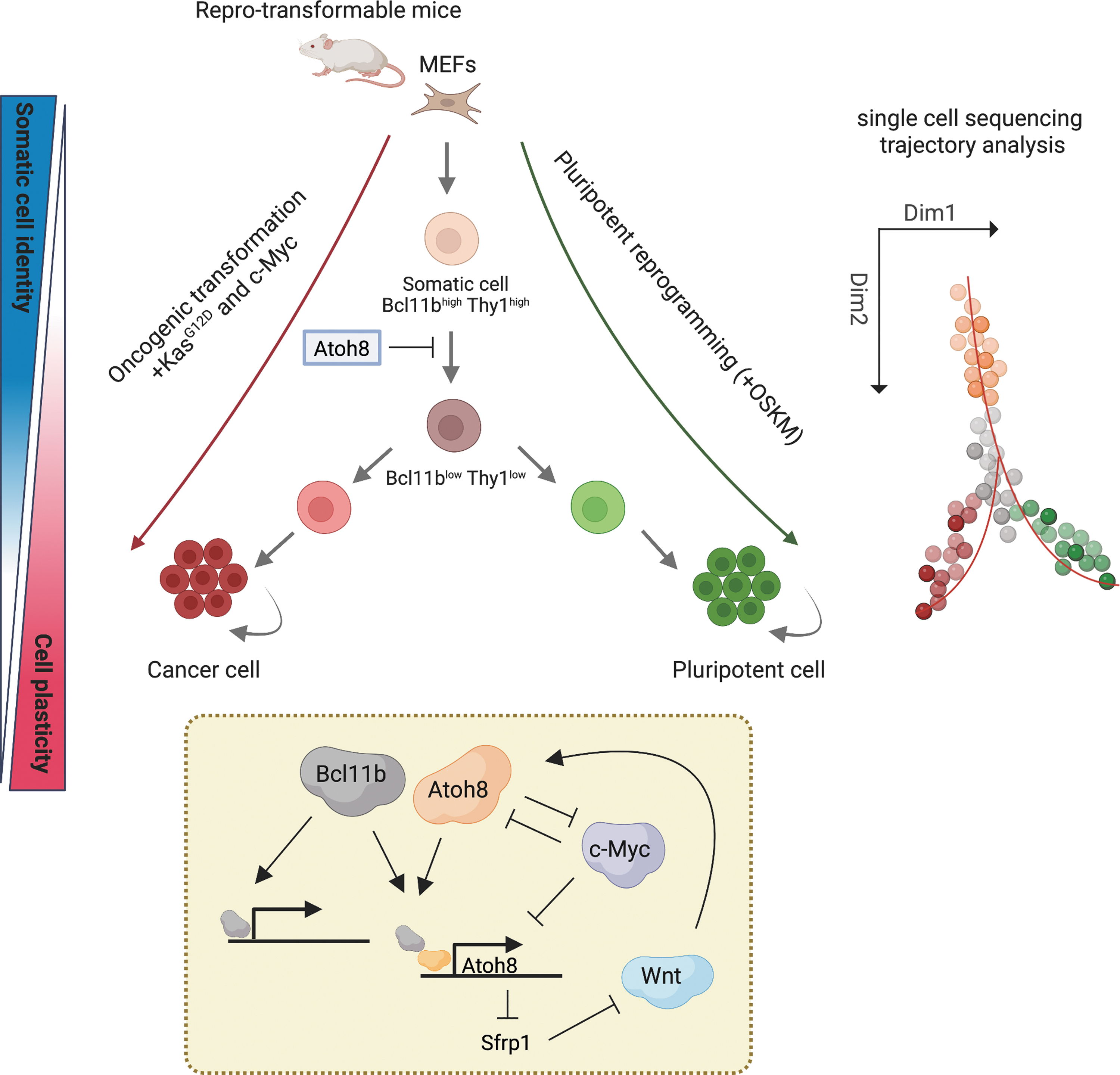
Schematic recapitulating this study to identify key regulators during the multistep processes of cell pluripotent reprogramming and oncogenic transformation through single-cell sequencing techniques. Loss of somatic cell identity and gain of cell plasticity can be induced from reprotransformable mice MEFs. Bcl11b and Thy1 serve as markers to distinguish cellular intermediates that tend to develop pluripotent cells or cancer cells during cell fate transitions. The transcription factor Atoh8 restrains the emergence of cell plasticity by transcriptional regulation of Wnt signaling activity and c-Myc self-antagonization. The figure is created using (BioRender.com). MEFs, mouse embryonic fibroblasts; Atoh8, atonal bHLH transcription factor 8.
They conducted single-cell RNA sequencing to measure RNA expression in mouse embryonic fibroblasts (MEFs) after both 5 days and 10 days of OSKM or K-rasG12D/c-Myc overexpression, as well as in fully reprogrammed induced pluripotent stem cells (iPSCs) and transformed cells. In addition, they performed principal component analysis and t-distributed stochastic neighbor embedding to define 12 clusters among 30,146 single cells and then utilized diffusion maps and slingshot to rebuild the pseudotemporal progression during pluripotent reprogramming and oncogenic transformation. Their designed single-cell trajectories allowed them to transiently identify the intersection of specific clusters of cells before the diversion of pluripotent reprogramming and oncogenic transformation.
By computing the marker genes predominantly found in MEFs, they selected the glycoprotein Thy1 (Polo et al, 2012), a known MEF marker during pluripotent reprogramming, and the transcriptional factor (TF) Bcl11b (Li et al, 2010; Wakabayashi et al, 2003), a hematopoiesis-associated TF that is downregulated during reprogramming and transformation and silenced in iPSCs and transformed cells, as a marker for further investigation. In comparison with other cell populations, they found that cells with Bcl11blow Thy1low acquired the cellular plasticity tendency to form pluripotent stem cells or aggressive tumors.
Suppression of Bcl11b and/or Thy1 facilitated MEF reprogramming to pluripotency and transformation to malignancy. Interestingly, Bcl11blow Thy1low cells still maintained their original cellular identities, suggesting a disengaged process of gain of new plasticity and loss of original identity during pluripotent reprogramming and oncogenic transformation.
They then comprehensively characterized the molecular signatures of reprogramming and transformation using Bcl11blow Thy1low intermediate cells and unveiled that atonal bHLH transcription factor 8 (Atoh8) is a negative regulator of cell plasticity and is downregulated during reprogramming and transformation. Depletion of Atoh8 enhances OSKM-induced reprogramming and KrasG12D/c-Myc-induced transformation. Mechanistically, inhibition of Atoh8 leads to the emergence of the Bcl11blow Thy1low cell population through activation of the Wnt pathway through Sfrp1 suppression and c-Myc upregulation, which are two critical signaling regulators for tissue regeneration and tumor formation.
Moreover, Atoh8 preferentially occupies inaccessible regions (ATAC-seq-positive) of enhancers and binds with Bcl11b on the Atoh8 locus for activation, whereas the binding of Atoh8-bound regions by c-Myc is finely switched from somatic cells (Atoh8highc-Myclow) to iPSCs and/or transformed cells (Atoh8lowc-Mychigh), indicating relocation of c-Myc during reprogramming and/or transformation.
In summary, the study by Huyghe et al (2022) discovered key regulators controlling cell plasticity during pluripotent reprogramming and oncogenic transformation by dissecting and comparing the transcriptional trajectories of reprogramming and transforming cells. Their findings provide a new insight into how TF-mediated regulatory networks finetune cellular plasticity during pluripotent reprogramming and oncogenic transformation.
Footnotes
Authors' Contributions
M.-F.H., R.S., and D.-F.L. wrote the article. M.-F.H. made the figure.
Author Disclosure Statement
The authors declare they have no conflicting financial interests.
Funding Information
D.-F.L. was supported by CPRIT RR160019 and NIH/NCI R01CA246130. D.-F.L. is a CPRIT scholar in cancer research.