




Abstract
A 6-year-old mare, a valuable polo horse, died of complications following postcolic surgery. To preserve its genetics, ear skin samples were collected immediately after death and stored in an equine embryo transfer medium at 4°C for 5 days. After trypsin digestion, monolayer fibroblast cultures were established, but signs of massive bacterial infection were found in all of them. As an ultimate attempt for rescue, rigorously and repeatedly washed cells were individually cultured in all wells of four 96-well dishes. New monolayers were established from the few wells without contamination and used for somatic cell nuclear transfer. Four of the six Day 7 blastocysts derived from 14 reconstructed zygotes were transferred in four naturally cycling mares on Day 5 after ovulation. The embryo transfers resulted in 2 pregnancies, one from a fresh and one from a vitrified blastocyst. The vitrified embryo transfer resulted in a healthy offspring, now 21 months old, genetically and phenotypically identical to the somatic cell donor animal.
Introduction
Among various potential uses of somatic cell nuclear transfer (SCNT) in domestic animals, preserving valuable genetics is an obvious but rarely used approach (Folch et al., 2009; Oh et al., 2008). There are very few reports of successful genetic rescue by SCNT following the unexpected or accidental death of an irreplaceable animal (Ahn et al., 2011; Hong et al., 2011; Kim et al., 2013), and, to our knowledge, equine postmortem genetic rescue has not been described previously. In contrast to human cloning, where ethical concerns dominate, concerns regarding animal cloning are mostly based on practical issues including the possible disproportional suffering of recipients and offspring, products with potential consumption hazards, and narrowing genetic diversity due to widespread application. While these are not the predominant issues in horses, technical and logistic problems have hampered advancement, including the regrettably low number of properly equipped laboratories and qualified scientists, and the lack of established, proven, and widely known procedures.
Microbial contamination is a frequent problem when establishing and culturing lines of cells derived from biopsied tissue. The addition of antibiotics to the medium and repeated washing are approaches to possibly salvage infected cultures of cells. This article describes a successful attempt to save the genetics of a valuable horse following its untimely death and involves persistent efforts in an almost hopeless case to recover donor fibroblasts from an infected monolayer culture. The outcome may encourage other teams to use SCNT even when the situation is seriously handicapped, and the odds seem very low.
Materials and Methods
The timeline of events is briefly summarized in Table 1. On October 23, 2017, the 6-year-old mare Easter, located at Catalina Genetics, North Richmond, New South Wales, Australia, was rushed to a local equine hospital for colic due to a twisted colon. During the following hours, the doctors decided to perform surgery to solve the problem, but on October 29, she had to be euthanized due to postoperative complications. Immediately after the death, three 2 cm in diameter pieces of ear skin were obtained with a surgical blade and stored in 15 mL Falcon tubes in 3 mL equine embryo transfer medium (EMCARE™, Agtech) at 5°C. The following days were spent with desperate attempts to find a laboratory in Sydney experienced in fibroblast monolayer preparation. Eventually, after 5 days of storage, the tubes were placed in an icebox with cold gel packs to keep the temperature between 5°C and 10°C and transported by flight to the laboratory of Reinclonation Pty, Southport, Queensland, Australia.
Timeline of Events During Rescue Cloning of the Valuable Polo Horse
OPU, ovum pick-up; SCNT, somatic cell nuclear transfer.
Immediately upon arrival, the samples were rigorously washed in phosphate buffered saline containing 50 μg/mL Gentamicin (G1397; Sigma), then cut into small pieces and digested in Trypsin-EDTA solution (T4049; Sigma) at 38.5°C for 60 minutes. After centrifugation, the pellet was resuspended in culture medium (DMEM, D5648; Sigma), supplemented with 15% fetal bovine serum (S6501; Sigma), 50 μg/mL Gentamicin, 1% nonessential amino acid (M7145; Sigma), seeded in 250 mL Falcon tissue culture flasks and incubated at 38.8°C in a 5% CO2 in air atmosphere with maximum humidity. After 3 days, the medium in the flasks with outgrowths was carefully changed, and the cells were further cultured. Unfortunately, on Days 5 to 7 after seeding, microscopic signs of bacterial contamination were detected in all flasks with growing monolayers. Trypsinization, repeated centrifugation, and dilution in 3 × concentration (150 μg/mL) of Gentamicin solution could not eliminate the presence of the infective agents. Hence, a new approach had to be used to rescue the fibroblasts.
Thus, the infected monolayer cultures were trypsinized, diluted in DMEM culture medium containing 3 × concentration of Gentamicin, and centrifuged at 250 × g for 10 minutes in 15 mL Falcon tubes. The pellet was resuspended in the same solution and centrifuged again. The procedure was repeated three times. Meanwhile, wells of four flat bottom 96-well culture plates (CLS3599; Corning) were filled with 200 μL culture medium. The centrifuged and resuspended fibroblasts were transferred into 60 mm in diameter Petri dishes filled with 10 mL culture medium. With a finely drawn and fire-polished glass Pasteur pipette, individual fibroblasts were collected from the dish and transferred to the 96-well plates, preferably one cell per well, by using the least possible volume of medium for transfer. The plates were then incubated at 38.8°C in 5% CO2 in air with maximum humidity for 72 hours.
In ∼20% of the wells, proliferation and spreading of fibroblasts were detected. Ten days after seeding, no infection and considerable growth were seen in six wells. These wells were trypsinized, and the cells were further cultured in a 33 mm in diameter Petri dish until they reached confluence. As the laboratory was not prepared for horse cloning, cultures were then disaggregated by trypsin, suspended in the above-mentioned cell culture medium containing 10% dimethylsulfoxide (D8418; Sigma) and stored in liquid nitrogen.
On September 15, 2019, samples were transferred back to Catalina Genetics, where a purpose-designed laboratory was subsequently established for equine SCNT. The laboratory has demonstrated success at producing cloned foals using variousnuclear donor cell lines and in vivo collected (ovum pick-up, OPU) as well as in vitro-matured oocytes as recipient cytoplasts (Cortez et al., 2023). Easter's frozen cells were thawed and, after two passages, used as nuclear donors for SCNT.
The process of SCNT has been described in detail recently (Cortez et al., 2023). Briefly, after OPU (Lazzari et al., 2020; Stout, 2020), cumulus cells were removed from the oocytes by pipetting in H-SOF medium supplemented with 1 mg/mL hyaluronidase. Denuded oocytes and donor cells were transferred to a droplet of H-SOF covered with mineral oil. The polar body and the metaphase plate of each mature oocyte were aspirated using an enucleation pipette attached to a Piezo drill (PMAS-CT150; PrimeTech, Japan). The donor cells were then loaded into an injection pipette, and a single cell was deposited into the perivitelline space of each cytoplast.
Couplets were held for 1 hour in H-SOF at 38°C before being transferred to fusion medium on a fusion chamber slide. A direct current pulse of 2.2 kV/cm strength and 15 μs duration was immediately applied to the couplets. The couplets were then washed three times in H-SOF, transferred to the embryo culture medium, which consisted of DMEM/F-12 medium supplemented with 10% fetal bovine serum, and incubated for 2 hours. Activation was carried out by incubation in 5 μM ionomycin for 5 minutes, then in 1 mM 6-dimethylaminopurine and 5 μg/mL cycloheximide in embryo culture medium for 4 hours. The reconstructed embryos were incubated in an atmosphere of 5% CO2, 5% O2, and 90% N2 with maximum humidity at 38.5°C. Day 7 blastocysts were evaluated morphologically according to the guidelines of the International Embryo Technology Society (IETS).
Grades 1 and 2 blastocysts were vitrified and warmed using the Cryotop method according to the manufacturer's instructions (Kitazato BioPharma, Shizuoka, Japan; for more detail see Cortez et al., 2023). After warming, the blastocysts were stored at 38.5°C in culture medium for 1 hour in a portable incubator (LabMix, WTA, Brazil) before embryo transfer. Day 7 blastocysts (fresh or cryopreserved) were transferred transcervically to recipient mares on Day 5 postovulation as described previously by Cortez et al. (2023). On Day 14 postovulation (Day 9 after ET), and on Days 42 and 90 of gestation, the mares were examined for pregnancy.
Results
Thawing and culture of the donor cells resulted in monolayers with cell shapes similar to those in common fibroblast cultures, but most cells were larger compared to other fibroblast cells obtained from live animals without decontamination treatment. These differences persisted after several passages. For cloning, only smaller cells were used. Out of the 22 oocytes obtained by OPU, 16 matured, allowing the generation of 14 reconstructed embryos. Two days after activation, 12 two-cell embryos were observed, and six blastocysts were obtained on Day 7. Two blastocysts were immediately transferred into two recipients, resulting in one pregnancy that was lost on Day 22 after ovulation.
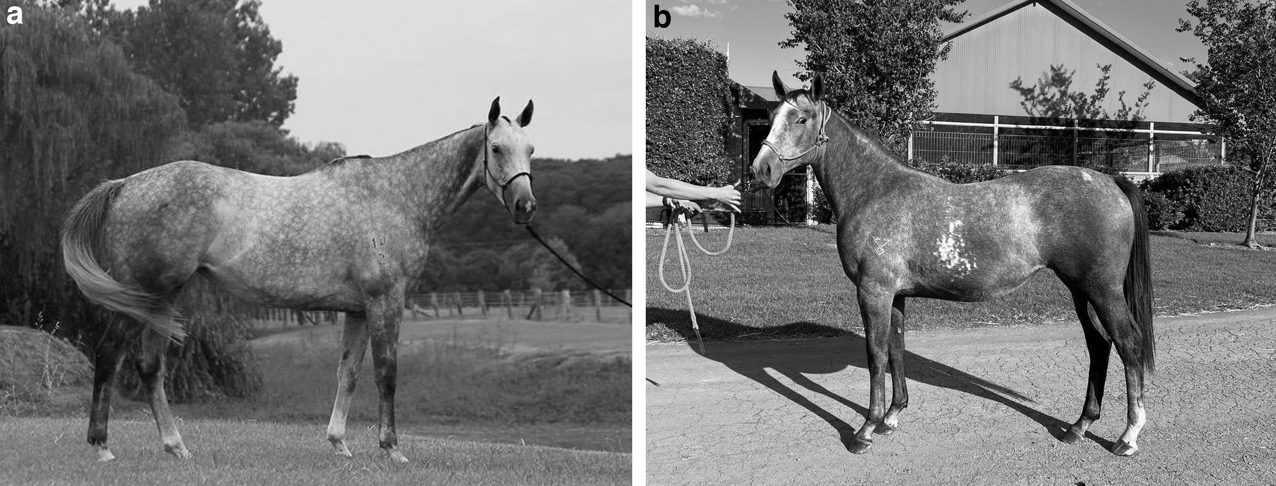
The remaining four blastocysts were vitrified. Out of these, two were transferred into two recipients, resulting in one pregnancy and a live birth on Day 247 of gestation. The resulting clone, called Samsara, was 21 months at the submission of this manuscript and in good health. Her morphological traits are identical to those of the donor animal (Fig. 1a and b). According to the Comparison Listing performed by the Racing Australia Equine Genetics Research Centre, Hunter Valley, NSW, Australia, the profiles of the somatic cell donor and the cloned offspring match at 12 of 12 ISAG approved microsatellite markers. The offspring has all the skills to become an excellent polo player like her somatic cell donor. Her training will start at around 4 years of age.
Discussion and Conclusion
Horse cloning by SCNT is now regarded as a routine procedure, although applied successfully at only a handful of laboratories worldwide. Recipient oocytes may be obtained from abattoir ovaries or from in vivo OPU (Cortez et al., 2023), and both traditional, micromanipulation-based methods and handmade, zona-free cloning (HMC) can be used successfully for embryo reconstruction (Salamone and Maserati, 2023). Due to the limited number of horses slaughtered in Australia, OPU-derived oocytes were used in our present work. To maximize the number of reconstructed embryos, we selected the micromanipulation-based procedure, where one oocyte is needed to make one embryo, whereas HMC requires two enucleated half-oocytes for the reconstruction of each embryo.
For rescue cloning, to save the genetics of a valuable animal, the nuclear donor cell preparation may require extraordinary efforts to accommodate unexpected death or other special situations. The sudden death of our donor on a continent without a successful equine cloning team made the task nearly impossible. Although some companies in North and South America offer pet or even horse cloning from tissues stored in refrigerators, to our knowledge, there is no scientific publication describing the possibilities and limitations of this approach. Okonkovo and Singh (2015) have derived fibroblast-like cells from tissues wrapped in sterile paper towels and stored at 4°C for up to 45 days, but the study did not examine the usability of these cells for SCNT.
Our attempt to prepare fibroblast monolayers after 5 days of storage of ear tissue pieces in a domestic household refrigerator was partially successful. The cells formed monolayers, but were infected massively by unidentified bacteria, and the infection could not be eliminated by the commonly suggested method using increased antibiotic concentration and repeated washing by centrifugation and resuspension. Accordingly, as an ultimate attempt, another approach was used; after repeated washings, individual cells were placed on the bottom of wells of 96-well dishes, with the faint hope that in some wells, the concentration of bacteria would not be enough to survive the antibiotic treatment.
Eventually, our attempt resulted in a contamination-free fibroblast-like culture that could be frozen and stored till the purpose-designed cloning laboratory was established and proven to be suitable for horse cloning. While most of the thawed cells were slightly larger than the cells of monolayers typically derived with standard procedures from live animals and were not used as nuclear donors, suitable in vitro and in vivo development was achieved using the smaller cells for embryo reconstruction. The 21-month-old healthy foal has all the genetic and morphological traits of the donor animal, demonstrating that SCNT can be successfully used under exceptional circumstances and nearly hopeless situations.
Footnotes
Authors' Contributions
J.V.C.: Investigation, Methodology, Validation, Writing—Original Draft. K.H.: Investigation, Supervision, Validation, Writing—Review and Editing. C.G.G.: Investigation, Methodology, Resources, Writing—Review and Editing. M.H.: Investigation, Methodology. Z.M.: Validation, Writing—Review and Editing. G.V.: Investigation, Methodology, Validation, Writing—Original Draft; Review and Editing.
Author Disclosure Statement
The authors declare they have no conflicting financial interests.
Funding Information
No funding was received for this article.