




Abstract
MicroRNAs are a class of small noncoding RNAs that function as critical gene regulators through targeting mRNAs for translational repression or degradation. In this study, we showed that miR-376c expression level was decreased while transforming growth factor-alpha (TGFA) mRNA expression levels were increased in osteosarcoma tissues and cell lines, and we identified TGFA as a novel direct target of miR-376c. Overexpression of miR-376c suppressed TGFA expression and the expression of its downstream signaling molecule such as epidermal growth factor receptor, and attenuated cell proliferation and invasion. Forced expression of TGFA could partly rescue the inhibitory effect of miR-376c in the cells. Taken together, these findings will shed light on the role and mechanism of miR-376c in regulating osteosarcoma cell growth via miR-376c/TGFA axis, and miR-376c may serve as a potential therapeutic target in osteosarcoma in the future.
Introduction
In a variety of cancers, miRNA expression is significantly altered, and it has the potential to be a prominent diagnostic and prognostic tool (Mishra and Merlino, 2009). It is important to elucidate the function of miRNAs in tumor pathogenesis and progression, because miRNAs have the potential to regulate various critical biological processes, including differentiation, progression, apoptosis, proliferation, and drug resistance of tumor cells (Ryan et al., 2010). However, not much is known about the expression and deregulation of miRNAs in osteosarcoma with very poor prognosis and high possibilities of tumor proliferation and migration. Although proliferation and migration have been acknowledged as the most lethal attributes of solid tumors, the molecular mechanism underlying them is still limited.
Bioinformatic algorithms assess that all the human miRNAs may regulate up to 30% of human genes that represent the majority of genetic pathways (Hwang and Mendell, 2006). Many studies have identified specific miRNAs expression profiles of multiple cancer types compared to those of normal adjacent tissues. Depending on cellular contexts and target genes that they regulate, miRNAs may function as tumor suppressors or oncogenes (Lu et al., 2005; Calin and Croce, 2006). Among these functional miRNAs, miR-376c has been demonstrated to significantly decrease in multiple cancer types and play a role of tumor suppressor. For instance, miR-376c leads to attenuation of melanoma proliferation and migration (Zehavi et al., 2012). Besides, a study found that miR-376c exhibited a lower expression in osteosarcoma cell lines compared with that in osteoblast cell lines (Duan et al., 2011), which implied the involvement of miR-376c in osteosarcoma proliferation and metastasis.
Transforming growth factor-alpha (TGFA) encodes a growth factor that is a ligand for the epidermal growth factor receptor (EGFR), which activates a signaling pathway for cell proliferation, differentiation, and development. This protein may act as either a transmembrane-bound ligand or a soluble ligand. This gene has been associated with many types of cancers. It has been reported that EGFR activation by its cognate ligand EGF, and by TGFA, triggers different biological responses, such as cell proliferation, differentiation, migration, and survival (Hsieh and Conti, 2005). TGFA is structurally related to EGF and widely expressed in developing embryos and normal adult tissues including the colon, skin, mammary gland, liver, and kidney (Booth and Smith, 2007). Indeed, high levels of TGFA were associated with an increased expression of EGFR, indicating autocrine, paracrine, and juxtacrine functions exerted during tumorigenesis (Kumar et al., 1995).
In the present study, we confirmed the regulatory relationship between miR-376c, a known tumor suppressive miRNA, and an oncogene, TGFA. We provided evidences that miR-376c could impede osteosarcoma cell proliferation and migration, at least partly by targeting TGFA.
Materials and Methods
Tissue samples, cell lines, and cell transfection
A total of 13 pairs of primary osteosarcoma and their matched adjacent normal bone and myeloid tissues were collected. All samples were obtained from patients who underwent surgical resections at The Second Xiangya Hospital of Central South University (Changsha, China) and snap-frozen in liquid nitrogen, then stored at −80°C for further use. This project was approved by the Ethic Committee of The Second Xiangya Hospital of Central South University. The permit number from the Ethics Committee is S167.
The human osteoblasts hFOB and three human osteosarcoma cell lines, including MG-63, Saos-2, and SW1353, were purchased from American Type Culture Collection. Cells were routinely grown in RPMI-1640 medium (Invitrogen), supplemented with 10% fetal bovine serum (Gibco), and cultured in a 37°C humidified atmosphere of 5% CO2. Ectopic expression of miR-376c in cells was achieved by transfection with miR-376c mimics (Genepharma) using Lipofectamine 2000 (Invitrogen). Overexpression of TGFA was performed using TGFA ORF expression clone (GeneCopoecia). Cells were plated in 6-well clusters or 96-well plates and transfected for 24 or 48 h. Transfected cells were used in further assays or RNA/protein extraction.
RNA extraction and SYBR green quantitative polymerase chain reaction analysis
Total RNA was extracted from cells using TRIzol reagent (Invitrogen). Mature miR-376c expressions in cells were detected using a Hairpin-it TM miRNAs qPCR kit (Genepharma). Expression of RNU6B was used as an endogenous control. TGFA expression was measured by SYBR green qPCR assay (Takara). Data were processed using 2−ΔΔCT method.
CCK-8 cell proliferation assay
Cell proliferation rates were measured using Cell Counting Kit-8 (CCK-8) (Beyotime). For 24 h, 0.5×10⁴ cells were seeded in each 96-well plate, transfected with the indicated miRNA or siRNA, and further incubated for 24, 48, 72, and 96 h respectively. About 10 μL CCK-8 reagent was added to each well at 1 h before the endpoint of incubation. The OD450 nm value in each well was determined by a microplate reader.
Cell invasion and migration assay
The invasive and migratory potential of cells was evaluated using transwell inserts with 8 μm pores (Coring). For invasion assay, at 24 h after transfection, 3.0×10⁵ cells in serum-free medium were added to each upper insert precoated with matrigel matrix (BD). About 500 μL 10% FBS medium was added to the matched lower chamber. After 48 h incubation, noninvaded cells were removed from the upper surface of the transwell membrane with a cotton swab, and invaded cells on the lower membrane surface were fixed in methanol, stained with 0.1% crystal violet, photographed, and counted. For migration assay, the procedures were similar, except that 2×10⁵ cells were added into the inserts without being matrix gel precoated. Six random fields at 100× magnification for each insert were counted. Inserts were conducted in triplicate in three separate experiments.
Western blot analysis
Immunoblotting was performed to detect the expression of TGFA and EGFR in osteosarcoma cell lines. Cultured or transfected cells were lysed in RIPA buffer with 1% PMSF. Protein was loaded onto an SDS-PAGE minigel and transferred onto PVDF membrane. The blots were probed with 1:1000 diluted rabbit polyclonal TGFA and EGFR antibody (Abcam) at 4°C overnight and subsequently incubated with HRP-conjugated secondary antibody (1:5000). Signals were visualized using ECL Substrates (Millipore). β-actin was used as an endogenous protein for normalization.
Luciferase reporter assay
A fragment of 3′UTR of TGFA (1089 bp) containing the putative miR-376c binding site (1510–1517) was amplified by polymerase chain reaction (PCR) using the following primers:
wt-TGFA (forward) 5′ CCGCTCGAGCACCAGTAAAACAAGGAACTTG3′
wt-TGFA (reverse) 5′ GAATGCGGCCGCTTTACAGAAACAAATGCAATGTTAC3′
The PCR product was subcloned into a psiCHECK-2 vector (Promega, Madison, WI) immediately downstream to the luciferase gene sequence. A psiCHECK-2 construct containing 3′UTR of TGFA with a mutant seed sequence of miR-376c was also synthesized using the following primers:
mut-TGFA (forward) 5′ TTAAAATTTCCTACTTGTGTATAGAAATGGAAAG 3′
mut-TGFA (reverse) 5′ ACAAGTAGGAAATTTTAATACAGTGGTTCTC3′
All constructs were verified by DNA sequencing. HEK293 cells were plated in 96-well clusters, then cotransfected with 100 ng constructs with or without miR-376c precursors. At 48 h after transfection, luciferase activity was detected using a dual-luciferase reporter assay system (Promega) and normalized to Renilla activity.
Statistical analysis
All data from three independent experiments were expressed as mean±SD and processed using SPSS17.0 statistical software. The expression of miR-376c in osteosarcoma tissues and their matched adjacent normal bone and myeloid tissues were compared by Wilcoxon's paired test. The difference among the groups in migration and invasion assay was estimated by Student's t-test or one-way ANOVA. A p value of <0.05 was considered to be statistically significant.
Results
miR-376c is frequently downregulated in osteosarcoma tissues and cell lines
We performed SYBR green quantitative PCR analysis to detect the expression levels of miR-376c and TGFA in osteosarcoma tissues and cell lines. In the large panel of 13 cases of primary osteosarcoma tissues and their adjacent normal bone and myeloid tissues, our results showed that miR-376c was significantly decreased in 11 (84.6%) osteosarcoma tissues and TGFA mRNA was increased in 10 (76.9%) when compared with that in the paired adjacent normal tissues (Fig. 1A, B). As shown in Figure 1C, Spearman's correlation test showed a significant inverse correlation of the miR-376c and TGFA mRNA expression levels in osteosarcoma tissues with Spearman's correlation, R 2=0.584 (p<0.001), confirming that lower expression levels of miR-376c were significantly associated with higher levels of TGFA mRNA expression in the same set of osteosarcoma tissues. In addition, we extended our test to one human osteoblast and three human osteosarcoma cell lines. The total three cell lines showed a notable loss of miR-376c and obvious upregulation of TGFA mRNA expression, in comparison with the control human osteoblast cell lines (Fig. 1D, E).
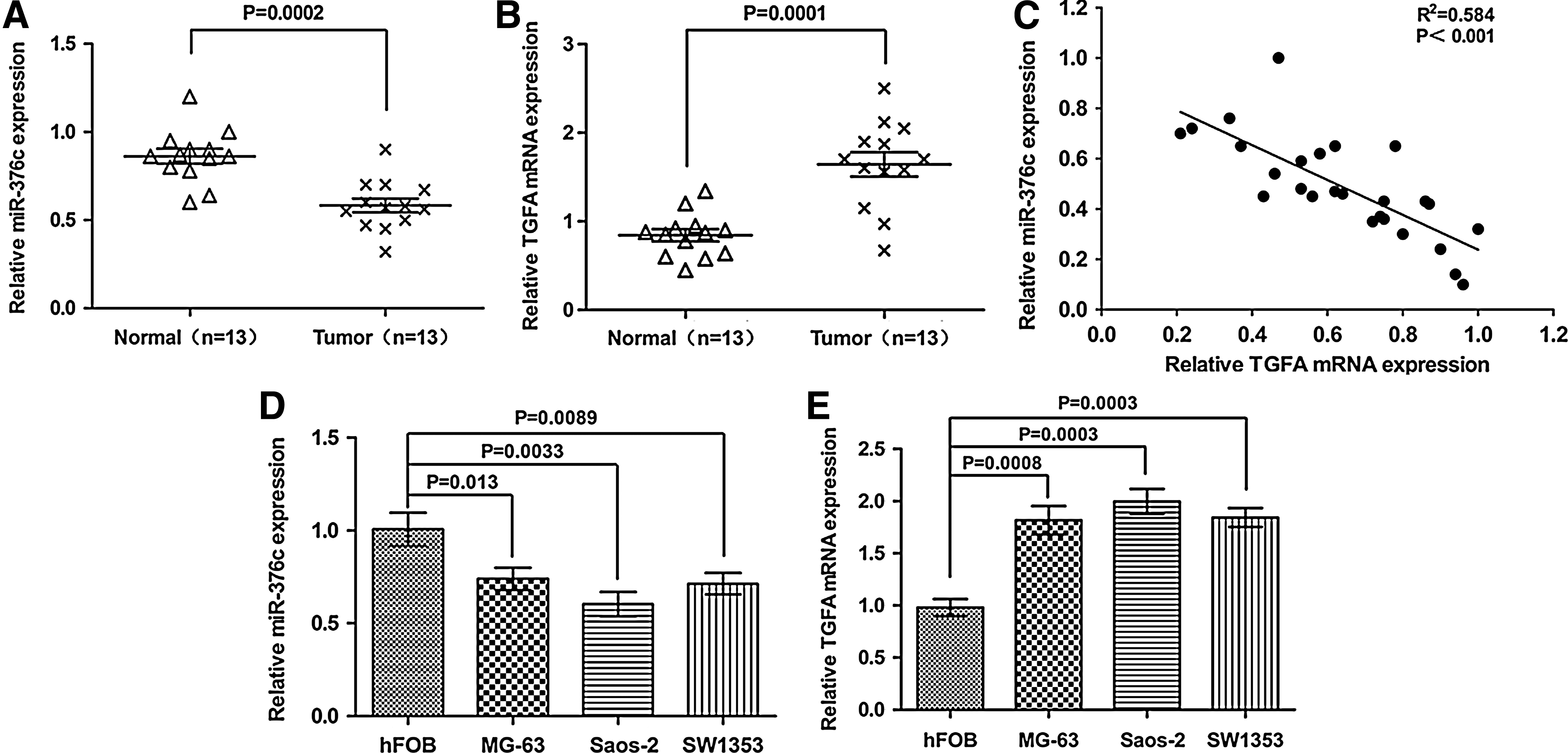
The transforming growth factor-alpha (TGFA) mRNA levels were negatively correlated with miR-376c in both primary osteosarcoma tissues and osteosarcoma cell lines.
MiR-376c directly targets TGFA
The combining sites of TGFA 3′UTR with miR-376c were predicted by Targetscan (
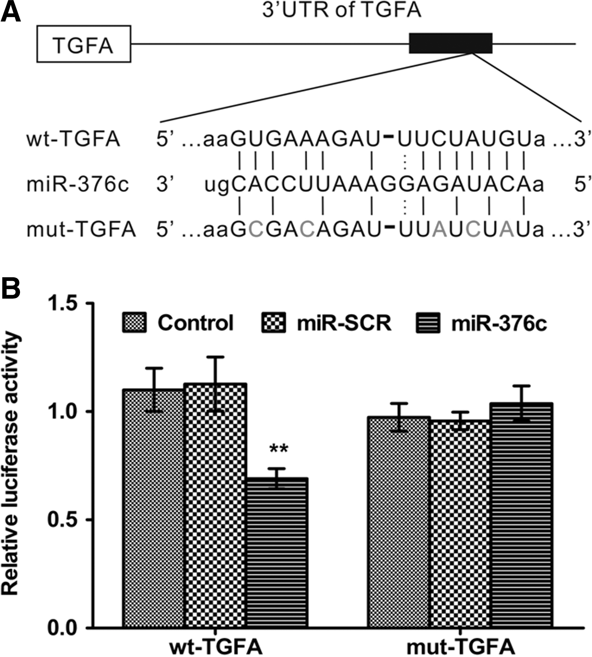
miR-376c directly targets TGFA by binding to its 3′UTR.
MiR-376c efficiently suppressed TGFA and EGFR
To further study the biological role of miR-376c in osteosarcoma tumor progression, we infected Saos-2 cells with lentivirus carrying miR-SCR and miR-376c. We routinely observed a highly efficient infection (>90%) 72 h after infection at multiplicity of infection of 10 through estimating EGFP expression under a fluorescent microscope (Fig. 3A). Quantitative RT-PCR showed that, at 72 h after infection, the expression of miR-376c was upregulated as compared with miR-SCR (Fig. 3B). Moreover, we observed the enhanced miR-376c in Saos-2 cells significantly repressed TGFA protein expression compared with cells transfected with scramble control by western blot (Fig. 3C, D). Meanwhile, apparent reduce of EGFR protein expression were also observed by western blot (Fig. 3E, F).
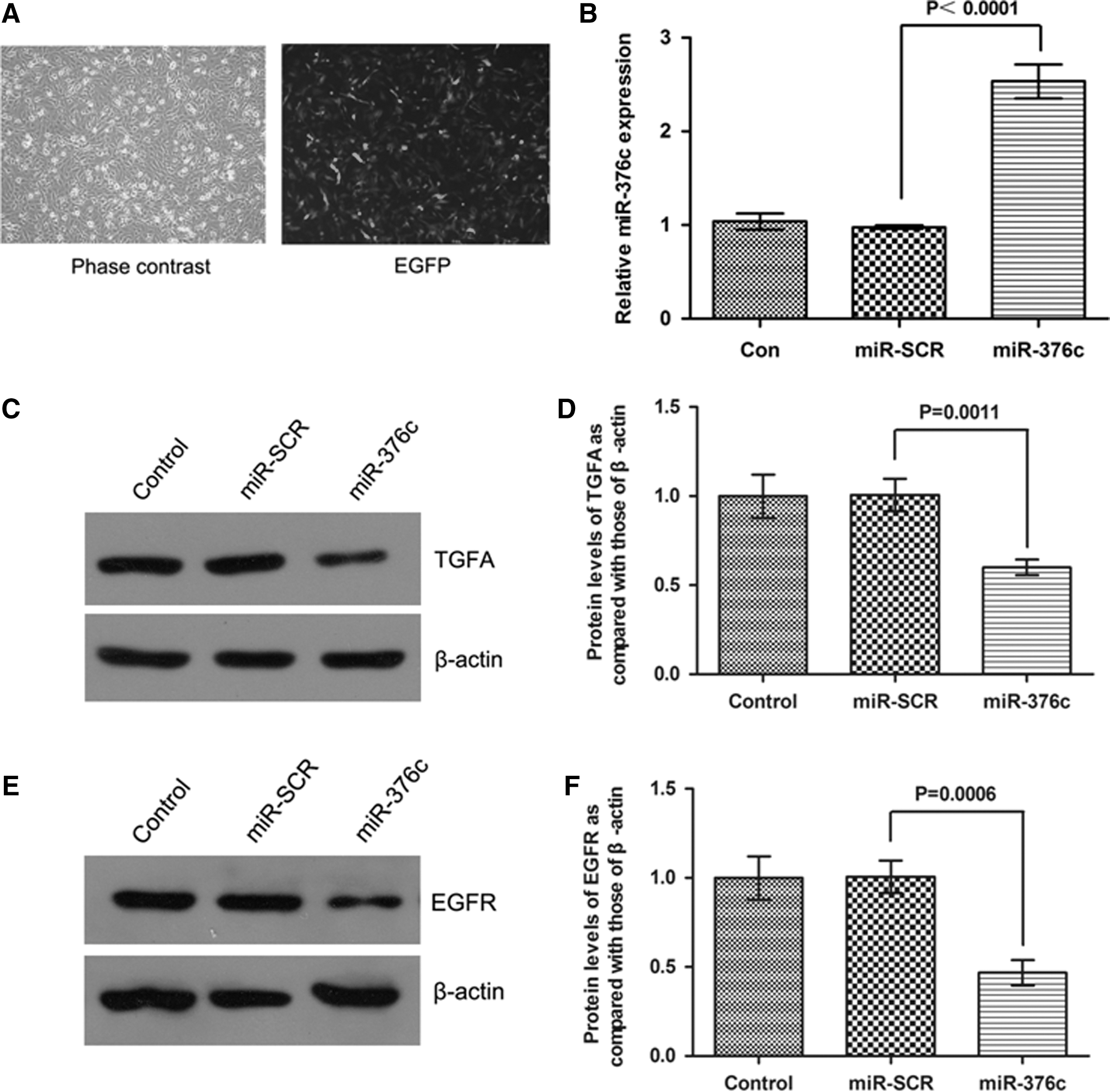
Overexpression of miR-376c inhibited TGFA and epidermal growth factor receptor (EGFR) expression.
Effect of miR-376c on osteosarcoma cell growth, migration, and invasion
To validate whether miR-376c regulates osteosarcoma cell growth, we performed a proliferation assay by transfecting miR-376c mimics or scramble control into Saos-2 cells. It showed that the increased expression of miR-376c induced significant inhibition on cell growth (Fig. 4A). As shown in Figure 4B, C, compared to the scramble control, miR-376c mimics transfected Saos-2 cells and exhibited significant impairment of migratory ability. The corresponding effect on invasive ability was also observed in parallel invasion assay.
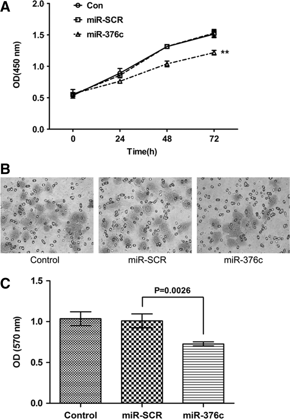
Effects of miR-376c on proliferation, migration, and invasion of Saos-2 cell lines.
Forced expression of TGFA increased osteosarcoma cell growth, migration, and invasion
To further confirm the potential relationship between miR-376c and the downstream gene TGFA, we tested cell growth and motility under the condition of overexpression of TGFA. Once TGFA expression was effectively upregulated by TGFA ORF clone (Fig. 5A), transfected Saos-2 cells exhibited increased cell growth (Fig. 5B), upregulated cell invasion ability (Fig. 5C, D), which was in contrast with the effects induced by upregulation of miR-376c. Taken together, these results indicated that miR-376c functions as a potent tumor suppressor through regulating TGFA expression.
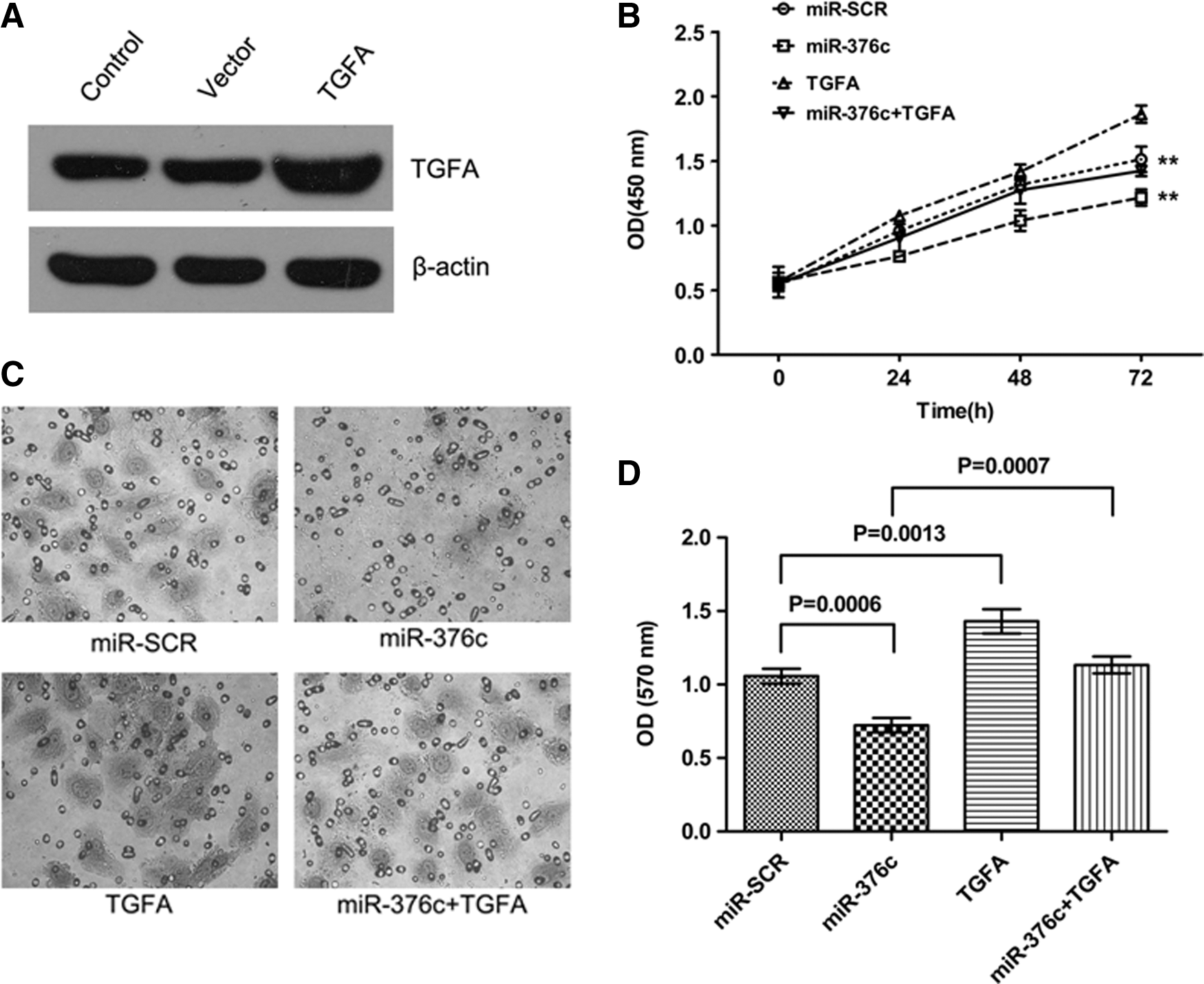
Overexpression of TGFA restored the inhibitory effects of proliferation, migration, and invasion of Saos-2 cell lines.
Forced expression of TGFA restored the effects of miR-376c
As we showed above, TGFA is a direct target of miR-376c. Therefore, we wondered whether forced expression of TGFA is enough to reverse the expression of miR-376c-inhibited downstream molecules. TGFA ORF plasmid was transfected into miR-376c- or miR-SCR-expressing cells. As shown in Figure 6, the decreased level of TGFA by miR-376c expression was rescued via the introduction of TGFA ORF clone. Similarly, ectopic expression of TGFA also restored miR-376c-inhibited EGFR expression in Saos-2 cells, indicating that miR-376c inhibits EGFR expression by targeting TGFA.

Overexpression of TGFA restored the inhibitory effects of miR-376c in TGFA and EGFR expression. Saos-2 cells stably expressing miR-SCR or miR-376c were transfected with TGFA ORF plasmid at 2 mg per well. After transfection for 48 h, the levels of specific proteins were analyzed by immunoblotting.
Discussion
Most deaths from cancer are caused by complications arising from metastasis. Therefore, targeting metastatic disease is a pivotal anti-cancer strategy. Studies on tumor invasion and metastasis have revealed the critical role of miRNA in these processes by the mechanism that miRNA could regulate a variety of genes pivotal for invasion or metastasis (Lim et al., 2005; Dalmay and Edwards, 2006). Quite recently, some miRNAs have been identified to promote (Gaziel-Sovran et al., 2011; Yang et al., 2011; Oneyama et al., 2012) or suppress (Li et al., 2009; Fang et al., 2011; Xu et al., 2012) tumor invasion or metastasis, providing potential therapeutic targets on anti-metastasis strategy.
In osteosarcoma, miR-199a-3p expression had a significant effect on osteosarcoma cell growth in vitro. Overexpression of miR-199a-3p by transfection significantly decreased osteosarcoma cell growth and migration (Duan et al., 2011). Besides, miR-199a-3p was reported to regulate mTOR and Met to influence the doxorubicin sensitivity in liver cancer cells (Fornari et al., 2010). In this study, we raised the hypothesis that miR-376c might be involved in osteosarcoma metastatic process. Finally, we confirmed the link between miR-376c and TGFA, a positive tumor metastasis-related gene, and found that miR-376c inhibited osteosarcoma cell invasion and migration via targeting TGFA. Our results obtained from quantitative RT-PCR validated that miR-376c was commonly downregulated in osteosarcoma cell lines, and in 10 out of 13 (76.9%) enrolled osteosarcoma patients, these results were consistent with previous studies. We restored miR-376c expression in Saos-2 cells and found that miR-376c inhibited cell proliferation and invasion. These findings suggested that miR-376c was involved in the processes of metastasis. Interestingly, the levels of miR-376c were negatively correlated with TGFA mRNA levels in osteosarcoma tissues. It was reported that TGFA was identified to be an independent prognostic indicator of metastasis formation and metastasis-free survival. Mechanically, TGFA transcriptionally promote EGFR signaling pathway (Vivacqua et al., 2009), which has been reported essential for tumorigenesis and metastasis. Our results confirmed a vital molecular relationship between miR-376c and TGFA/EGFR. We showed that, at both mRNA and protein level, upregulation of miR-376c expression in Saos-2 cells effectively suppressed TGFA and EGFR expression, and forced expression of TGFA could reverse the expression of EGFR. It suggested a potential inverse relevance of miR-376c and TGFA in osteosarcoma and the main effect of TGFA of the cells is autocrine affect, because the miR-376c downregulated the level of the cellular TGFA mRNA and protein. Additionally, these results suggested miR-376c might inhibit EGFR expression via TGFA signaling.
Further, by luciferase reporter assay study, we verified that miR-376c directly target TGFA gene through binding to specific complementary site within its 3′ untranslated region. These results verified that miR-376c inhibited the expression of EGFR via TGFA signaling. Taken together, these findings sufficiently consolidated that miR-376c played a suppressive role in cellular proliferation, migration, and invasion, at least, in part due to directly inhibiting TGFA expression. Activation of EGFR signaling pathway leads to multiple malignant processes, including cell growth, angiogenesis, cell motility, invasiveness, and metastasis (Vivacqua et al., 2009).
Our study revealed the inhibitory effect of miR-376c on TGFA, and partly elucidated a potential molecular mechanism by which miR-376c participated in osteosarcoma agrressiveness. More recently, Hurst et al. (Chan et al., 2005) proposed a novel field of cancer-related miRNAs termed metastamir that are associated with metastatic processes. For example, miR-21 is a mastermind of metastasis that promotes cell survival, migration, invasion, in vivo intravasasion, and metastasis (Asangani et al., 2008; Gregory et al., 2008; Wang et al., 2009), whereas miR-200 family is delinquent whose absence contributes to EMT phenotype (Park et al., 2008; Korpal et al., 2008). MiR-204 has recently been recognized as a direct post-transcriptional repressor of Snai1 mRNA and consistent with its postulated tumor suppressive role, it was found to be underexpressed in different cancer cell lines. Attenuated expression of miR-204 resulted in loss of cell–cell adhesion supporting the EMT-related properties of Snai1 (Wang et al., 2010). These metastamir represent potential candidate cancer prognostic markers and therapeutic targets for metastatic cancers. Our findings suggest that miR-376c could function as a metastamir via targeting TGFA.
In conclusion, we newly described miR-376c/TGFA link and provided a potential mechanism for TGFA deregulation and contribution to osteosarcoma cell proliferation and invasion. As a result, restoration of miR-376c expression could have an important implication for the clinical management of osteosarcoma.
Footnotes
Disclosure Statement
No competing financial interests exist.