




Abstract
Chronic rhinosinusitis with nasal polyps (CRSwNP) is one of the most frequently encountered chronic nasal diseases with a significant impact on patient quality of life. The aim of our study was therefore to investigate the association between the POSTN, IL-4, and IL-13 gene expression and the nasal polyp development. The objective of this study was to determine differential expression of POSTN, IL-4, and IL-13 genes in the mucosa and polyps of 63 patients with CRSwNP and 23 chronic rhinosinusitis (CRS) without nasal polyps (CRSsNP) when compared with patients with nasal septum deviation (n=18) who were used as controls. The expression level was investigated using reverse transcription–polymerase chain reaction assays in the polyp tissue and the mucosa of paranasal sinus collected while undergoing functional endoscopic sinus surgery. Expression of the mRNAs of all three genes, IL-4, IL-13, and POSTN, was significantly greater in the paired tissues of CRS patients with NPs or without NPs than in control subjects, with highest levels of POSTN and IL-13 seen in CRSwNP. An increased level of POSTN, IL-4, and IL-13 gene expression may be related to the development of chronic rhinosinusitis with nasal polyps, but polyp formation seemed to be associated especially with POSTN and IL-13 expression.
Introduction
T
Nowadays, it is important to identify new clinical biomarkers that enable more accurate diagnostics and monitoring of CRS disease activity. It may serve to develop new effective treatments based on the principles of personalized medicine. For this reason, in our study, we sought to further investigate expression of POSTN and its immunity mediators, IL-4 and IL13, in a large group of patients with CRS and to elucidate the differential expression between patients with CRSwNP and CRS without nasal polyps (CRSsNP) when compared with controls. Furthermore, we aimed to explore which one, IL-4 or IL13, stimulates the production of POSTN during CRS development.
Materials and Methods
All 104 subjects were treated surgically at the Department of Otolaryngology and Laryngological Oncology, Medical University of Lodz, in 2010–2012. The diagnosis of CRSwNP was based on the history, clinical examination, nasal endoscopy, and computed tomography according to the current EP3OS guidelines. A total of 63 patients with CRSwNP included 24 women and 39 men (mean age 48.7±13.3 years). The reference groups were as follows: patients with CRS without NPs (n=23, including 14 women and 9 men, mean age 46.4±13.2 years) and patients with nasal septum deviation (n=18, including 9 women and 9 men, mean age 42.8±13.9 years).
During surgery, samples of polyps and adjacent mucosa were taken from the same side of CRSwNP patients. Fragments of the mucosa from paranasal sinuses with CRS without NPs and fragments of the mucosa from the paranasal sinuses in patients with nasal septum deviation were collected during the planned endoscopic procedures. Immediately after resection, the tissue fragments were placed in test tubes with 1 mL of RNAlater (Qiagen) and frozen at −70°C until they were processed for RNA extraction. The studies were approved by the Bioethics Commission at the Medical University of Lodz (Decision no. RNN/40/09/KB of 6th January 2009), and consent of each patient was obtained.
RNA extraction and quantitative reverse transcription–polymerase chain reaction
RNA for reverse transcription–polymerase chain reaction (RT-PCR) was extracted from polyps and mucosa samples using the TriPure Isolation Reagent (Roche Diagnostics GmbH) according to the manufacturer's recommendations. Briefly, frozen tissue specimens (∼50 mg) were placed in 1 mL cold TriPure reagent and homogenized in a tissue homogenizer on wet ice (0–4°C); the time of homogenization was limited to 30-s intervals for up to 2 min and submitted to centrifugation at 20,000 g for 10 min at 4°C. Following the addition of 0.2 mL chloroform to the supernatants, the mixture was incubated at room temperature for 20 min with occasional stirring. The samples were then submitted to centrifugation again at 12,000 g for 15 min at 4°C. The resulting supernatants (colorless aqueous phase containing RNA) were precipitated by mixing with 0.5 mL isopropanol, incubated for 5–10 min at room temperature, and centrifuged as aforementioned. The pellet containing total RNA was washed with 1 mL ethanol (75%) and centrifuged at 7500 g for 5 min at 4°C. The RNA pellet was air-dried, and resuspended in RNase-free water (incubated at 55–60°C, 10–15 min).
First-strand cDNA was synthesized using a commercial RevertAid First Strand cDNA Synthesis Kit (Thermo Scientific) with Moloney murine leukemia virus reverse transcriptase. Synthetic oligonucleotide primers based on the cDNA sequences of POSTN, IL-4, IL-13, SDHA, and GAPDH were prepared as follows: for POSTN: 5′-GCC ATC ACA TCG GAC ATA-3′ and 5′-CTC CCA TAA TAG ACT CAG AAC A-3′; for IL-4: 5′-ATG GTT GCT GTC TCA TCA GC-3′ and 5′-ACG TAC TCT GGT TGG CTT C-3′; for IL-13: 5′-CCT CAA TCC TCT CCT GTT GG-3′ and 5′-TTT GGT GTC TCG GAC ATG C-3′; for SDHA: 5′-CTC CAT GTT CCC CAG AGC AG-3′ and 5′-GCA TTT GGC CTT TCT GAG GC-3′; and for GAPDH: 5′-TCT TTT GCG TCG CCA GCC GA-3′ and 5′-CCA GGC GCC CAA TAC GAC CA-3′ (Invitrogen). RNA (1 μg) and random hexamers (2.5 mM) were denatured at 65°C for 10 min and added to the reverse transcription mixture as instructed by the manufacturer. After incubation at 37°C for 1 h, cDNA from the reverse transcription mixture was subjected to PCR in a 10 μL volume containing 5 pmol of the primer pair and DreamTaq Green PCR Master Mix (Thermo Scientific) using a MultiGene™ Gradient thermal cycler (Labnet International, Inc.). The DNA was denatured for 5 min at 95°C, followed by 35 PCR cycles. Each cycle included a 30-s denaturation at 94°C, 30-s primer annealing at 57°C, and 45-s polymerization at 72°C. An 8 μL aliquot of each RT-PCR reaction mixture was analyzed by electrophoresis on a 1.5% agarose gel and stained with ethidium bromide (Fig. 1). The density of the ethidium bromide luminescence was measured by a Quantity One in Gel Doc 2000 (Bio-Rad). The results of gel densitometry were used to specify the relative amount of mRNA of the POSTN, IL-4, and IL-13. The level of analyzed genes was standardized against two references genes, GAPDH and SDHA.
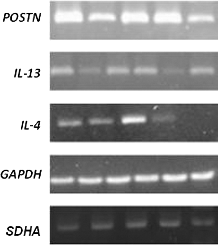
Polymerase chain reaction analysis of POSTN, IL-13, and IL-4 gene expression and two reference genes, GAPDH and SDHA, in nasal polyp tissue of CRSwNP patients with the products separated by electrophoresis on 1.5% agarose gels. CRSwNP, chronic rhinosinusitis with nasal polyps.
Statistic methods
Statistical analysis was performed using a commercial statistical software package STATISTICA 8.0. PL (StatSoft, Inc.). The distribution of the data was examined first by Shapiro–Wilk tests. The significance of the differences in the expression levels among the different groups was analyzed with the Mann–Whitney test. Spearman's rank correlation test was used to correlate the results of cytokines and POSTN expression in NPs and adjacent mucosa of CRSwNP patients. Significance was determined by the conventional 0.05 alpha level.
Results
The analysis of POSTN, IL-13, and IL-4 gene expression was performed in pairs of NP tissue and middle turbinate samples from the same side of CRS with NPs patients, in mucosa from paranasal sinuses patients with CRS without NPs, and in the mucosa from the paranasal sinuses of controls. We used two different reference genes (GAPDH and SDHA) to improve analysis of gene expression data. Expression of the mRNAs of all three genes, POSTN, IL-13, and IL-4, was significantly greater in the paired tissues of CRS patients with NPs or without NPs than in control subjects. The POSTN expression level was markedly higher in the NP tissues compared with the adjacent mucosa of those patients with CRS with NPs and mucosa of CRS patients without NPs, whereas no differences in the POSTN expression were observed while comparing the mucosa of these two groups of patients. These differences were observed in both reference genes, but they were more significant in the case of the SDHA gene (Table 1). Similarly, it was observed that the IL-13 mRNA level was significantly higher in polyps compared with adjacent mucosa of patients with CRS with NPs and without NPs. No differences in the IL-13 expression level were observed in comparisons of the mucosa of these two groups of patients when the expression was calculated relative to the GAPDH gene, whereas in the case of SDHA reference, the IL-13 mRNA level was significantly higher in mucosa of CRS with NPs than CRS without NPs (Table 2).The analysis of the IL-4 gene expression indicated a significantly higher level of this cytokine in the polyp tissue and nasal mucosa of the patients with CRS with NPs compared with the level in the nasal mucosa from those patients with CRS without NPs. However, the IL-4 expression did not differ between NP tissues and adjacent mucosa. The same differences were observed relatively in both reference genes, GAPDH and SDHA (Table 3).
CRSsNP, chronic rhinosinusitis without nasal polyps; CRSwNP, chronic rhinosinusitis with nasal polyps; DNS, nasal septum deviation.
Additionally, we aimed to identify which of the POSTN potential inductors, IL-4 or IL-13, is responsible for the production of POSTN in CRS. A significant correlation was detected between the quantitative expression of IL-13 and the POSTN expression in the polyp tissue (r=0.446, p=0.000216 relative to GAPDH, r=0.422, p=0.000502 relative to SDHA), but not in adjacent mucosa (r=0.133, p=0.291 relative to GAPDH, r=0.200, p=0.110 relative to SDHA) of the same patients using Spearman's rank correlation test (Fig. 2). We did not observe similar correlations between IL-4 and POSTN gene expression in polyp tissue and adjacent mucosa (r=0.242, p=0.0517 relative to GAPDH, r=0.179, p=0.154 relative to SDHA and r=0.0822, p=0.514 relative to GAPDH, r=0.085, p=0.494 relative to SDHA, respectively) (Fig. 3).
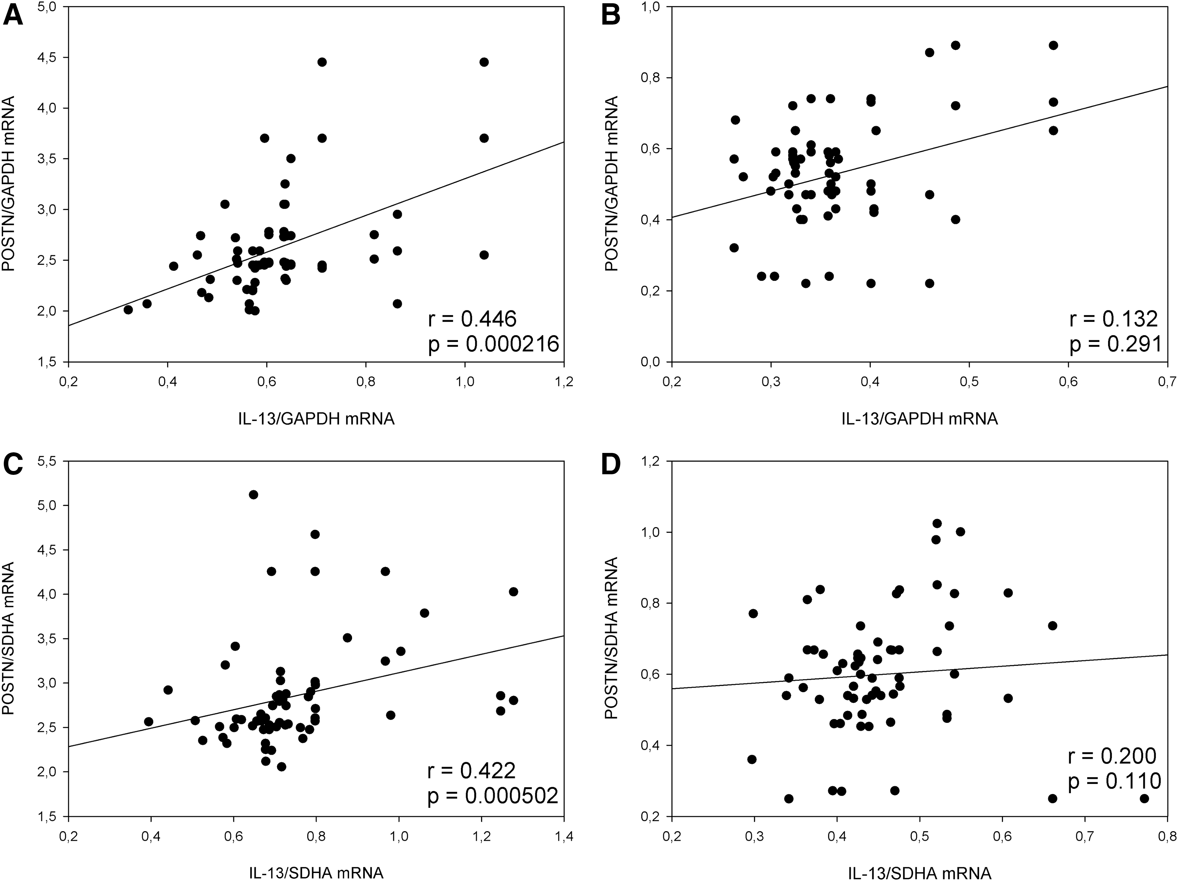
Correlation analysis between IL-13 and POSTN mRNA expression levels relative to GAPDH and SDHA in the nasal polyps
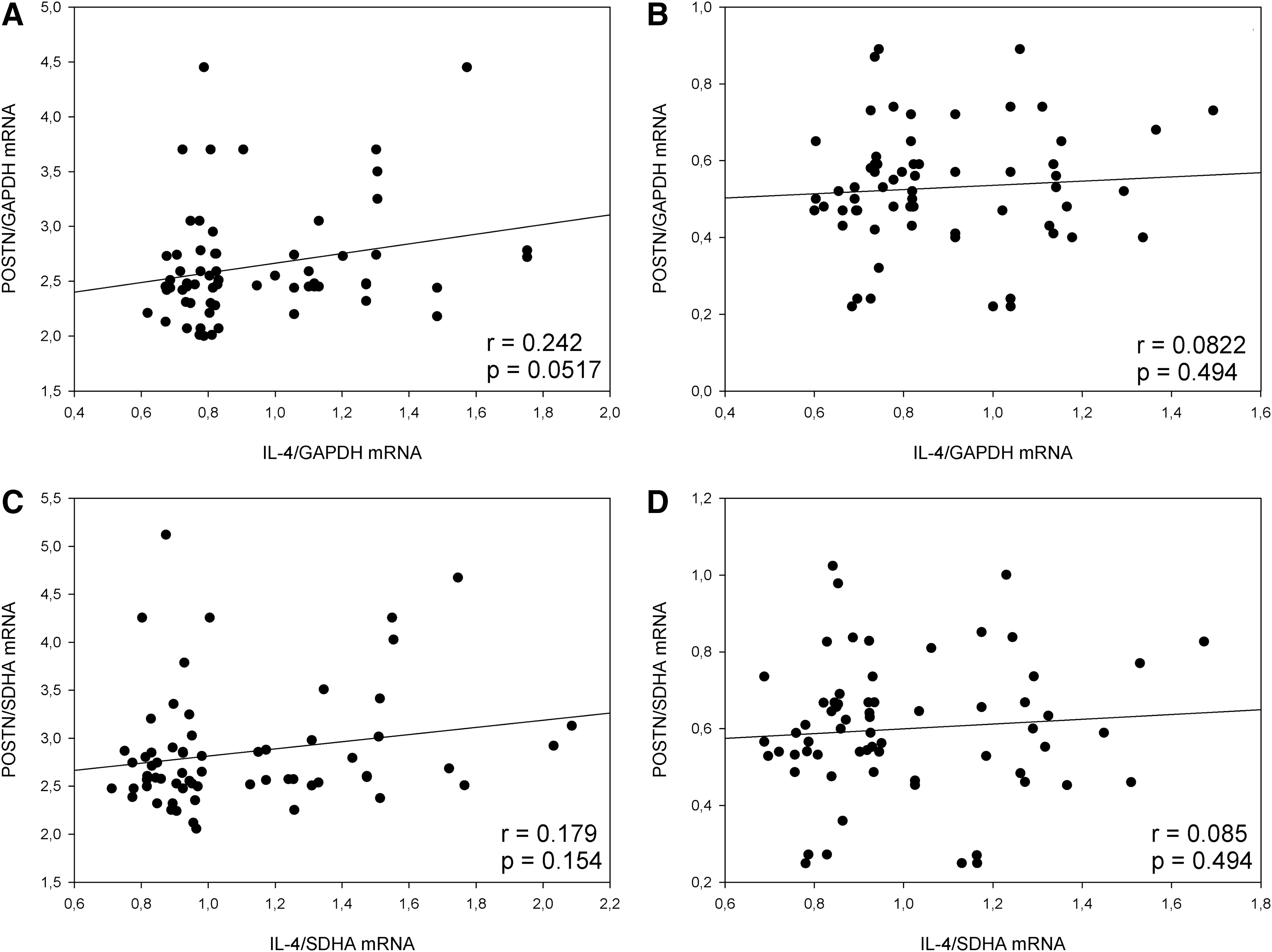
Correlation analysis between IL-4 and POSTN mRNA expression levels relative to GAPDH and SDHA in the nasal polyps
Discussion
The CRSwNP tissue is known to express high levels of multiple inflammatory genes as well as the ECM genes, which are potentially important, but relatively neglected, components of CRS mucosal immunity. In our study, we have shown that POSTN, IL-4, and IL-13 expression was significantly increased in the sinonasal tissue of patients with CRSwNP as well as with CRSsNP when we compared them to controls. However, we found that the pattern of POSTN, IL-4, and IL-13 expression in NPs and the paired adjacent mucosa and also in CRS mucosa is different, suggesting a diverse role of these inflammatory molecules in either type of CRS disease.
POSTN is observed in normal nasal tissue, but it is expressed more strongly in the basement membrane of patients, CRS with or without NPs, especially when they are complicated by allergic rhinitis and/or aspirin-induced asthma (Ishida et al., 2012). The Stankovic et al. study of comparing biopsy samples from patients with nasal polyps and biopsy samples from control subjects showed an increase in POSTN expression in diseased tissue, as did a second study comparing sinus samples with nasal floor of 15 CRS patients (Stankovic et al., 2008). Moreover, it was shown that both the POSTN gene expression and protein were elevated in active CRS and decreased following successful surgical and medical therapy (Zhang et al., 2012). In addition, it is recommended that measurement of POSTN in the sera and nasal lavage fluids may be useful for distinguishing patients with CRS from healthy controls (Kanemitsu et al., 2013). In this study, we observed that samples of tissues obtained from both CRS with NPs and without NPs showed markedly increased expression of the POSTN gene compared with control subjects, with highest levels of POSTN seen in CRSwNP. The increase in the expression of POSTN in CRS, with and without polyps, tissue is not surprising because POSTN plays multiple potential roles in epithelial structure and immune regulation. Several lines of evidence have suggested the importance of this matricellular protein in accelerating lung inflammation by enhancing chemokine production in fibroblasts, eosinophil recruitment, or TGF-beta activation in airway epithelial cells (Wen et al., 2010; Conway et al., 2011). What turned out to be more interesting in our analysis was that POSTN expression was greater in CRSwNP than CRSsNP, and especially that similar results were obtained by Zhang et al. in previous studies (Zhang et al., 2012). Taken together, these findings suggest that POSTN is associated with pathogenesis of CRS; however, it seems that POSTN acts in a completely different way in both subgroups of this disease. The CRS is related with remodeling, but distinct remodeling features differentiate both subgroups of CRS. The POSTN may be implicated as an important factor in remodeling processes through its effect on the activity of TGF-beta and MMPs.
The CRSsNP is characterized mainly by Th1-driven inflammation with high levels of active TGF-beta 1 signaling with subsequent excessive collagen deposition with thickening of the collagen fibers in the ECM and fibrosis formation (Daines et al., 2011). POSTN secreted by airway epithelial cells is able to activate TGF-beta-mediated and increase type I collagen production in fibroblasts, and it induces the release of TIMP-1, inhibiting the proteolytic activity of ECM. Therefore, persistent upregulation of POSTN in the sinonasal epithelium is likely to contribute to mechanisms of increased airway fibrosis in CRSsNP (Sidhu et al., 2010).
On the contrary, as far as CRSwNP is concerned, there is a low TGF-beta 1 protein concentration, a decreased expression of TGF-beta RII, and a low number of phospho-smad 2 positive cells, all of which indicate a low level of TGF-beta signaling in CRSwNP (Van Bruaene et al., 2009). In consequence, the POSTN in CRSwNP might induce the production of MMP-9, MMP-10, and MMP-13, but not TIMP-1, due to the relative lack of TGF-b1, resulting in degradation of the ECM and edema formation, which is the hallmark of nasal polyps (Watelet et al., 2004). Within the pseudocysts present in CRSwNP, the inflammatory cells showed positive staining for MMP-9, suggesting a direct degradative function (Watelet et al., 2004). Zhang et al. suggested that greater POSTN expression in CRSwNP than CRSsNP may be a result of this particular ECM destruction and more severe epithelial damage leading to activation of the healing process in the CRSwNP group (Zhang et al., 2012). Several studies have recently shown that POSTN has a physiological role in cutaneous wound repair. It was observed that delayed reepithelialization after a scratch wound is seen in a POSTN−/− knockout mouse model as opposed to the wild type (Nishiyama et al., 2011). In addition, in nasal tissue of CRS patients, stronger POSTN staining was most frequently seen in areas with epithelial hyperplasia or breaks in the epithelium (Zhang et al., 2012). Moreover, during tissue repair, POSTN helps secretion of MMP-9 and expression of Notch1 on the cell surface, activities which are probably regulated by the association of precursor MMP-9 and Notch1 with POSTN for effective proteolysis by a furin-like proteinase to generate their secreted mature forms (Kudo, 2011; Romanos et al., 2014).
Based on the knowledge that POSTN is a highly inducible product of IL-4 and IL-13, we wished to explore the potential interaction of these molecules with POSTN in CRS polyp formation. In CRSwNP patients, the presence of Th2 cytokines, which most importantly include IL-4, IL-5, and IL-13, is documented (Pietruszewska et al., 2008; Pawankar, 2001). The IL-4 and IL-13 are associated with eosinophilic inflammation and strongly support the survival and activation of eosinophils and eosinophilia through induction of eotaxin production. Furthermore, the increased presence of IL-4 and IL-13 can play a role in upregulating the vascular cell adhesion molecule (VCAM)-1, and this facilitates the further infiltration of eosinophils into the tissue (Kumagai et al., 2003; Provost et al., 2012). It was also shown that IL-4 exposure impairs sinonasal epithelial wound healing and may contribute to prolonged healing in Th2 inflammatory rhinosinusitis, while IL-13 did not significantly impair sinonasal epithelial wound resealing in vitro (Wise et al., 2013). Eosinophilia is believed to be the hallmark of CRSwNP establishment either in early or late phases, with TH-2 biased cytokines. Indeed, it was recently demonstrated that the three major T-effector cell cytokines, Th1, Th2, and Th17, described until recently can coexist in the upper airway mucosa within one patient; their pattern in nasal polyp tissue affects and differentiates mucosal inflammation, demonstrating a mixed osinophilic/neutrophilic activation pattern when present concurrently in these subjects, whereas polyp tissues from other subjects did not express any of the key T-cell cytokines (Van Bruaene et al., 2008). Moreover, the significantly elevated levels of IL-13 were found in a predominant pattern of neutrophilia instead of eosinophilia in nasal specimens from patients with CRSwNP (Nabavi et al., 2014). In another study, it was shown that the number of cells expressing IL-13 was significantly higher in subjects with CRS compared with normal controls independently on the allergy status. In contrast, the numbers of IL-13, but not IL-4, mRNA-positive cells were increased in the CRS subjects without atopic symptoms compared with the normal controls. Results of these investigations suggest that the relationship between the two cytokines IL-4/IL-13 and the polyp formation is not necessarily associated with the induction of eosinophilic inflammation (al Ghamdi et al., 1997; Ayers et al., 2011; Bhakta and Woodruff, 2011). In our study, similarly to POSTN, we found that the IL-13 expression level was markedly higher in the NP tissues compared with the adjacent mucosa. Additionally, IL-13 expression in NP tissue was significantly correlated with POSTN expression. It is possible that the locally increased levels of IL-13 lead to upregulation of POSTN and therefore to polyp growth.
Relatively little is known concerning the expression of IL-4 in sinus disease. Our studies have demonstrated elevated levels of IL-4 expression at the mRNA in CRSwNP in comparison with CRSsNP as well as control sinus tissue. However, the IL-4 expression did not differ between NP tissues and adjacent mucosa. Nabavi et al. observed that the serum level of IL-13 in the patients with CRSwNP was significantly higher than the controls, but IL-4 did not differ significantly between the two groups (Nabavi et al., 2014). On the other hand, the subjects with chronic hyperplastic eosinophilic sinusitis and NPs showed that the IL-4 was highly expressed in the tissue of the allergic subgroup when compared with either healthy controls or the nonallergic subgroup (Hamilos et al., 1995). Investigation of nasal secretions from subjects with CRS found higher levels of IL-4 protein when compared with controls (Riechelmann et al., 2005). In another study, it was shown that IL-4 transcripts were found to be high in the ethmoid sinus mucosa and nasal turbinate tissue of allergic CRS subjects (Kamil et al., 1998). Moreover, Steinke et al. have demonstrated elevated levels of IL-4 expression at the mRNA and protein levels in aspirin-exacerbated respiratory disease in comparison with control sinus tissue. In summary, reports of IL-4 elevation in CRS are less frequent; however, elevated levels of IL-4 have been reported in CRSwNP and allergy and also with aspirin-exacerbated respiratory disease (Steinke et al., 2012). Thus, IL-4 seems to play an important pathogenic role only in subgroups of CRSwNP with the AERD phenotype or when complicated by allergy.
Conclusion
Our findings suggest that an increased level of POSTN, IL-4, and IL-13 gene expression may be related to the development of CRSwNP, but the polyp formation seems to be associated especially with POSTN and IL-13 expression.
Footnotes
Disclosure Statement
No competing financial interests exist.