




Abstract
This study aimed to explore the efficacy of propofol to treat malignant pheochromocytoma (PCC) in vitro and in vivo. In vitro, PC12 cells were treated with different concentrations of propofol (0, 1, 5, and 10 μg/mL) for specific times followed by a MTT assay to detect cell proliferation. Transwell assays were performed to assess the function of propofol on the migration and invasion of PC12 cells, and flow cytometry to analyze cell apoptosis and cell cycle progression. Quantitative real-time polymerase chain reaction was carried out to analyze the expression level of mRNA (Bcl-2, Bax, and CyclinE). The levels of Bcl-2, Bax, CyclinE, FOXO1, FOXO3, Bim, procaspase-3, and active caspase-3 were determined by western blotting. In vivo, the effects of propofol on PCC tumor growth were detected by transplanted mouse model. Transferase dUTP nick-end labeling was performed to detect tissue cell apoptosis. The results indicated that propofol inhibited PC12 cell proliferation, prevented cell migration and invasion, and induced the apoptosis of PC12 cells in a dose- and time-dependent manner. Propofol treatment increased the expression of Bax and decreased that of Bcl-2. In addition, propofol significantly induced the G1/S phase arrest in PC12 cells, and the expression of Cyclin E was reduced. Moreover, the levels of FOXO1, FOXO3, Bim, procaspase-3, and active caspase-3 were enhanced by propofol treatment. In vivo, propofol treatment significantly reduced the PCC tumor growth and induced tissue cell apoptosis. In conclusion, propofol has potent anti-PCC activity in vitro and in vivo, and is a potential small-molecule drug for treating malignant PCC.
Introduction
P
However, it can also follow a course with such atypical symptoms as epigastric distress, nausea, and vomiting (Beattie and Heasman, 1958). An uncontrolled PCC may cause life-threatening hypertensive crisis and cardiac arrhythmia (Voros et al., 1996). Therefore, it is important for PCC to be specified in the preoperative period, and suitable pharmacotherapy to be implemented (Shinn et al., 2012; Björklund and Backman, 2017; Zhang et al., 2017). In recent years, research on PCC has drawn more and more attention. For instance, small activating RNA induces dsP53-285
Proteasome inhibitors were considered as a potential medical therapy for malignant
Propofol is an anesthetic. Recently, many studies have demonstrated that propofol has a variety of other effects, including possible anticancer actions. As the complexity of endoscopic surgery increases, the desire to use propofol and deep sedation are becoming more common in endoscopy kits. Targeted antitumor therapy has been considered a promising new treatment strategy for malignant PCC (Lowery et al., 2013; Mohammed et al., 2014; Fishbein, 2016).
This study aimed to investigate the effects of propofol on PCC in vivo and in vitro.
Materials and Methods
Materials
Rat PCC PC12 cells were obtained from American Type Culture Collection ATCC (Manassas, VA). Dulbecco's modified Eagle's medium (DMEM) and fetal bovine serum (FBS) were from Gibco. All primary antibodies (anti-β-actin, anti-cyclinE, anti-Bax, anti-CyclinE; anti-FOXO1, anti-FOXO3, anti-Bim, anti-procaspase-3, and anti-active caspase-3) were acquired from Cell Signaling Technology (Boston, MA).
Cell culture and treatment
Rat PCC PC12 cells were cultured in DMEM supplemented with 10% FBS and 1% penicillin/streptomycin (Sigma-Aldrich). Cells were incubated in a humidified incubator at 37°C with 5% CO2. Propofol was dissolved in dimethyl sulfoxide (DMSO) and stored at −20°C.
Rat PCC PC12 cells were treated in the following manner: (1) Cells were treated with different concentrations (0, 1, 5, and 10 μg/mL; 0 μg/mL as control) of propofol for 24, 48, and 72 h at 37°C with 5% CO2, and subsequently analyzed by the MTT assay. (2) Cells were treated with different concentrations (0, 1, 5, and 10 μg/mL; 0 μg/mL as control) of propofol for 48 h, and subsequently detected by flow cytometry and Transwell assay.
Western blotting
Following treatment, cells were collected and total proteins were extracted with 40 mM Tris–HCl (pH 7.4) containing 150 mM NaCl and 1% (v/v) Triton X-100, supplemented with protease inhibitors. The protein concentration was determined using the bicinchoninic acid protein assay (Pierce, IL). Equal amounts of protein were resolved on 10% sodium dodecyl sulfate–polyacrylamide gel electrophoresis gels, and then transferred to a polyvinylidene difluoride membrane (Millipore, MA). The blots were blocked with 5% skimmed milk in Tris Buffered Saline with Tween-20 (TBST) and then probed with antibodies. The blots were washed three times and subsequently incubated with horseradish peroxidase-conjugated secondary antibodies.
Immunoreactive bands were visualized using the enhanced chemiluminescence detection system, and images were captured with a Fuji LAS3000-mini imaging system (Fujiflm, Tokyo, Japan). Furthermore, the immunoreactive bands were quantified by ImageJ software (v1.41; NIH, Bethesda, MD). The protein levels of the stripes were normalized based on the gray value of β-actin.
RNA isolation and quantitative real-time polymerase chain reaction
Total RNA was extracted from the PC12 cells (blank and propofol groups) using TRIzol reagent. The concentration of RNA was detected by NanoDrop 2000. The RNA samples were stored at −80°C for future use. Then, cDNAs were synthesized with the miScript Reverse Transcription Kit (GmbH, Qiagen) according to the manufacturer's protocol. The QuantiFast SYBR Green PCR Kit (Qiagen) was used to perform quantitative real-time polymerase chain reaction (qRT-PCR) under a CFX Connect Real-Time System (Bio-Rad). Glyceraldehyde-3-phosphate dehydrogenase was used as the internal control. The 2−ΔΔCq method was applied for the quantification of relative gene expression. Primer sequences were obtained from GenScript (Nanjing) as required.
MTT assay
A total of 5 × 10³ cells were seeded into the wells of 96-well plates in triplicate and incubated for 24 h before treatment to determine cell growth. MTT (20 μL; 5 mg/mL; Sigma-Aldrich) was added to each well 24, 48, and 72 h after propofol treatment. Subsequent to incubation for 4 h, the MTT medium was removed, and 150 μL DMSO was added. After shaking for 15 min at room temperature, the optical density (OD) of each sample was determined with an Enzyme Immunoassay Instrument. The cell inhibition rate = (1 − OD value of treatment/OD value of control) × 100%.
Cell migration and invasion assays
The effects of propofol on cell migration and its invasive capability were detected using a 24-well Transwell plate (8-mm pore size; NY). Briefly, chamber inserts were coated with or without 200 mg/mL of Matrigel and dried overnight under sterile conditions. The cells (1 × 10⁴ cells) were suspended in different concentrations of propofol–1640 medium (0, 1, 5, and 10 μg/mL), and then seeded into the top chamber; 1640 medium supplemented with 20% FBS was added to the lower chamber. Following incubation for 48 h at 37°C, the top chambers were wiped with cotton wool to remove the nonmigratory or noninvasive cells and subsequently fixed with 100% methanol for 10 min. Following Hematoxylin–Eosin staining, the migratory and invasive cells on the underside of the membrane were counted at five random fields under a microscope (Olympus).
Flow cytometry analysis
Following treatment with propofol for 48 h, PC12 cells were harvested and washed with phosphate-buffered saline (PBS). The cells were subsequently fixed with 70% methanol at −20°C overnight and then washed with PBS twice. Then, the cells were stained with RNase A Propidium Iodide (PI), and flow cytometry was used to detect cell cycle distribution.
Cells were stained with Annexin V and PI to detect cell apoptosis (Apoptosis Detection Kit, BD Biosciences). After incubation for 15 min in the dark, apoptotic cells were detected by flow cytometry according to the manufacturer's protocol.
Establishment of a PCC mouse xenograft model
The present study was performed according to the principles and procedures of the National Institutes of Health's Guide for the Care and Use of Laboratory Animals. This study was approved by the Animal Care and Use Committee of the 3rd Affiliated Hospital of Harbin Medical University. A PCC mouse xenograft model was established as previously described (Lian et al., 2017). In brief, PC12 cells (1 × 10⁶ cells/100 μL) were subcutaneously implanted into the left posterior abdomen of 4- to 5-week-old female athymic nude mice. Vernier calipers were used to measure the volume of the tumor.
When the tumor volume reached 100–200 mm3 (∼8 days after injection), the mice were randomly divided into two groups (n = 5 per group). (1) Sham group: the mice were intraperitoneally injected with saline; (2) propofol group: the mice were intraperitoneally injected with propofol (35 mg/kg). Mice were treated every 2 days for 14 days. All mice were sacrificed at the end of the experiment, and the tumor was stripped and measured.
Terminal deoxynucleotidyl transferase dUTP nick end labeling assay
The tumors were taken out, fixed with 4% neutral-buffered paraformaldehyde, and then paraffin embedded. Paraffin blocks were cut into 4-μm sections and histologically examined after Hematoxylin and Eosin staining. Paraffin slices were deparaffinized and then rehydrated (Shim et al., 2017). The cell death detection kit was used for staining with terminal deoxynucleotidyl transferase dUTP nick end labeling. ProLong Gold Antifade Mountant was performed and DAPI was used to mount the samples.
Statistical analysis
All experiments were repeated at least for three times. Results were expressed as mean values ± SD. Statistical analysis was performed by Student's t-test. A level of p < 0.05 was considered to be significant.
Results
Propofol inhibited PC12 cell proliferation, migration, and invasion
The present study showed that propofol inhibited PC12 cell proliferation and prevented PC12 cell migration and invasion in a dose-dependent and time-dependent manner (Fig. 1). The results obtained from the Transwell assays showed that the number of migratory and invasive PC12 cells significantly decreased in propofol-treated groups (Fig. 2).

Effect of propofol on PC12 cell proliferation. PC12 cells were treated with different concentrations of propofol (0, 1, 5, and 10 μg/mL) for 24, 48, 72 h, respectively. Then the proliferation inhibition rate of cells from different groups was assessed by MTT assay. Data are reported as mean ± SD. *p < 0.05, **p < 0.01 versus control group.
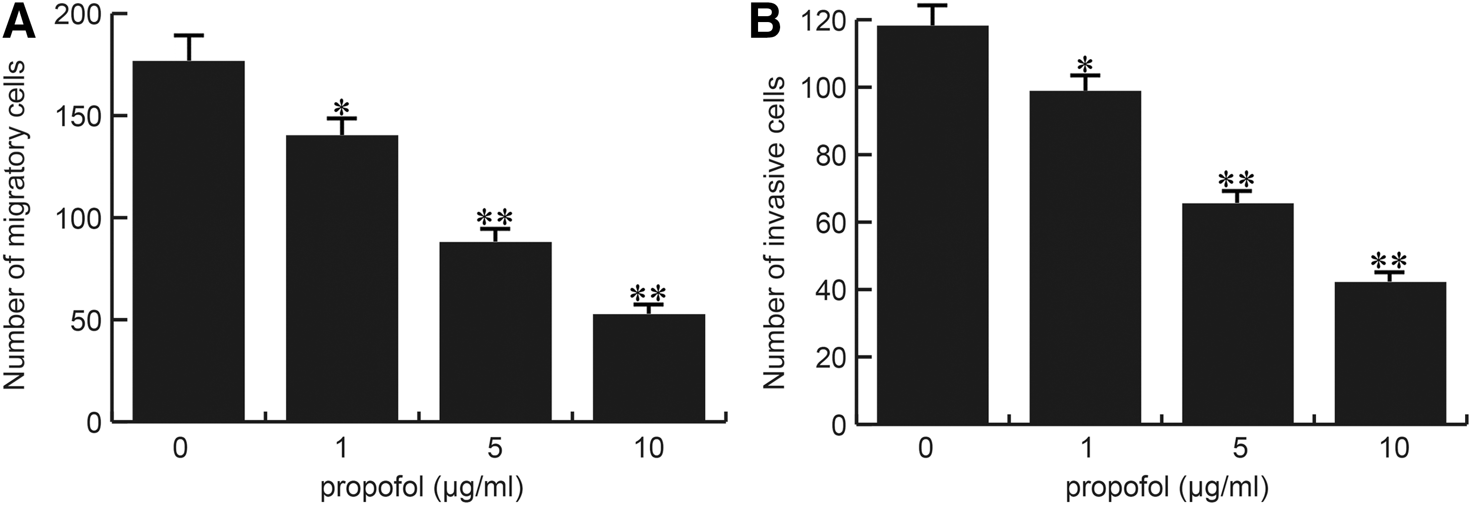
Effect of propofol on PC12 cell migration and invasion. PC12 cells were treated with different concentrations of propofol (0, 1, 5, and 10 μg/mL) for 48 h, respectively. Then the migration and invasion of cells in different groups were determined by transwell assay. Mean number of migrated and invasive cells was determined in triplicate.
Propofol induced cell cycle arrest in PC12 cells
The cell cycle analysis was used to explore the possible mechanism by which propofol inhibited PC12 cell proliferation. The result demonstrated that treatment with propofol for 48 h significantly increased the percentage of PC12 cells in the G1 phase and decreased that in the G2 phase in a dose-dependent manner (Fig. 3A, B). Meanwhile, the western blotting results showed that the expression of cyclin E, a cell cycle-associated protein, was blocked by propofol treatment (Fig. 3C, D).
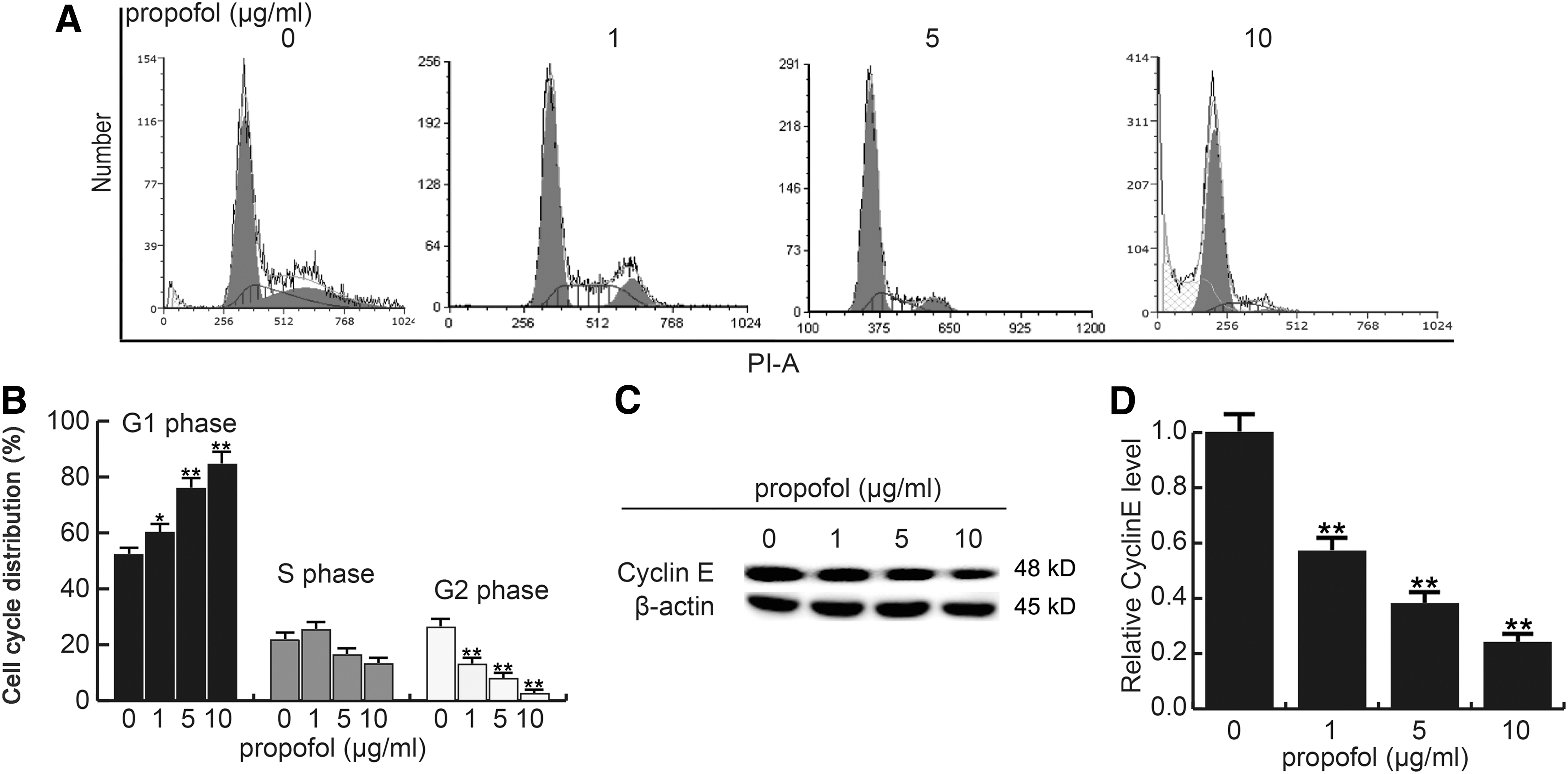
Effect of propofol on PC12 cell cycle. Forty-eight hours after treatment with propofol (0, 1, 5, and 10 μg/mL), PC12 cell cycle was determined.
Propofol induced apoptosis in PC12 cells
The effect of propofol on cell apoptosis was measured using the Annexin V-FITC/PI staining method. The results demonstrated that the apoptosis rate of PC12 cells significantly increased after 48 h of propofol treatment (Fig. 4A, B). Consistent with the results of apoptosis, the protein and mRNA expression of Bax increased and the Bcl-2 level decreased dose dependently in response to propofol treatment for 48 h (Fig. 4C, D). Thus, the findings of the present study clearly revealed that propofol induced apoptosis in PC12 cells.
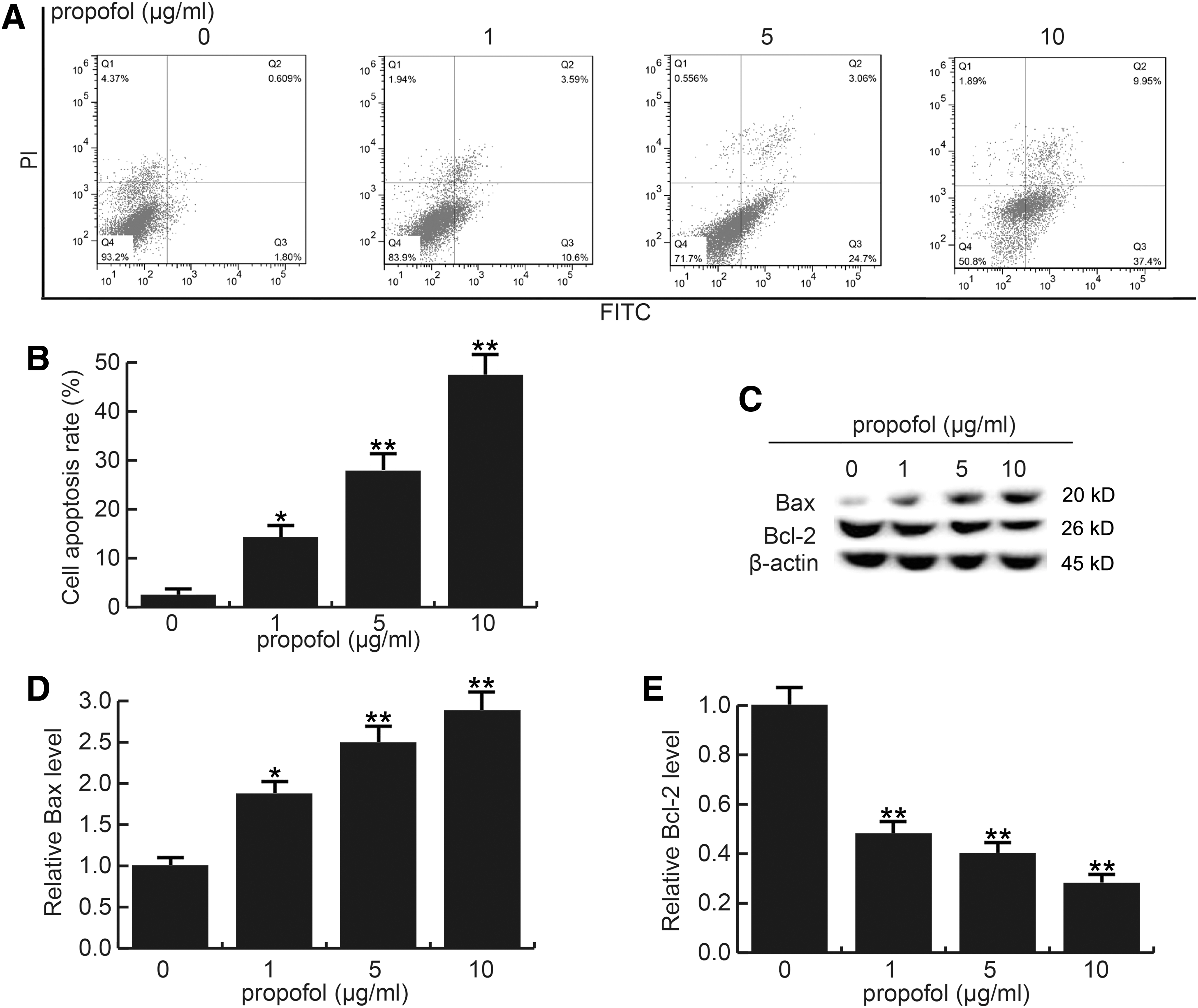
Effect of propofol on PC12 cell apoptosis. Forty-eight hours after treatment with propofol (0, 1, 5, and 10 μg/mL), PC12 cell apoptosis was measured.
Propofol enhanced the expression of FOXO1, FOXO3, Bim, procaspase-3, and active caspase-3 in PC12 cells
Studies have reported that FOXO transcription factors play critical roles in regulating various cell functions such as cell survival, cell proliferation, apoptosis, DNA repair, and metabolic processes (Furukawa-Hibi et al., 2005; Paik et al., 2007; Liu et al., 2009). FOXO is involved in cell growth and apoptosis by directly inducing FOXO3a-dependent apoptotic protein Bim expression (Gilley et al., 2003) and by activating caspase family (Brunet et al., 1999). A previous study showed that propofol significantly increased the protein levels of FOXO1, FOXO3, Bim, procaspase-3and activated caspase-3 in lung cancer cells (Yang et al., 2017). Therefore, in the present study, we explored whether propofol could affect the protein levels of FOXO1, FOXO3, Bim, procaspase-3, and activated caspase-3 in PC12 cells. Our data indicated that the expression levels of FOXO1, FOXO3, Bim, procaspase-3, and active caspase-3 were found to increase in a dose-dependent manner in response to propofol treatment for 48 h (Fig. 5).
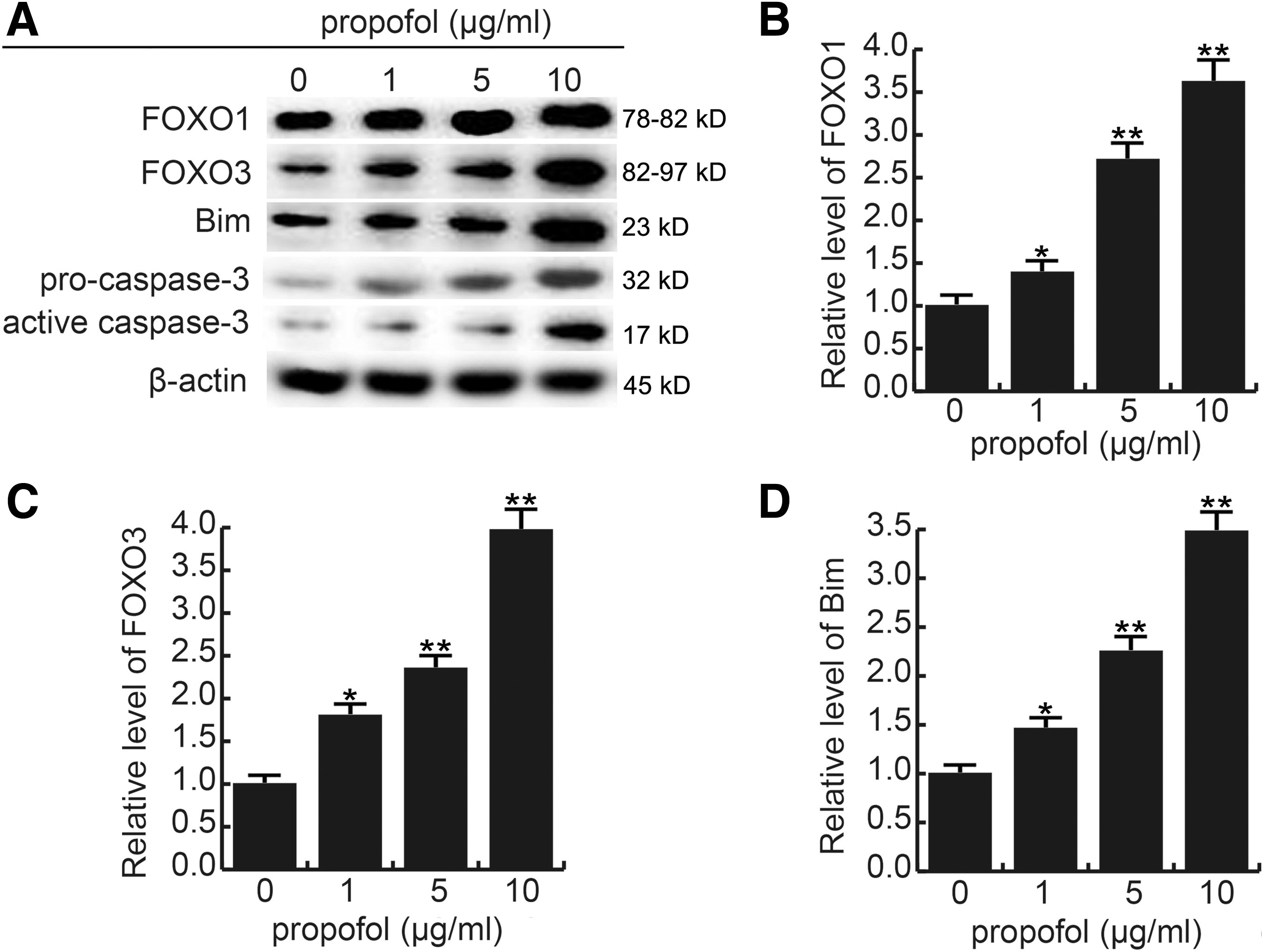
Effect of propofol on FOXO1, FOXO3, Bim, procaspase-3, and active caspase-3 expression in PC12 cells. Forty-eight hours after treatment with different concentrations of propofol (0, 1, 5, and 10 μg/mL), mRNA
Propofol reduced the growth of PCC tumors in a PC12 xenograft model
A previous study indicated that propofol has potential anticancer activity in vitro, and the present study hypothesized that propofol could inhibit PCC tumor growth in vivo. A PC12 cell xenograft model was established in this study. The PCC tumor volume and tumor weight were detected 14 days after treatment, and the results showed that the PCC tumor volume and weight of the propofol treatment group were significantly lower than that of the sham group (Fig. 6A–C). Furthermore, propofol was found to significantly induce cell apoptosis in PCC tumors compared with the sham group (Fig. 6D).
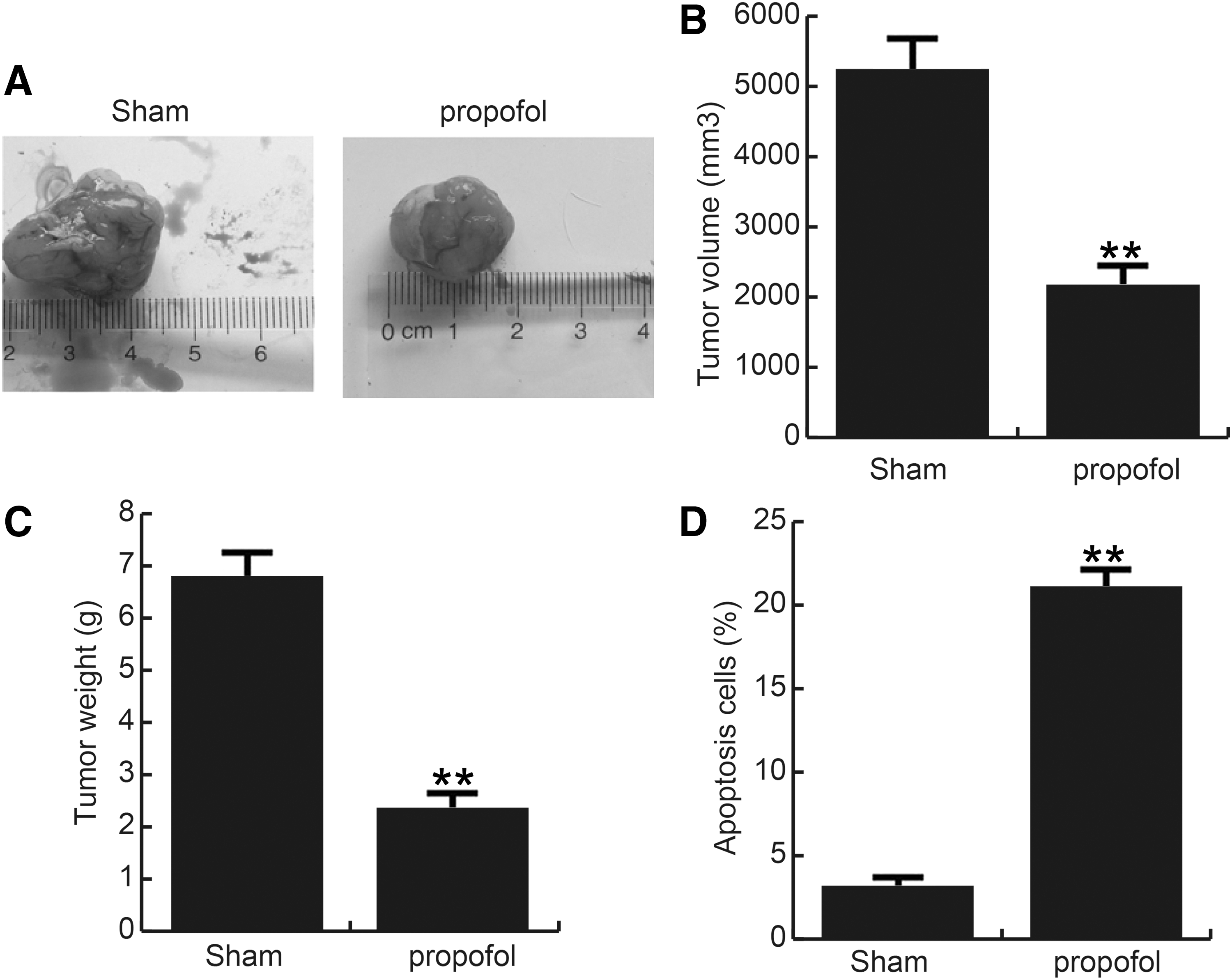
Propofol inhibits the growth of pheochromocytoma tumors.
Discussion
Due to the low incidence of PCC tumors and its complex pathogenesis, the treatment of malignant PCC is quite limited (Nilsson et al., 2009; Lian et al., 2017). Therefore, a few effective chemotherapeutic drugs are used to treat malignant PCC (Casaccia et al., 2017; Caza et al., 2017). Finding a new effective diagnosis and treatment method has been a hot issue in the study of malignant PCC. Chromogranin A and plasma-free metanephrine or normetanephrine determined by radioimmunoassay have been considered as the effective biomarkers of PCC or paraganglioma (Bílek et al., 2017). The report of Yu (2017) suggested that the use of proteasome inhibitors may be a potential medical therapy for malignant PCC. Bortezomib alone or in combination with salinosporamide A can promote
Studies have shown that propofol has not only an anesthetic effect but also an anticancer function. Propofol is one of the most common intravenous anesthetics used in the tumor resection operation (Siddiqui et al., 2005; Spangenberg et al., 2008; Venook et al., 2010). The inhibitory effect of propofol on the growth and metastasis of tumor cells and its effect on tumor immunity have been gradually noticed (Wang et al., 2015; Liu et al., 2016). The purpose of this study was to investigate the effect of propofol on malignant PCC in vivo and in vitro.
This study demonstrated that propofol could effectively suppress tumor growth and induced cell apoptosis in malignant PCC in vitro and in vivo. In view of the effect of propofol drug, an alternative therapy for malignant PCC may be considered using propofol alone or combined with other methods. First, the proliferation ability of PC12 cells was found to be significantly inhibited by propofol in a dose- and time-dependent manner. In addition, the data suggested that propofol dose dependently repressed PC12 cell migration and invasive capability. Furthermore, the findings of this study indicated that propofol prevented PC12 cell proliferation and metastasis through inducing cell cycle arrest and cell apoptosis through regulating cell growth and cell apoptosis-related genes, which were consistent with previous studies (Hsu et al., 2017; Meng et al., 2017; Yang et al., 2017).
Finally, a PC12 xenograph model, which has been used in a variety of studies on PCC, was established to perform our in vivo study, and the results from animal experiment were consistent with results from cell experiments.
PC12 cells are widely used in in vitro studies of neurological diseases. However, using PC12 cell xenograft model to study the PCC in vivo has some limitations. (1)
Taken together, this study showed that propofol effectively inhibited PCC cell proliferation, migration, and invasion; induced cell cycle arrest and apoptosis; and suppressed PCC tumor growth in the xenograft mouse model. In addition, these effects were mediated by the downregulation and inactivation of proteins involved in cell survival and cell apoptosis. Propofol is a promising new chemotherapeutic small molecule for treating malignant PCC.
Conclusion
Propofol prevents the progression of malignant PCC in vitro and in vivo, and it is a promising new chemotherapeutic small molecule for the treatment of malignant PCC.
Footnotes
Disclosure Statement
No competing financial interests exist.