
Abstract
Substantial research has revealed that peroxisome proliferator-activated receptor-gamma (PPARG) plays a critical role in glucose homeostasis and lipid metabolism, and recent studies have shown different effects in the progression of different tumors. However, the role of PPARG and its target gene in clear cell renal cell carcinoma (ccRCC) are incompletely understood. Clinical data revealed abnormal glucolipid metabolism in primary ccRCC samples. In addition, transcriptional profiling indicated that PPARG expression was positively correlated, whereas Six2 expression was negatively correlated with the overall survival of ccRCC patients. Staining showed that PPARG was mainly expressed in tumor cell cytoplasm, and Six2 was localized to the nuclei. In a ccRCC cell line, PPARG activation promoted cell apoptosis, inhibited cell migration and proliferation, and reduced Six2 expression. Mechanistically, overexpressing Six2 downregulated E-cadherin expression and cell apoptosis, but PPARG activation reversed those effects. Taken together, PPARG promotes apoptosis and suppresses the migration and proliferation of ccRCC cells by inhibiting Six2. These findings reveal that the PPARG/Six2 axis acts as a central pathobiological mediator of ccRCC formation and as a potential therapeutic target for the treatment of patients with ccRCC.
Introduction
Renal cell carcinoma (RCC) is among the 10 most common cancers in both men and women worldwide. Every year, >60,000 new kidney cancer cases are diagnosed (CEUR-WS, 2018). It is also the most common type of kidney cancer and accounts for nearly 90% of cases (National Comprehensive Cancer Network®, 2018). The clear cell RCC (ccRCC) is the major histomorphological subtype that contributes to the majority of deaths from kidney cancer. Despite noticeable advances in partial or completely resection, ablation, and active surveillance, disease recurrence and metastasis cannot be prevented in nearly 30% of patients suffering from nephrectomy (Duran et al., 2017; Farber et al., 2017 Hsieh et al., 2017). Thus, ccRCC remains lethal, and exploring the genomic and molecular mechanisms of ccRCC is necessary.
Peroxisome proliferator-activated receptor-gamma (PPARG) is a ligand-activated transcription factor that belongs to the nuclear receptor superfamily. PPARG is part of a subfamily with PPARα and PPARβ/δ. PPARs predominantly regulate lipid metabolism and adipogenesis. Thiazolidinediones are potent insulin sensitizers that act as activating ligands, and rosiglitazone (RGZ) is one of the common ligands for PPARG (Ahmadian et al., 2013; Grygiel-Gorniak, 2014). Several researchers have recently aimed to elucidate its significant role in lipid metabolism, energy homeostasis, immunity, and inflammation, although the function of PPARG in cancer progression remains controversial (Sabatino et al., 2012). Many studies have indicated that PPARG may serve as a tumor suppressor. In endocrine-resistant breast cancer, anti-PPARG therapy is a potential strategy to improve the curative efficacy of estrogen-induced apoptosis (Fan et al., 2018). In human oral cancer cells, inducing PPARG by the binding ligand can result in growth arrest (Wang et al., 2016). Conversely, when neuroblastoma SK-N-SH cells are used to construct a metastatic xenograft mouse model, the effect of the PPARG agonist is not obvious (Krieger-Hinck et al., 2010). In ccRCC, PPARG mRNA and protein levels are abundant (Sanchez et al., 2018). However, the role of PPARG in ccRCC has not yet been elucidated.
Sine oculis-related homeobox 2 (Six2) belongs to the Six family, which is composed of six homeobox genes (Six1–Six6) (Armat et al., 2016; Xu et al., 2016). Six2 is a key gene during embryonic development whose main function is maintaining the self-renewal of renal progenitor cells. Deletion of Six2 caused premature differentiation of mesenchymal cells (Mao et al., 2017; Combes et al., 2018). Recent studies have highlighted Six2 in Wilms tumor (WT). In a human WT cell line, Six2 enhanced cell survival by regulating the WNT/β-catenin pathway (Pierce et al., 2014). A role of Six2 in other cancers has also been explored. It is known that loss of E-cadherin is related to malignancy, and recent studies have proposed that Six2 promotes breast cancer and hepatocellular cancer through epigenetic control of E-cadherin (Wang et al., 2014; Li et al., 2018). However, the role of Six2 in ccRCC is still unclear.
In this study, we demonstrated that PPARG is a novel regulator of Six2 in ccRCC and explored their relationship with E-cadherin; our results complement this study in ccRCC.
Materials and Methods
Patient information
The basic patient information of 152 subjects come from the First Affiliated Hospital of Chongqing Medical University (Yuzhong, Chongqing, China), including 102 ccRCC patients and 50 healthy controls. Diagnosis was made based on pathological and cytogenetic analysis according to the 2016 World Health Organization criteria. The healthy controls had no ccRCC or other clinical signs of disease. To analyze each group of clinicopathological parameters, we constructed a table corresponding to Fuhrman grades I, II, and III (Table 1). We also compared the main glucolipid metabolism features of ccRCC patients with those of healthy controls. The diagnostic procedures were completed before this study was performed. During the analysis, the researchers were fully blinded to the patients' data.
Summary of Clinicopathological Parameters
Immunohistochemistry
Paraffin-embedded tissues were a gift from Chongqing Medical University. After deparaffinizing, dehydrating, and immersing in sodium citrate buffer (pH 6.0), the slides were blocked with blocking solution (Zhongshan Golden Bridge Bio-technology, China) and treated with primary antibodies (Six2 [1:100; Proteintech, China], PPARG [1:100; Wanleibio, China]) at 4°C overnight. Next, Poly-horseradish peroxidase anti-rabbit IgG (Zhongshan Golden Bridge Bio-technology) was added to the slides. The slides were stained with diaminobenzidin for visualization (Zhongshan Golden Bridge Bio-technology), and counterstaining was performed with hematoxylin. The specimens then underwent a repeated dehydration step. Histological sections incubated with phosphate-buffered saline (PBS) were used as a negative control. This study was approved by the ethical committee of the Chongqing Medical University, and the approved protocol was followed in the supplemental materials.
Cell culture and transient transfection
The ccRCC cell lines, 786O cells, and OSCR2 cells were cultured in RPMI-1640 Medium (Gibco, Grand Island, NY) supplemented with 10% fetal bovine serum (Gibco) and 1% penicillin/streptomycin (Gibco). ccRCC cells were cultured under conditions of 37°C and 5% CO2. Lipofectamine 2000 reagent (Invitrogen, Grand Island, NY) was used for siRNA transfection according to the manufacturer's instructions.
Plasmid construction
The coding sequence of Six2 was cloned from cDNA by polymerase chain reaction (PCR) with the following primers: CMVHIS-GFP-Six2 (F: 5′-GGATCCGAATTCATCTCCATCTCCATGCTGCCCACCTTCGGC-3′; R: 5′-ATGGTGGTGCTCGAGGATCCTCGGGTCCAGGTGCTCCAA-3′). The target fragment was ligated into the expression vector (CMVHIS-GFP; Invitrogen, Carlsbad, CA) at the site of BamHI and EcoRI by ligation-independent cloning. Finally, all plasmids were verified by sequencing. The siRNA was purchased from GenePharma (Shanghai, China).
Lentivirus packaging and infection of cells
Lentivirus packaging was based on a laboratory system. First, one expression vector (CMVHIS-GFP-Six2/CMVHIS-GFP-Control) and two packaging vectors (pMD2.G and psPAX2) were transfected into HEK 293 T cells. After 48 h, the supernatant containing the recombinant lentiviruses was collected and filtered. The supernatant was added to six-well plates containing WT 786O or OS-RC-2 cells, and 8 mg/mL polybrene was added to each well. After 24 h, the puromycin (Invitrogen) concentration should reach 10 mg/mL. One to 2 weeks were required to select stable cell lines.
Scratch wound healing assay
786O cells and OS-RC-2 cells were seeded in six-well plates in complete medium overnight. When the cell density reached nearly 90%, a wound was made by creating a perpendicular scratch with a 200 μL pipette tip. After washing the wound track three times with PBS, cells were cultured in serum-free medium in the absence or presence of 50 μM RGZ. Photographs were taken at 0, 12, and 24 h with a fluorescence microscope (ECLIPSE Ti-s; Nikon, Tokyo, Japan) and a POT Diagnostic (Sterling Heights, Michigan) CCD camera. Scratch width was assessed by NIH ImageJ software (National Institutes of Health, Bethesda, MD).
5-Ethynyl-21-deoxyuridine assay
786O and OS-RC-2 cells were incubated in 96-well plates treated with DMSO or RGZ for 24 h, and 5-ethynyl-21-deoxyuridine (EdU) kit (RiboBio, China) was then used according to the manufacturer's instructions. Photographs were taken at 24 h with a fluorescence microscope (ECLIPSE Ti-s; Nikon) and a POT Diagnostic CCD camera.
Flow cytometry assay
786O and OS-RC-2 cells were treated with 50 μM RGZ for 24 h. The apoptosis rate was detected by an Annexin V-FITC/7-AAD or PI staining (KeyGEN BioTECH, China). After another 72 h, the cells were collected into centrifuge tubes and washed three times. The remaining procedures were completed in the flow cytometry room, College of Life Sciences, Chongqing Medical University.
RNA extraction and real-time PCR
Total RNA was extracted from 786O and OSRC2 cells after Trizol® (Invitrogen) methods' protocol. Two microgram total RNA was used to reverse transcription by using the First Strand cDNA Synthesis kit (Thermo Fisher Scientific, Waltham, MA) based on the manufacturer's instruction. The 1 μL cDNA, which is taken as a template, was mixed into standard PCR reactions containing Ultra SYBR Mixture (Com Win Biotech, Bei Jing, China). And the expression levels of Six2 with 18S mRNA. List of primers used for real-time PCR: F: 5′-GCCGAGGCCAAGGAAAGGGAG-3′; R: 5′-GAGTGGTCTGGCGTCCCCGA-3′.
Western blot
The 786O and OSCR2 cell proteins were extracted by RIPA lysis buffer (Beyotime, Haimen, China). The sample loading quantity was determined by a BCA protein assay reagent kit (Thermo Scientific, Waltham, MA). The protein (30 μg) was separated onto SDS-PAGE gels and then transferred onto PVDF membranes. Then, the membranes were blocked for 2 h by 5% skimmed milk powder solution dissolved in TBST. After blocking, the membranes were incubated with primary antibodies for E-cadherin (1:1000; Bioworld, China), N-cadherin (1:1000; Bioworld), Six2 (1:1000; Proteintech), PPARG (1:500; Wanleibio), and β-actin (1:5000; CWBIO, Guangzhou, China). The membranes were washed three times with TBST and exposed to HPR-conjugated goat anti-mouse IgG and goat anti-rabbit IgG (1:5000; CWBIO). Bands were visualized through a chemiluminescent substrate (Merck, Billerica, MA).
Statistical analysis
All experiments were repeated independently three times. The results are presented as the mean ± SEM. Significant differences were measured by GraphPad Prism 5 (GraphPad, Inc., La Jolla, CA).
Results
Six2 expression is increased in ccRCC tissues
We first collected the basic clinical information of 102 ccRCC patients and 50 healthy subjects from the First Affiliated Hospital of Chongqing Medical University. Table 1 shows the detailed clinicopathological characteristics of the ccRCC patients. PPARG activation mainly regulates adipogenesis and insulin sensitization. As shown in Table 2, glucolipid metabolism features, total cholesterol, high-density lipoprotein cholesterol, and glucose were higher in ccRCC patients than in healthy controls. The PPARG signaling pathway mainly regulates adipogenesis and insulin sensitization. A KM-plotter assay of PPARG and Six2 in the cancer genome atlas kidney renal clear cell carcinoma (TCGA-KIRC) data set, which contains expression data from 531 ccRCC samples, indicated that PPARG expression was positively correlated with the overall survival of ccRCC patients, but Six2 expression was negatively correlated with overall survival (Fig. 1C, D). Next, we detected the expression of PPARG and Six2 in ccRCC patients (Table 3). Compared with adjacent nontumor tissues, Six2 expression was markedly increased in ccRCC tissues. No difference was observed in kidney PPARG expression between the two groups (Fig. 1A, B).Together, our data suggest that Six2 may contribute to ccRCC progression.
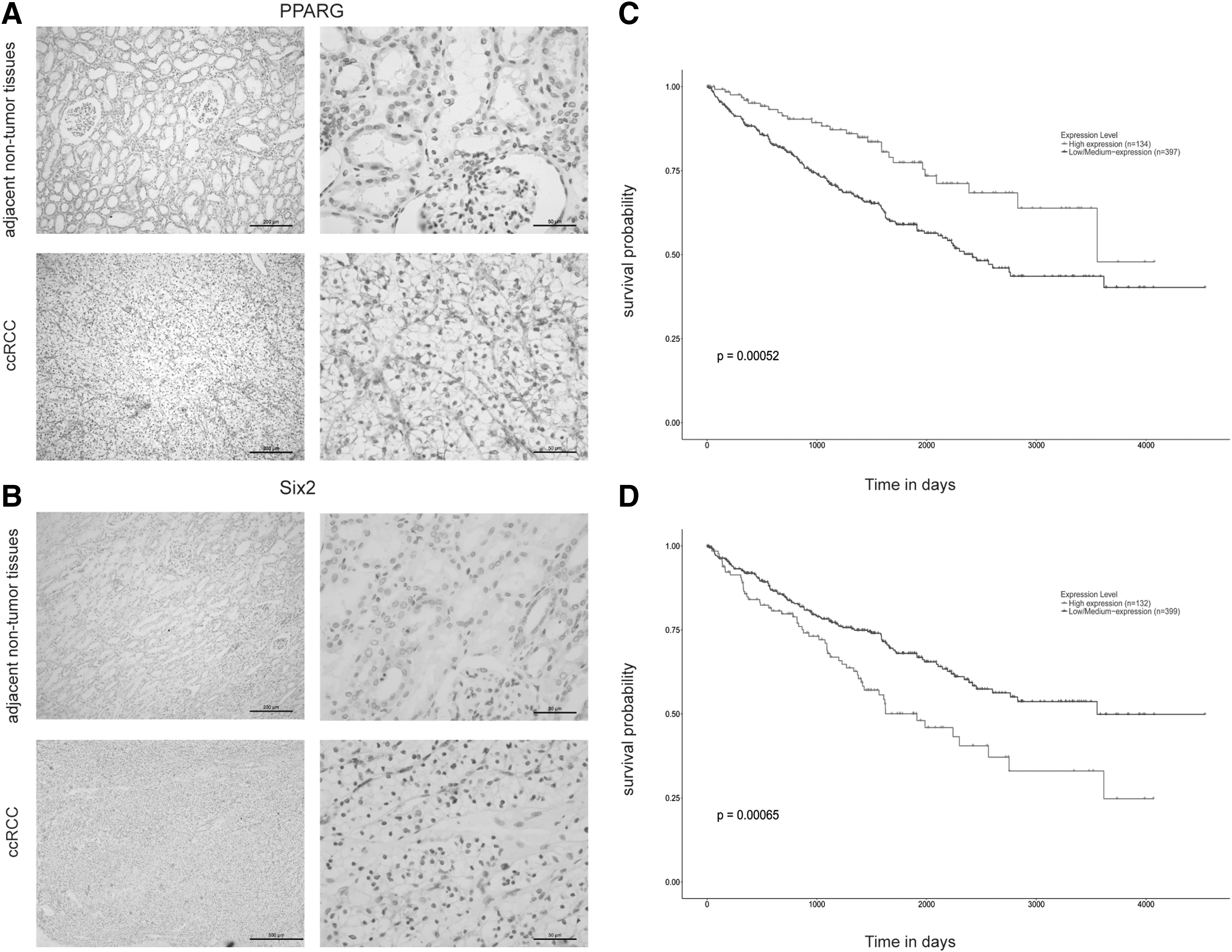
PPARG expression exhibited no obvious difference, whereas Six2 expression was increased in ccRCC tissues.
Main Glucolipid Metabolism Features in ccRCC Patients and Control Subjects
ccRCC, clear cell renal cell carcinoma; GLU, glucose; HDL-C, high-density lipoprotein cholesterol; LDL-C, low-density lipoprotein cholesterol; TC, total cholesterol; TG, triglycerides.
The Detailed Individual Data for Each Human Subject
BMI, body mass index.
PPARG activation regulates migration, apoptosis, and proliferation of 786O and OS-RC-2 cells
Migration, apoptosis, and proliferation are three crucial hallmarks of tumor development. We further investigated the effects of PPARG on migration, apoptosis, and proliferation of ccRCC cells; 786O and OS-RC-2 cells were treated with 50 μM RGZ, a PPARG agonist, for 24 h. A scratch wound healing assay was conducted to test the effect of RGZ on the migration of 786O and OS-RC-2 cells. As shown in Figure 2A, B, activating PPARG obviously inhibited the migration of 786O and OS-RC-2 cells. In addition, the cell apoptosis assay demonstrated that activating PPARG significantly increased the apoptosis ratio of 786O and OS-RC-2 cells (Fig. 2C, D). Furthermore, the Edu assay illustrated that the proliferation of 786O and OS-RC-2 cells was inhibited by RGZ (Fig. 2E, F). Collectively, our results demonstrated that PPARG could negatively modulate migration and proliferation and positively regulate apoptosis in ccRCC cells.

PPARG activation regulates the migration, apoptosis, and proliferation of 786O and OSRC2 cells.
PPARG activation decreases the expression of Six2 and maintains the E-cadherin level
To investigate the potential molecular mechanism, we detected the RNA and protein expression of Six2 after applying a PPARG agonist. The RNA and protein expression of Six2 were significantly decreased (Supplementary Fig. S1), and protein expression of E-cadherin was increased in ccRCC cells after treatment with the PPARG agonist RGZ for 24 h (Fig. 3A, B). To confirm the interrelationship between PPRAG and Six2, SiRNA-PPRAG (100 nM) or siRNA-Control was transfected into 786O and OS-RC-2 cells. The siRNA-PPARG-transfected 786O and OS-RC-2 cells exhibited downregulated Six2 protein. Regulation of E-cadherin by Six2, which promotes breast cancer metastasis, has been proposed. E-cadherin expression was increased in the siRNA-PPARG-transfected group, OS-RC-2OS-RC-2, whereas there was no obvious change in N-cadherin level (Fig. 3C, D). As expected, when 786O and OS-RC-2 cells, which stably express Six2, were treated with RGZ, the reduction in E-cadherin was alleviated (Fig. 3E, F), suggesting that PPARG is sufficient to inhibit the ability of Six2 to repress E-cadherin.
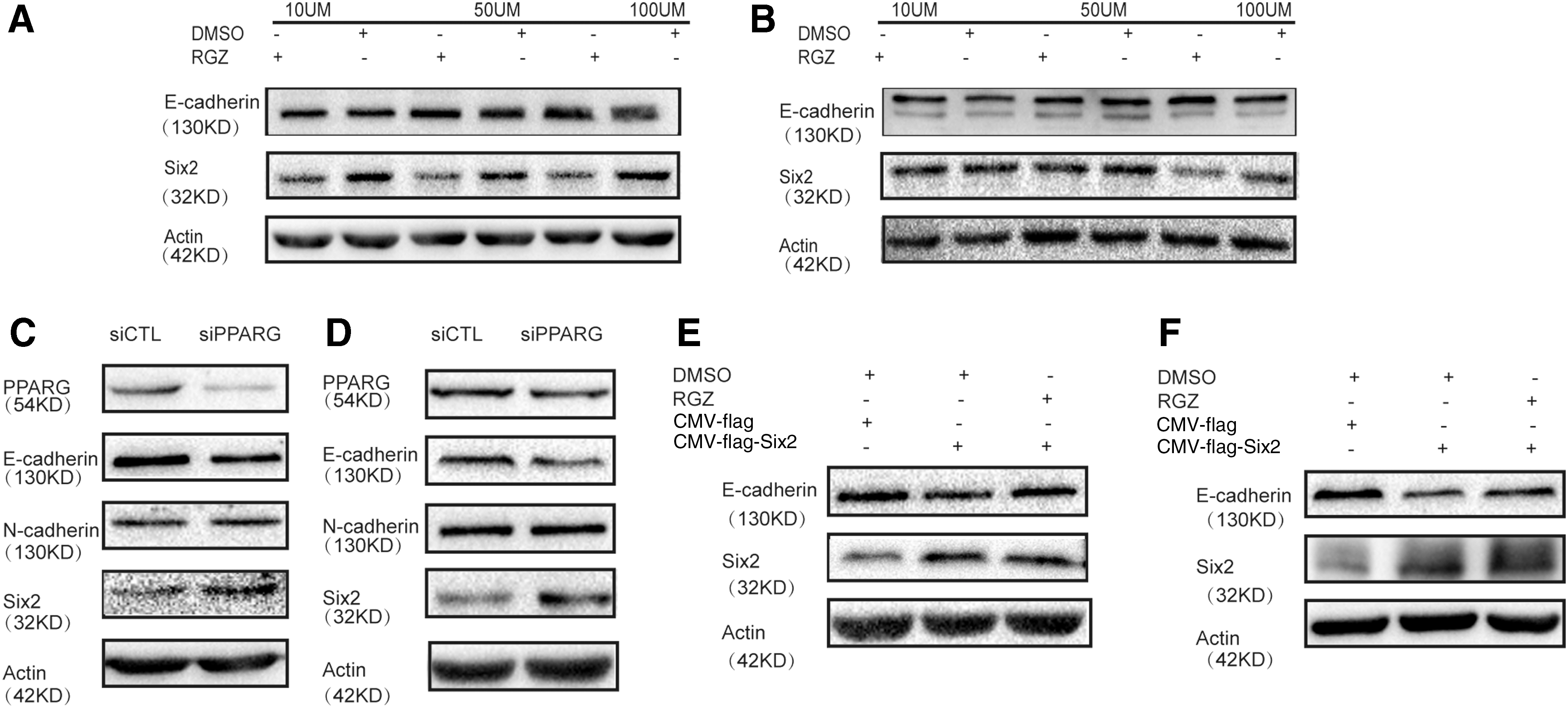
PPARG decreased the expression of Six2 and maintained the E-cadherin protein level.
Inhibition of Six2 suppresses proliferation and induces apoptosis of 786O and OSCR2 cells
Finally, we explored the potential effects of Six2 on proliferation and apoptosis in PPARG-activated ccRCC cell lines. 786O and OS-RC-2 cell lines transfected with control vector or stably expressing Six2 OS-RC-2 were dosed with 50 μM RGZ or DMSO for flow cytometry and EdU assays. Overexpression of Six2 reduced apoptosis, which was reversed by RGZ treatment (Fig. 4A, B). Similarly, overexpressing Six2 increased cell proliferation, and RGZ inhibited this proliferation ability (Fig. 4C, D). Collectively, these results indicated that PPARG activation could inhibit proliferation and induce apoptosis of 786O and OS-RC-2 cells by inhibiting Six2.
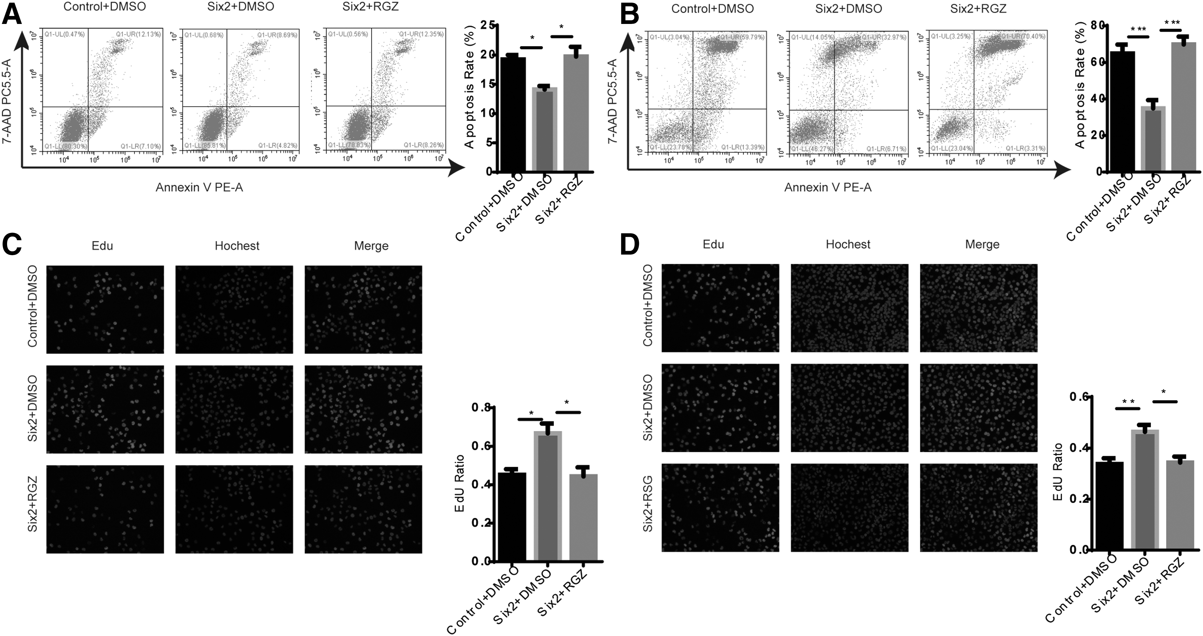
PPARG activation reduced the proliferation and apoptosis of 786O and OSRC2 cells by overexpression of Six2. 786O and OSRC2 stable cell lines transfected with empty vector or Six2 were treated with 50 μM RGZ, a PPARG agonist, or DMSO for 24 h, and the apoptosis rate was detected using flow cytometry
Discussion
It is generally accepted that the main functions of PPARG are regulating adipogenesis, lipid metabolism, and inflammation (Kroker and Bruning, 2015). Recently, an increasing number of studies have indicated that PPARG has tumorigenic or antitumorigenic functions. Accumulating evidence suggests that activation of PPARG by agonists can confer an inhibitory effect against cancer. However, the underlying mechanisms in ccRCC remain unclear.
Six2 serves as an important regulator of embryonic development. Interestingly, this gene, which plays a critical role in embryogenesis, is often reinstated in tumors; however, the roles and related mechanisms of Six2 in tumor progression have not been completely elucidated. In our study, we first collected ccRCC clinical information and samples. Analysis of the clinicopathological characteristics of the patients indicated that ccRCC patients were concentrated in the Fuhrman grade II group and that the morbidity of men was higher than that of women. Furthermore, histological examination suggested that PPARG may not be activated and that it is mainly expressed in cytoplasm. Interestingly, Six2, the downstream gene of PPARG, was highly expressed in the nuclei of ccRCC tissues.
At the cellular level, PPARG activation inhibited migration and proliferation and induced apoptosis of 786O and OS-RC-2 cellsOS-RC-2. In contrast, we also found that activation PPARG downregulated Six2 and upregulated E-cadherin at the cellular level, whereas PPARG knockdown induced the opposite effects. We also detected a reduction in E-cadherin after overexpression of Six2, which was rescued by activation of PPARG. In addition, overexpressing Six2 increased the proliferative capacity of cells and decreased apoptosis, although activation PPARG prevented this result.
In summary, our findings revealed that PPARG activation attenuated the migration and invasion of 786O and OS-RC-2 cells. It also induced apoptosis of 786O and OS-RC-2 cells. The phenotype of tumor formation in ccRCC was presented as negative modulation of Six2. Therefore, a better understanding of the underlying mechanism might provide novel insight for developing anticancer therapies.
Conclusions
Our study suggests that PPARG can negatively modulate Six2 in ccRCC tumor formation. This mechanism may also act at the E-cadherin protein level. PPARG activation is closely related to proliferation, apoptosis, and migration of 786O and OS-RC-2 cells. Therefore, it can be speculated that uncovering the potential mechanism may shed light on novel anticancer therapies.
Footnotes
Acknowledgments
This study was supported by the National Natural Science Foundation of China (grant nos. 81572076 and 81873932), the Chongqing Special Postdoctoral Science Foundation (grant no. XmT2018074), and the China Postdoctoral Science Foundation (grant no. 2018M633327).
Disclosure Statement
No competing financial interests exist.
Supplementary Material
Supplementary Figure S1
References
Supplementary Material
Please find the following supplemental material available below.
For Open Access articles published under a Creative Commons License, all supplemental material carries the same license as the article it is associated with.
For non-Open Access articles published, all supplemental material carries a non-exclusive license, and permission requests for re-use of supplemental material or any part of supplemental material shall be sent directly to the copyright owner as specified in the copyright notice associated with the article.