




Abstract
Advanced nonsmall-cell lung cancer (NSCLC) patients with mutated epidermal growth factor receptor (EGFR) can remarkably benefit from target therapy of EGFR-tyrosine kinase inhibitors (TKIs). However, increasing drug sensitivity and improving outcomes of NSCLC patients to EGFR-TKI therapy remains a challenge. Several studies have shown a link between microRNAs and drug resistance in cancer. In this study, we hypothesized that the rs12740674 single nucleotide polymorphism in the enhancer of miR-1262 may affect its expression, which may impact the outcome of NSCLC patients treated with EGFR-TKIs. The rs12740674 polymorphism was genotyped in two independent cohorts, including 319 EGFR-TKI treated stage IIIB/IV NSCLC patients. The allele-specific regulation on miR-1262 transcription by rs12740674 and impacts of miR-1262 on gefitinib sensitivity were evaluated in vitro and in vivo. Cox regression analyses indicated that the rs12740674 T allele was significantly associated with short survival time in both cohorts (p < 0.05). Luciferase assays demonstrated that the rs12740674 T allelic enhancer showed weaker capability to promote miR-1262 transcription compared with the C allelic enhancer, which may be due to reduced transcription factor binding according to electrophoretic mobility shift assays. Furthermore, significantly decreased miR-1262 expression in NSCLC and nontumor lung tissues of T allele carriers was observed compared with levels in C allele carriers. Moreover, miR-1262 expression enhanced the anticancer effects of gefitinib on NSCLC cells. Our data indicate that miR-1262 may be a potential therapeutic target for NSCLC.
Introduction
Advanced nonsmall-cell lung cancer (NSCLC) patients with tumors carrying mutations in the mutated epidermal growth factor receptor (EGFR) gene, such as the EGFR exon 19 deletion mutation (Ex19del) or the Leu858Arg mutation (L858R), can remarkably benefit from target therapy with EGFR-tyrosine kinase inhibitors (TKIs) (Mok et al., 2009; Maemondo et al., 2010; Mitsudomi et al., 2010; Rosell et al., 2012; Soria et al., 2018). Presently, oral administration of EGFR-TKIs such as gefitinib, erlotinib, afatinib, or osimertinib is the standard therapeutics for locally advanced or metastatic NSCLC harboring mutated EGFR (Hanna et al., 2017; Planchard et al., 2019). However, some patients with NSCLC harboring EGFR-TKI-sensitizing mutations show no responses to EGFR-TKIs. Moreover, it discourages future clinical applications that most NSCLC patients with ideal disease control after initial EGFR-TKI therapy would develop acquired drug resistance. Hence, conquering drug resistance to EGFR-TKIs continues to be a key issue in NSCLC management.
microRNAs (miRNA) are a class of evolutionarily highly conserved small noncoding single-stranded RNAs that post-transcriptionally regulate gene expression by suppressing translation or inducing mRNA degradation (Bartel, 2004). miRNAs interact with their target mRNAs by recognizing specific sequences in the mRNA 3′-untranslated region with help from the RNA-induced silencing complex (Bartel, 2004). Recent studies have shown that miRNAs have are involved in the development and progression of human malignancies (Calin and Croce, 2006), with some miRNAs potentially functioning as promising cancer therapeutic targets and biomarkers (Calin and Croce, 2006; Esquela-Kerscher and Slack, 2006). Frequently dysregulated expression of miRNAs has been observed in cancers due to germline genetic polymorphisms, genomic gain or loss of cancer-related gene loci, DNA methylation or promoter/enhancer histone modification, as well as alternations in miRNA processing (Esquela-Kerscher and Slack, 2006; Bader et al., 2010).
Xie et al. (2017) performed a systematic annotation of miRNA-related single nucleotide polymorphisms (SNPs) with the Encyclopedia of DNA Elements (ENCODE) data and identified the rs12740674 SNP located at 61,743 bp downstream from miR-1262 that maps to a strong enhancer. miR-1262 was found to significantly inhibit lung cancer cell proliferation (Xie et al., 2017). HaploReg prediction revealed that rs12740674 fell into the DNase peak region with several transcriptional factors (TFs) binding, such as FOS, a crucial regulator of mammalian gene expression (Chinenov and Kerppola, 2001; Tempé et al., 2014; Xie et al., 2017). These results suggest that allelic binding of TFs may lead to different miR-1262 expression levels in NSCLC cells and, thus, diverse effects on prognosis of NSCLC patients.
Herein, we specifically examined the role of the miR-1262 rs12740674 enhancer genetic variant in the outcome of advanced lung adenocarcinoma patients treated with EGFR-TKIs. We found that the enhancer SNP showed allelic regulation of miR-1262 expression in vitro and in vivo. Our findings show that the rs12740674 T allele associated with decreased miR-1262 expression significantly contributes to poor prognosis of advanced lung adenocarcinoma patients treated with EGFR-TKIs.
Materials and Methods
Study subjects
In this study, we enrolled two independent cohorts (Hubei cohort and Shandong cohort), including 319 patients with stage IIIB or IV lung adenocarcinoma (see Supplementary Data). All cases have prevalent EGFR activating somatic mutations and treated with EGFR-TKIs. A total of 46 NSCLC and normal tissues adjacent to the tumors were obtained from another set of patients who received surgery in Shandong Cancer Hospital and Institute. The detailed characteristics of all patients have been reported previously (Zhang et al., 2019a, 2020). This study was approved by the Institutional Review Board of Shandong Cancer Hospital and Institute (Protocol No. 201605005). At recruitment, written informed consent was obtained from each patient. The methods were carried out in accordance with the approved guidelines.
Genotyping
We collected blood samples from all patients in both the Hubei cohort and Shandong cohort with informed consent. We then extracted genomic DNA from each blood sample and genotyped the miR-1262 rs12740674 SNP using these genomic DNA samples. Genotypes of the miR-1262 rs12740674 polymorphism were determined using the iPLEX Sequenom MassARRAY platform (Sequenom) as previously reported (Zhang et al., 2019a,b, 2020). The miR-1262 rs12740674 polymerase chain reaction (PCR) primers are 5′-ACGTTGGATGTGACATCCTGATGAGTCAGC-3′ (Forward) and 5′-ACGTTGGATGGACTAATCCCCATCTCTCTC-3′ (Reverse). The miR-1262 rs12740674 UEP_SEQ primer is 5′-CCAAATAACTCAGGGTCA-3′. The miR-1262 rs12740674 EXT1_SEQ primer is 5′-CCAAATAACTCAGGGTCAC-3′. The miR-1262 rs12740674 EXT2_SEQ primer is 5′-CCAAATAACTCAGGGTCAT-3′. We reciprocally tested 15% DNA samples and the reproducibility was 100%.
Quantitative reverse transcription-PCR
Total cellular RNA was isolated from NSCLC tissues, normal lung tissues as well as lung adenocarcinoma H1299 and PC9 cells with Trizol (Invitrogen). Each RNA sample was treated with RNase-free DNase (Invitrogen) to avoid contamination of genomic DNA. The cDNAs were generated from RNA samples using the Revert Ace kit (Toyobo, Osaka, Japan). Human miR-1262 and U6 small RNA were detected with specific stem-loop RT-PCR primers (Ribobio, Guangzhou, China) using SYBR-Green qRT-PCR (quantitative reverse transcription-PCR) as described previously (Zhang et al., 2019a; Xiong et al., 2020).
miR-1262 luciferase reporter gene constructs
A potential 491 bp enhancer region around the rs12740674 genetic polymorphism (rs12740674C or rs12740674T) was cloned from human genomic DNA. We used specific PCR primer pairs with XhoI and KpnI restriction sites are p-491F, CGG
Dual-luciferase reporter assays
Luciferase reporter constructs (pGL3-Promoter, p-C, and p-T) were transfected into H1299 and PC9 cells. pRL-SV40 (1 ng; Promega) expressing renilla luciferase was cotransfected to standardize transfection efficiency. Luciferase activities were detected at 48 h after transfection using a luciferase assay system (Promega). Each luciferase construct was transfected in triplicate. Each independent transfection was also performed in triplicate.
Electrophoretic mobility shift assays
Double-stranded and 5′ biotin-labeled oligonucleotides corresponding to the miR-1262 rs12740674C or rs12740674T sequences were synthetized (Thermo). Nuclear extracts were obtained from lung adenocarcinoma PC9 cells and incubated with rs12740674C or rs12740674T 5′ biotin-labeled oligonucleotides at 25°C for 15 min using the Light Shift Chemiluminescent EMSA (electrophoretic mobility shift assay) Kit (Pierce). As described previously, the reaction mixture was separated on 6% PAGE (polyacrylamide gel electrophoresis), and the products were examined by Stabilized Streptavidin-Horseradish Peroxidase Conjugate (Pierce) (Xiong et al., 2020). In competition assays, unlabeled miR-1262 rs12740674C or rs12740674T probes at 10-, 50-, or 100-fold molar excess were also added to the reaction mixture before the addition of biotin-labeled C allele probes or T allele probes.
Cell proliferation and gefitinib drug sensitivity assays
Human PC9 and H1299 cells were seeded in a 96-well plate and transfected with 10 nM miR-1262 mimics, or NC RNA (Genepharma) with INTERFERin® (Polyplus). Gefitinib (EY0917; Amquar Co., Shanghai, China) diluted in dimethyl sulfoxide was added to the wells to achieve the desired final concentrations (H1299 cells: 1, 5, 10, 25, and 50 μM; PC9 cells: 1, 5, 10, and 20 μM) as described previously (Zhang et al., 2019a, 2020). After 48 h treatment, the survival rates of PC9 and H1299 cells were measured using the MTT assay (Sigma, Temecula, CA).
Statistics
The Kaplan–Meier method was used to examine the impacts of the rs12740674 genotypes on progression-free survival (PFS) or overall survival (OS). Survival curves of different genotypes were compared using the log-rank test. Multivariate Cox regression analyses were performed to identify prognostic factors of PFS or OS. The association between the miR-1262 rs12740674 polymorphism and recurrence risk or death risk was estimated through hazard ratios (HRs) and 95% confidence intervals (CIs) after adjustments with age of onset, gender, and smoking status, where it was appropriate. Student's t-test was used to compare differences between two groups, such as miR-1262 expression levels in NSCLC tissues or normal tissues. A p-value of <0.05 was considered as statistically significant. All statistical tests were two-sided and performed with Graphpad or SPSS software package.
Results
Effects of the miR-1262 rs12740674 genetic variant on PFS and OS in NSCLC
As shown in Table 1, the miR-1262 rs12740674 C > T genetic polymorphism was significantly associated with PFS in Hubei discovery cohort or Shandong validation cohort. Cox regression analyses indicated that the miR-1262 rs12740674 CT or TT genotype significantly contributed to unfavorable PFS (the CT genotype: HR = 2.52, 95% CI = 1.54–4.13, p = 2.5 × 10⁻⁴; the TT genotype: HR = 1.87, 95% CI = 1.29–2.69, p = 0.001) compared with the CC genotype in Hubei cohort. Similarly, the miR-1262 rs12740674 CT and TT genotypes were significantly associated with increased recurrence risk after gefitinib treatment, compared with the CC genotype in Shandong cohort (HR = 1.56, 95% CI = 1.01–2.40, p = 0.044). Kaplan–Meier survival estimates showed that patients with the miR-1262 rs12740674 CT or TT genotype had a worse PFS than patients carrying the CC genotype (Hubei cohort: log-rank p < 0.001; Shandong cohort: log-rank p = 0.046) (Fig. 1A, B).

Kaplan–Meier curve of PFS
Multivariate Cox-Regression Analyses of the miR-1262 rs12740674 Genetic Variation for Progression-Free Survival
HRs and 95% CIs for the association between clinical variables and recurrence or death risk was adjusted for age of onset, gender, smoking status, stage, and EGFR status, where it was appropriate.
CI, confidence interval; EGFR, epidermal growth factor receptor; HR, hazard ratio; N.C., not calculated.
The miR-1262 rs12740674 T allele was also significantly associated with worse OS compared with the miR-1262 C allele. As shown in Table 2, lung adenocarcinoma patients carrying the miR-1262 rs12740674 CT or TT genotype had increased death risk than patients carrying the CC genotype in Hubei cohort (HR = 1.92, 95% CI = 1.31–2.82, p = 0.001; or HR = 2.47, 95% CI = 1.44–4.24, p = 0.001). Furthermore, the miR-1262 CT and TT genotypes were correlated to shorter OS (HR = 2.27, 95% CI = 1.39–3.69, p = 0.001) compared with the CC genotype in Shandong cohort. Individuals with the miR-1262 rs12740674 CT or TT genotype had a worse survival than patients carrying the CC genotype (Hubei cohort: log-rank p < 0.001; Shandong cohort: log-rank p = 0.002) (Fig. 1C, D).
Multivariate Cox-Regression Analyses of the miR-1262 rs12740674 Genetic Variation for Overall Survival
HRs and 95% CIs for the association between clinical variables and recurrence or death risk was adjusted for age of onset, gender, smoking status, stage, and EGFR status, where it was appropriate.
The rs12740674 SNP-mediated allelic regulation of miR-1262 expression
The miR-1262 rs12740674 C > T genetic variant is located in a potential gene enhancer region and may lead to differential binding of multiple transcription factors, such as FOS, leading to dysregulated miR-1262 expression in cancer cells (Xie et al., 2017). Therefore, we evaluated how the rs12740674 genetic polymorphism impacts miR-1262 expression using dual-luciferase reporter gene assays in human H1299 and PC9 cells (Fig. 1A, B). Cells transfected with the rs12740674T allelic luciferase reporter construct (p-C) showed significantly higher luciferase activities compared with cells expressing the rs12740674C allelic reporter construct (p-T) (H1299 cells: p = 0.006; PC9 cells: p = 0.012). This indicates that the enhancer carrying the rs12740674C allele promoted increased miR-1262 transcription compared with the rs12740674T allelic enhancer.
We then conducted EMSA to distinguish the differences in binding capacity of nuclear extracts to the rs12740674T and C alleles (Fig. 2C). We detected increased binding of nuclear proteins from PC9 cells to the C allele biotin-labeled oligonucleotide probe compared with the T allele probe. In addition, a 10-, 50-, and 100-fold excess of unlabeled C allele oligonucleotides or unlabeled T allele oligonucleotides competed with the binding between nuclear proteins and the biotin-labeled C allele and T allele probes.
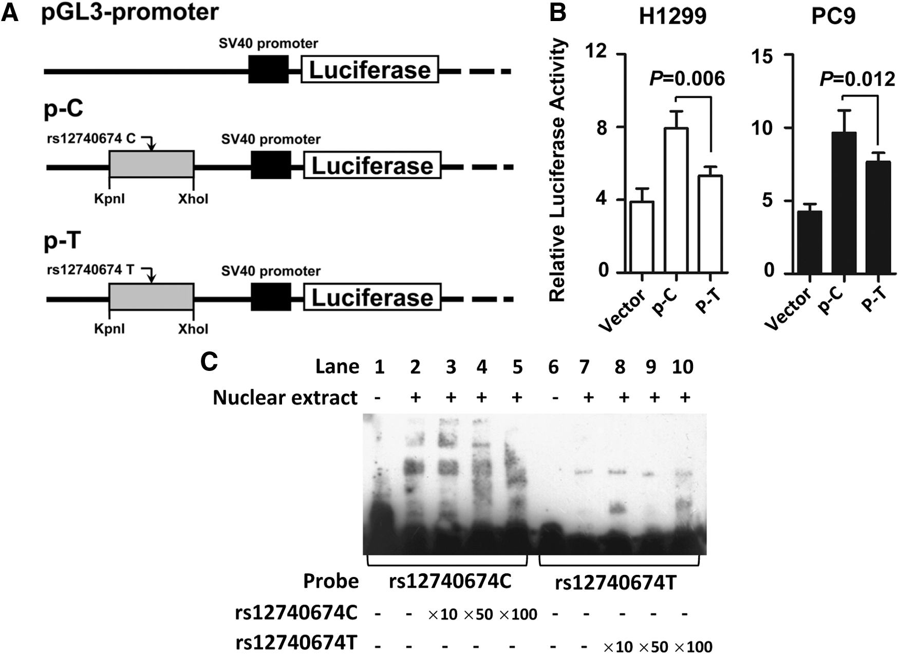
Decreased binding of transfactors in the miR-1262 enhancer by the rs12740674 C > T genetic variant impacts enhancer activities.
Decreased miR-1262 expression in human NSCLC tissue specimens
To validate the allele-specific effect of the rs12740674 SNP on miR-1262 expression in vitro, we detected miR-1262 expression in 46 NSCLC tissues and paired nontumor tissues. Decreased miR-1262 expression was detected in NSCLC tissues compared with levels in normal tissues (p = 0.022) (Fig. 3A). This indicates that miR-1262 may act as a tumor suppressor in NSCLC development. We then evaluated the allelic differences of miR-1262 expression in cancerous tissues and nontumor tissues. As shown in Figure 3B, individuals with the rs12740674 CC genotype had significantly higher miR-1262 levels in NSCLC tissues (mean ± standard error) than those with the CT and TT genotypes (2.338 ± 0.348 [n = 28] vs. 0.321 ± 0.064 [n = 18], p = 0.001). Similarly, much higher miR-1262 expression in the CC genotype carriers was observed compared with the CT or TT genotype carriers in normal tissues (0.502 ± 0.075 [n = 18] vs. 0.204 ± 0.047 [n = 28], p = 0.001) (Fig. 3C). These data are in line with the results of reporter gene assays.
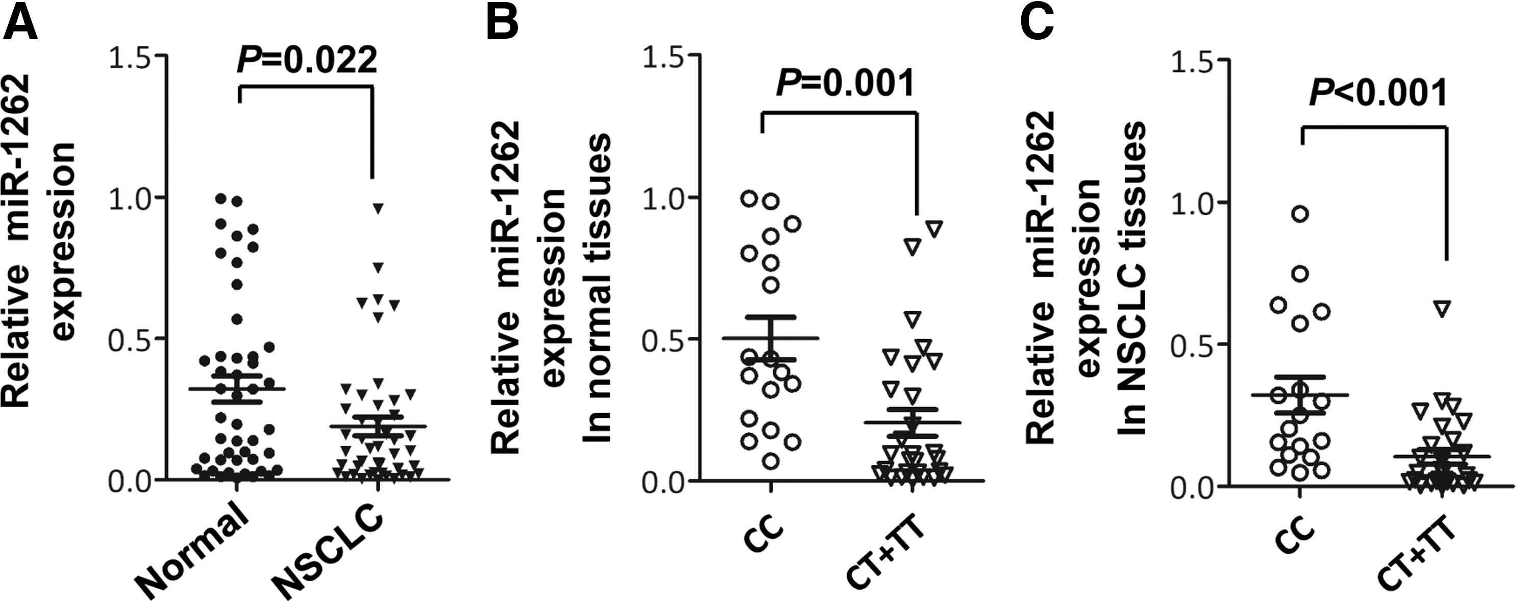
miR-1262 expression in NSCLC and normal lung tissues.
miR-1262 significantly enhances sensitivity of NSCLC cells to gefitinib
To investigate whether miR-1262 is involved in the response to EGFR-TKI therapy, we examined how miR-1262 impacts the sensitivity of lung adenocarcinoma PC9 and H1299 cells to gefitinib. Overexpression of miR-1262 significantly enhanced the antiviability effects of gefitinib in H1299 lung adenocarcinoma cells compared with control transfected cells (25 μM gefitinib group: p = 0.002; 50 μM gefitinib group: p < 0.001) (Fig. 4A). Ectopic miR-1262 expression also significantly decreased cell proliferation of PC9 cells treated with gefitinib compared with controls (25 μM gefitinib group: p < 0.001) (Fig. 4B).

miR-1262 significantly promoted the antiproliferation effects of gefitinib in H1299 cells
Discussion
Genetic mutations in tumors contribute to the drug sensitivity of NSCLC to EGFR-TKIs (Ng et al., 2012; Wheeler et al., 2013; Nelson et al., 2016; Chang et al., 2017). Germline polymorphisms located in the promoter and/or enhancer regions of the miRNA genes may lead to dysregulated miRNA expression. We found that the rs12740674 genetic variation in the miR-1262 enhancer resulted in allelic differential expression of miR-1262 in NSCLC. Associations between miR-1262 rs12740674 SNP and disease recurrence or death risk of lung adenocarcinoma patients treated with EGFR-TKI were examined through a two-stage cohort study approach. We found that the miR-1262 rs12740674 T allele was significantly associated with poor prognosis after EGFR-TKI therapy in Chinese Han populations, and low miR-1262 expression showed a similar trend. Furthermore, ectopic expression of miR-1262 significantly enhanced the sensitivity of lung adenocarcinoma cells to gefitinib.
We observed reduced expression of miR-1262 in NSCLC tissues in our cohort, suggesting a tumor suppressor role of miR-1262 in lung cancer. Similarly, we found downregulated miR-1262 expression levels in 39 TCGA lung adenocarcinoma (LUAD) tissues compared with paired nontumor tissues (Xie et al., 2017). We also found that individuals with the rs12740674 CC genotype showed significantly higher expression of miR-1262 in both NSCLC tissues and nontumor lung tissues. miR-1262 expression was associated with genotypes of rs12740674 in 410 TCGA lung adenocarcinoma tissues, with highest expression in carriers of the CC genotype (Xie et al., 2017).
The expressions of miRNAs are transcriptionally regulated by multiple TFs (O'Donnell et al., 2005; Chang et al., 2007; Yamakuchi et al., 2010; Ren et al., 2017; Anuj et al., 2019). For example, P53 transcriptionally transactivates expression of miR-34a, leading to dramatic reprogramming of gene expression as well as increased apoptosis (Chang et al., 2007). P53 also regulates hypoxic signaling through the transcriptional regulation of miR-107 in colon cancer (Yamakuchi et al., 2010). c-Myc activates transcription of a cluster of six miRNAs (miR-17-5p, miR-18, miR-19, miR-20, miR-92, and miR-106) and modulates E2F1 expression (O'Donnell et al., 2005). Notably, rs12740674 is located in a genomic region that binds several proteins, including FOS, MAFF, MAFK, and EP300 (Xie et al., 2017). Among these TFs, FOS is an important component of Activating Protein 1 (AP-1) and involved in regulating transcription of several miRNAs, such as miR-551a. Previous studies showed that FOS activates miR-551a expression and blocks tumorigenesis (Anuj et al., 2019).
Previous studies indicate that miR-1262 might play an essential role in drug response and drug-induced toxicity. Zhan et al. (2017) characterized the antimetastasis mechanisms of curcumin in lung cancer by constructing a miRNA gene network and identified miR-1262 as one of six key miRNA regulators in the network. miR-1262 may also be a biomarker for hepatocellular carcinoma (HCC) (Abd El Gwad et al., 2018). Abd El Gwad et al. (2018) found that a panel of three exosomal RNAs (miR-1262, lncRNA-RP11–513I15.6, and RAB11A) showed excellent sensitivity and specificity in discriminating patients with HCC from patients with chronic hepatitis C virus and healthy controls. Freitas et al. (2016) demonstrated that the differential expression of miRNAs, including miR-1262 in platelets and interactions with their target mRNAs, was associated with variability in platelet reactivity, clopidogrel response, and drug-induced toxicity. Similarly, we found that miR-1262 is also involved in regulating EGFR-TKI drug response in NSCLC cells.
Conclusion
To the best of our knowledge, this study is the first to elucidate the involvement of miR-1262 and the rs12740674 genetic variation in EGFR-TKI therapy in NSCLC. These results underline the complexity of the genetic regulation of miRNAs during cancer drug treatments. In view of our results showing that elevated miR-1262 expression in NSCLC may enhance sensitivity to EGFR-TKIs, we hypothesize that miR-1262 may be a potential therapeutic target for NSCLC.
Availability of Data and Material
All data generated or analyzed during this study are included in the article.
Footnotes
Authors' Contributions
M.Y. and N.Z. designed the study. T.L. and N.Z. performed the experiments. T.L., N.Z., and L.Z. performed data analyses. L.Z., B.W., Y.S., and Y.S. were responsible for the collection of specimens. M.Y. supervised the project. M.Y. and N.Z. contributed to drafting the article. All authors have read and approved the final submitted article.
Disclosure Statement
No competing financial interests exist.
Funding Information
This study was financially supported by National Natural Science Foundation of China (31671300, 31871306) and Taishan Scholars Program of Shandong Province (tsqn20161060). The study sponsor had no role in the study design, conduct, or interpretation of the data or the writing of the report, and decision to submit for publication.
Supplementary Material
Supplementary Data
References
Supplementary Material
Please find the following supplemental material available below.
For Open Access articles published under a Creative Commons License, all supplemental material carries the same license as the article it is associated with.
For non-Open Access articles published, all supplemental material carries a non-exclusive license, and permission requests for re-use of supplemental material or any part of supplemental material shall be sent directly to the copyright owner as specified in the copyright notice associated with the article.
