




Abstract
This study aimed to evaluate the role of HOXC11 in progression and prognosis in colon adenocarcinoma (COAD) patients. The COAD patient data were downloaded from “The Cancer Genome Atlas (TCGA)” database. The Wilcoxon rank-sum test or Kruskal-Wallis test was used to analyze the correlation between HOXC11 expression and clinicopathologic characteristics. The significance of difference in overall survival between different groups was determined by log-rank test. The HOXC11 expression was verified from mRNA and protein level by conducting real-time quantitative PCR, Western blot, and immunohistochemistry analysis. Significantly enriched Kyoto Encyclopedia of Genes and Genomes (KEGG) pathways were screened after gene set enrichment analysis. As a result, high HOXC11 expression was closely related to the occurrence of COAD based on the data in TCGA, which was then successfully validated in cell lines and clinical tissues. Enhanced HOXC11 expression was significantly associated with tumor-node-metastasis (TNM) and M stage. Prognosis of highly expressed HOXC11 COAD patients was significantly worse than those with low HOXC11 expression. GRAFT_VERSUS_HOST_DISEASE and other signaling pathways were significantly activated in high HOXC11 expression COAD patients. In conclusion, high expression of HOXC11 was closely associated with the progression of COAD, and HOXC11 was a promising prognostic biomarker in COAD patients.
Highlight
High expression of HOXC11 was closely related to the occurrence and progression of COAD.
The prognosis of COAD patients with high expression of HOXC11 was significantly inferior than those with low HOXC11 expression.
Introduction
Colon cancer is one of the most prevalent malignancies all around the world (Zhou et al., 2019) and it is the fourth most death-related cancer (Hatano et al., 2017). Colon adenocarcinoma (COAD) is the most common pathological type of colon cancer, which accounts for nearly 90% of colorectal cancer (CRC) cases (Fleming et al., 2012). At present, the prognostic predictions and treatment decisions of most colon cancer cases mainly depend on tumor-node-metastasis (TNM) staging system and other common clinicopathologic features (Hu et al., 2011). Meanwhile, it has been reported that patients with COAD usually exhibited varied responses to the treatment (Zhai et al., 2017) as COAD often resulted from complex genetic and epigenetic alterations (Harrison and Benziger, 2011; Puerta-Garcia et al., 2015). Coincidentally, it has been demonstrated that there were usually variable survival outcomes of patients assigned to the same TNM category (Nagtegaal et al., 2011), which indicated that these traditional methods may have limited prognosis value to some extent. Promisingly, prognostic biomarkers can be used as prognostic indicators and molecular predictive factors in targeted therapy and precision treatment (Garcia-Alfonso et al., 2015; Mody and Bekaii-Saab, 2018), based on which, our group has devoted to find new more reliable biomarkers of COAD, and we have found several possible genes and miRNAs (Min et al., 2019a, 2019b; Li et al., 2020). Thus, it is necessary to find novel targeted progression and prognostic biomarkers in COAD patients.
Recently, homeobox (HOX) genes have been widely explored in different kinds of cancers, which included HOX genes (HOXA, HOXB, HOXC, and HOXD) and non-HOX genes (for example, Cdx, Evx, Dlx, and so on) (Haria and Naora, 2013; Joo et al., 2016). Numerous studies have documented directly or indirectly the correlation between the HOX gene expression and various tumor cells' invasion and metastasis (de Bessa Garcia et al., 2020; Paco et al., 2020). A study in esophageal squamous cell carcinoma has reported that HOXC6, HOXC10, and HOXC13 were upregulated in tumor tissues compared to normal tissues (Takahashi et al., 2007). Another research has suggested that HOXD10 mRNA was significantly downregulated in stomach cancer tissues, which led to tumor proliferation (Wang et al., 2012). In addition, many HOX genes have been studied in CRCs, such as HOXCDX1, HOXCDX2, and HOXALX4 were significantly downregulated in CRC (Ebert et al., 2006; Baba et al., 2009; Olsen et al., 2016), while HOXB13, HOXB7, and HOXC6 were observed to be upregulated in CRC (Liao et al., 2011; Sanz-Pamplona et al., 2011). Moreover, HOXC11 has been documented to play an important role in tumorigenesis in many cancers. A vitro study has reported that HOXC11 was upregulated in kidney renal clear cell carcinoma (KIRC) and it was associated with patient overall survival (OS) (Cui et al., 2020). Another research has indicated that HOXC11 was involved in negative consequences to endocrine therapy in breast cancer (McIlroy et al., 2010). A study has suggested that HOXC11 may contribute to the leukemogenesis indirectly (Gu et al., 2003). However, to the best of our knowledge, the potential effects of HOXC11 in COAD progression and prognosis have hardly been studied.
In this study, by analyzing COAD-related data obtained from “The Cancer Genome Atlas (TCGA)” database and further experiments, we proposed not only to evaluate the correlation between HOXC11 expression and clinicopathologic characteristics but also to evaluate the prognostic value of HOXC11 expression in COAD.
Materials and Methods
Data collection
The mRNA expression data and corresponding clinical information of 456 COAD (COAD) patients were downloaded from TCGA (
Clinical Characteristics of Patients with Colon Adenocarcinoma Based on the The Cancer Genome Atlas Database
BMI, body mass index; TNM, tumor-node-metastasis.
COAD tissue samples
All 15 pairs of COAD tissues and normal paracancer tissues were obtained from patients who met NCCN diagnostic criteria (Benson et al., 2018) in Beijing Friendship Hospital from February 2020 to November 2020 [ethic code: 2017-P2-013-03]. All the experiments were approved by the Ethics Committee of Beijing Friendship Hospital, and informed consents were obtained from subjects.
Cell and reagents
Human colonic mucosa cell line NCM460 and colon cancer cell line SW480 were obtained from Cell Bank of the Chinese Academy of Sciences (Shanghai, China). For both cell line culture, complete Dulbecco's Modified Eagle's Medium (DMEM) was used to cell grow, consisting in DMEM (Gibco, Massachusetts) supplemented with 10% fetal bovine serum (Gibco), and 100 mg/mL antibiotics (Gibco).
For total RNA extraction, Trizol reagent (Cat# 15596-026; Invitrogen, Grand Island, CA) was used. TIAN Script RT kit (Cat# KR104-02; TIANGEN, Beijing, China) was used to reverse transcribe to complementary DNA. And SuperReal PreMix Plus kit (Cat# FP205; TIANGEN) was for the real-time quantitative PCR.
For Western blot and immunohistochemistry (IHC) analysis, antibodies against HOXC11 were purchased from Affinity Biosciences (Cat# DF9580, 1:500; Changzhou, China) and Abcam (ab243721, 1:100; Shanghai, China), respectively. And Tubulin antibody was from Ray Antibody Biotech (Cat# RM2007, 1:500; Beijing, China) as a control. Goat anti-Mouse IgG (H+L)–HRP and Goat anti-Rabbit IgG (H+L)–HRP were obtained from Bioworld (Cat# BS12478 and BS13278, 1:1000; Nanjing, China).
Survival analysis
Based on the Kaplan-Meier method, R language survival package and survminer package (
RNA extraction and the real-time quantitative PCR
The extracted total RNA was quantified by BioPhotometer (Eppendorf). The real-time quantitative PCR was performed for 40 cycles, and GAPDH was used as an endogenous control. All primers of this research used are listed in Table 2. Each sample was repeated thrice by the Connect CFX™ (BIO-RAD). The mRNA relative expression level was calculated by the 2–△△Ct method.
Primer Sequences
Western blot and IHC analysis
Total proteins were extracted from cell lines, and clinical details of all subjects are shown in the Supplementary Table S1. Western blot and IHC analysis were conducted as common process (Zeng et al., 2017). Image Pro Plus 6.0 software was used to analyze intensity.
Gene set enrichment analysis
R language ClusterProfiler package (Yu et al., 2012) and the gene set
Statistical analysis
The Wilcoxon rank-sum test was used to compare the expression level of HOXC11 in cancer samples and adjacent samples. The correlation between HOXC11 mRNA expression and clinicopathological characteristics was analyzed by Wilcoxon rank-sum test or Kruskal-Wallis test. Multivariate Cox regression proportional hazard model was used to determine the influence of HOXC11 mRNA expression and clinicopathological characteristics (age, gender, TNM Stage, etc.) on OS. The difference with p-value <0.05 is considered to be statistically significant. R software v3.6.2 was used for all statistical analyses.
Results
High expression of HOXC11 was closely related to the occurrence of COAD
The HOXC11 expression levels in colon cancer tissues and adjacent normal tissues were compared using the Wilcoxon rank-sum test. The paired COAD tissues (N = 41) exhibited significantly higher HOXC11 expression levels than the corresponding adjacent normal tissues (p = 0.0016) (Fig. 1A). Moreover, in the comparison between unpaired COAD samples and normal samples, significantly upregulated HOXC11 expression in COAD samples was also observed (p = 1.3e-5) (Fig. 1B). The analysis suggested that high expression of HOXC11 may be closely related to the occurrence of COAD.
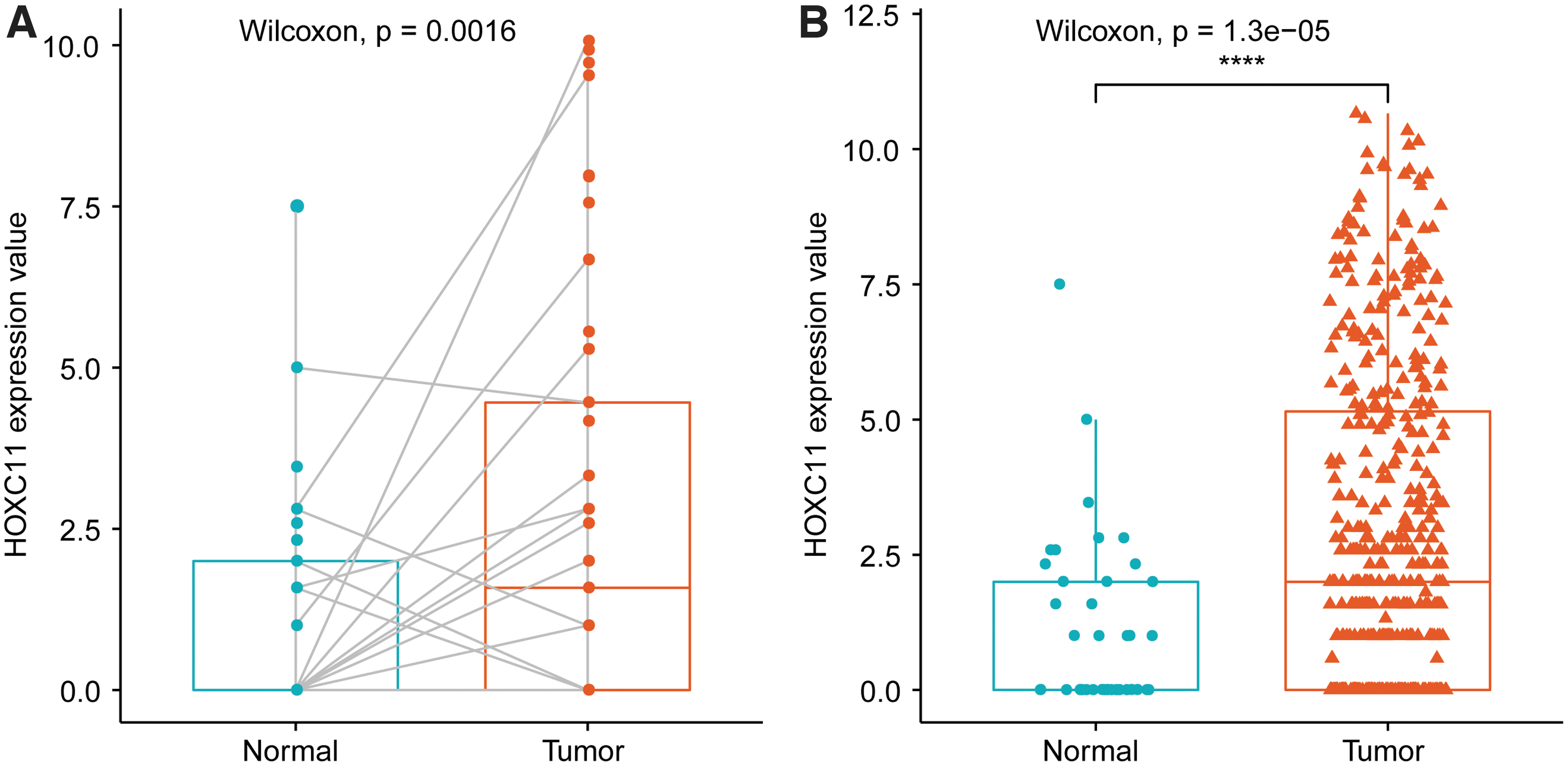
The expression of HOXC11 in colon cancer samples and adjacent samples.
Association of HOXC11 mRNA expression with clinicopathological characteristics
The association of HOXC11 mRNA expression in 433 COAD samples with the corresponding patients' clinicopathologic characteristics was analyzed by Wilcoxon rank-sum test or Kruskal-Wallis test. The results showed that enhanced HOXC11 mRNA expression was significantly associated with TNM stage (p = 0.045) (Fig. 2A) and M Stage (p = 0.0066) (Fig. 2D). Besides, through Wilcoxon rank-sum test, HOXC11 mRNA expression was statistically different in the following groups: Stage IV versus Stage I, Stage IV versus Stage II, Stage IV versus Stage III, T4 versus T2, and N2 versus N0 (Fig. 2A–D). However, there was no significant association between the enhanced HOXC11 mRNA expression and age and gender.

Association of HOXC11 with clinicopathological characteristics.
COAD patients with high HOXC11 expression had poor prognosis
According to the median expression of HOXC11, tumor samples were divided into two groups: high HOXC11 expression and low HOXC11 expression. To determine the prognostic value of HOXC11 expression in COAD patients, survival analysis was performed in patients with high HOXC11 expression and low HOXC11 expression, respectively. The results revealed that COAD patients exhibiting high HOXC11 expression had worse OS than patients exhibiting low HOXC11 expression (p = 0.02, hazard ratio [HR] = 1.66, 95% confidence interval [CI]: 1.11–2.49) (Fig. 3A).
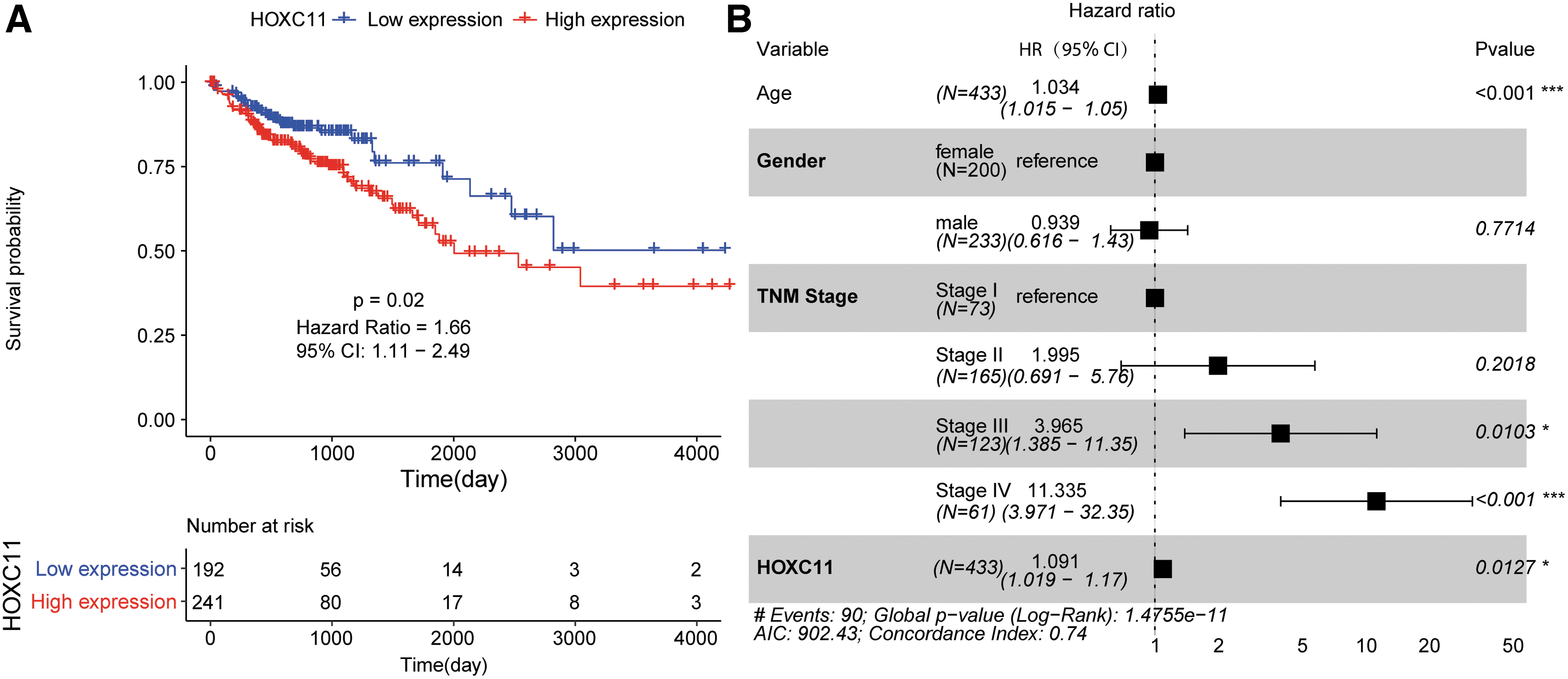
The prognosis of COAD patients was poor in high HOXC11 expression group.
Furthermore, to determine whether HOXC11 expression is an independent prognostic indicator, multivariate Cox regression analysis was performed, which included age, gender, TNM stage, and HOXC11. The results suggested that HOXC11 expression was still significantly correlated with OS of COAD patients. High HOXC11 expression was associated with higher death risk and it was a poor prognostic factor for COAD patients (HR = 1.091, 95% CI: 1.019–1.17, p = 0.0127) (Fig. 3B).
HOXC11 was at a high expression level in colon cancer cell lines and clinical COAD tissues
To further validate the HOXC11 expression level in colon cancer cell lines, mRNA and protein expression were determined in NCM460 and SW480. The results indicated that both relative mRNA (Fig. 4A, p < 0.001) and protein (Fig. 4B, C, p < 0.05) expression of HOXC11 were remarkably high in colon cancer cell lines, which was consistent with the analysis of public dataset. Moreover, the subsequent IHC analysis suggested that HOXC11 was at high expression level in all 15 COAD tissues, compared to the corresponding normal paracancer tissues (Fig. 5A, B). Thus, the experiment data showed that abnormally expressed HOXC11 was related to COAD progress.
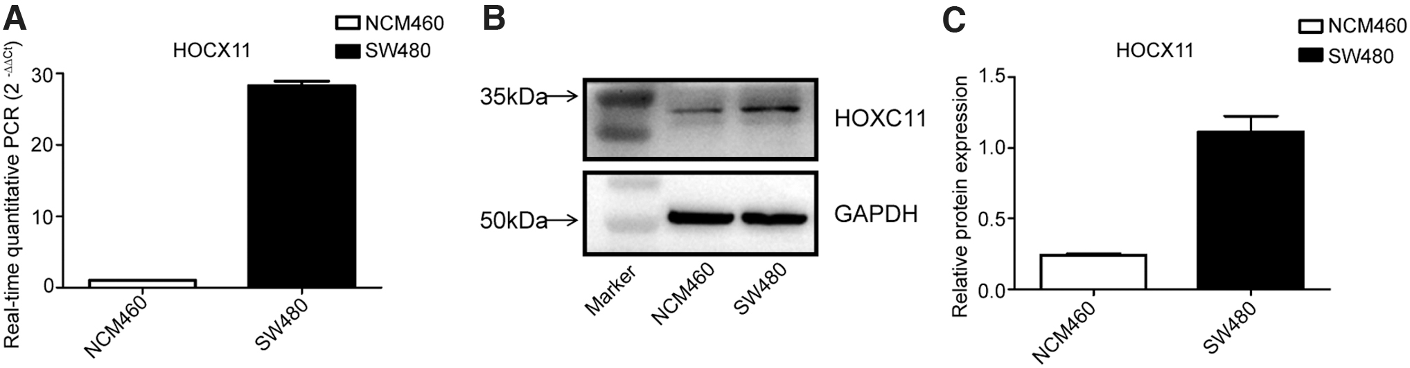
Upregulation of HOXC11 in colon cancer cell lines.
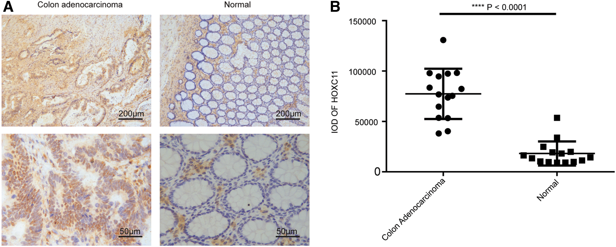
Upregulation of HOXC11 in COAD subjects.
HOXC11-related signaling pathways based on GSEA
GSEA was conducted to identify the potential signaling pathways involved in COAD between high and low HOXC11 expression datasets. KEGG pathways significantly enriched with p-value <0.05 were selected, see Table 3. According to the Normalized enrichment score, the top 5 enriched pathways are displayed in Figure 6. The results showed that KEGG_GRAFT_VERSUS_HOST_DISEASE, KEGG_CYTOKINE_CYTOKINE_RECEPTOR_INTERACTION, and other six pathways were activated in high HOXC11 mRNA expression group, and KEGG_LYSINE_DEGRADATION, KEGG_STARCH_AND_SUCROSE_METABOLISM, and four other pathways were inhibited in low HOXC11 expression group. And our results indicated that HOXC11 might influence these pathways, by affecting other genes involved in the pathways.
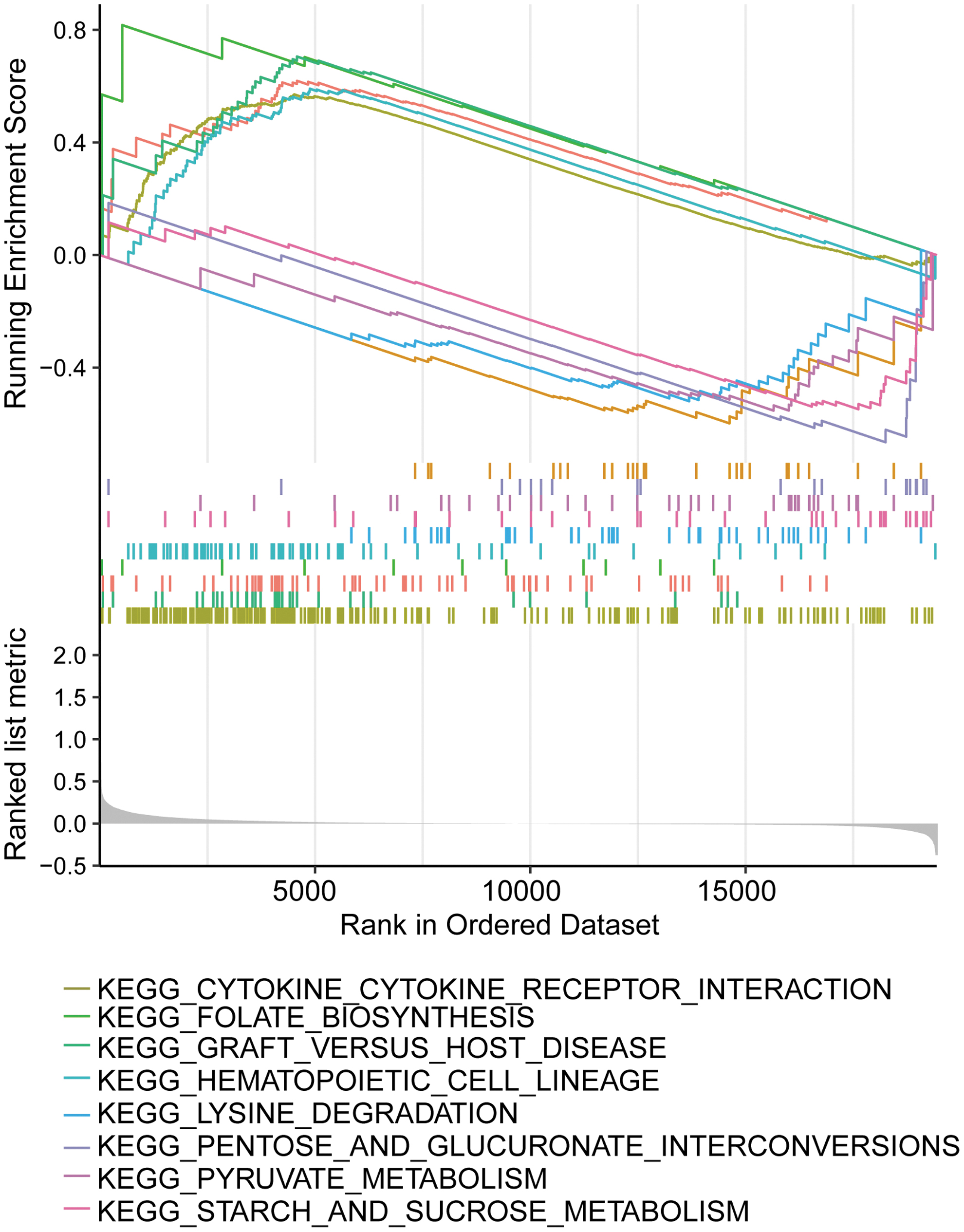
The results of GSEA enrichment of HOXC11 mRNA expression. GSEA, gene set enrichment analysis. Color images are available online.
Gene Set Enrichment Analysis of The Cancer Genome Atlas Colon Adenocarcinoma Tumors with the HOXC11 High Expression and Low Expression by Using Kyoto Encyclopedia of Genes and Genomes Pathway Gene Sets
KEGG, Kyoto Encyclopedia of Genes and Genomes; NOM, normal.
Discussion
In this study, based on COAD-related data obtained from TCGA database and further validation experiments, we explored the correlation between HOXC11 expression and COAD. On the one hand, we found that high expression of HOXC11 was probably closely associated with the occurrence of COAD. On the other hand, prognosis of COAD patients with high expression of HOXC11 was relatively poor, which indicated that HOXC11 might be a promising prognostic biomarker. In addition, we have identified several potential signaling pathways involved in HOXC11 expression in COAD patients based on GSEA results.
First, we have compared HOXC11 expression in COAD specimens and normal samples to confirm whether HOXC11 expression was related to COAD. And upregulated HOXC11 expression was observed not only in COAD but also in colon cancer tissues compared with normal tissues, which was then successfully verified in cell lines and tissues. Our data were in line with some previous similar researches. Numerous studies have demonstrated that aberrant expression of HOX genes was involved in carcinogenesis (Shah and Sukumar, 2010; Bhatlekar et al., 2014; Joo et al., 2016). HOX genes could be generally classified as HOX genes and non-HOX genes (Joo et al., 2016). At present, 39 HOX genes have been identified in humans and they were clustered into four groups named HOXA, HOXB, HOXC, and HOXD, respectively (Platais et al., 2016). It has been reported in hepatocellular carcinoma that HOX11 was also upregulated in cancer tissues (Kanai et al., 2010). Besides, another study suggested that HOXC13 was overexpressed in colon cancer (Tatangelo et al., 2018). In addition it has been documented that HOXC11 may function as a tumor driving gene in KIRC (Cui et al., 2020). Moreover, another study has reported that HOXC11 was involved in a biomolecular interaction network in breast cancer (McIlroy et al., 2010). And to the best of our knowledge, we have first explored the role of HOXC11 in COAD patients.
Furthermore, the association of HOXC11 mRNA expression with clinicopathological characteristics in COAD was then analyzed. Indeed, our data indicated that enhanced HOXC11 mRNA expression was significantly associated with TNM stage and M Stage, which suggested that HOXC11 was probably involved in COAD invasion and metastasis (Wu et al., 2020). Not only that, the results of survival analysis and multivariate Cox regression analysis also suggested that high HOXC11 expression was associated with higher death risk in COAD patients. Collectively, the results were consistent with most cancer-related deaths that resulted from distal metastases and recurrence (Villalba et al., 2017). Thus, HOXC11 should be an independent prognostic indicator for COAD patients.
In addition, HOXC11-related signaling pathways based on GSEA in COAD between high and low HOXC11 expression datasets were identified. We found that KEGG_GRAFT_VERSUS_HOST_DISEASE and several other pathways were significantly activated in high HOXC11 expression COAD patients, which may be related to the crucial role of intestinal homeostasis in graft-versus-host diseases (Teshima et al., 2016), as colon is a part of the intestine. Furthermore, it has been revealed that graft-versus-host diseases were usually characterized by the development of inflammation in the colon and liver (Bryson et al., 2004), which may be one of the colon cancers' similar clinical characteristics. However, few studies were found to demonstrate the correlation between GRAFT_VERSUS_HOST_DISEASE pathway with colon cancer and further researches were still needed.
Conclusions
In conclusion, in this study, our results have showed that high expression of HOXC11 is probably closely associated with the progression of colon cancer and there is relatively poor prognosis in high HOXC11 expression COAD patients, which indicates that HOXC11 may be a promising prognostic biomarker. In addition, we have also identified GRAFT_VERSUS_HOST_DISEASE and several potential signaling pathways involved in COAD between high and low HOXC11 expression patients, which may contribute to further study of the COAD.
Ethical Approval
All procedures followed were in accordance with the Ethics Committee of Beijing Friendship Hospital according to the Declaration of Helsinki (ethic code: 2017-P2-013-03).
Consent to Participate and Publication
Informed consents were signed by all patients.
Availability of Data and Materials
The datasets generated and analyzed during this study are available in the TCGA repository,
Footnotes
Disclosure Statement
No competing financial interests exist.
Funding Information
No funding was received for this article.
Supplementary Material
Supplementary Table S1
References
Supplementary Material
Please find the following supplemental material available below.
For Open Access articles published under a Creative Commons License, all supplemental material carries the same license as the article it is associated with.
For non-Open Access articles published, all supplemental material carries a non-exclusive license, and permission requests for re-use of supplemental material or any part of supplemental material shall be sent directly to the copyright owner as specified in the copyright notice associated with the article.
