




Abstract
Multidrug resistance (MDR) is the biggest challenge in cancer therapy. In this study, we explored the molecular mechanism of MDR in human liver cancer and explored the related diagnostic and prognostic values of the targeted genes in patients with hepatocellular carcinoma. We constructed a multidrug-resistant liver cancer cell line, HepG2/Dox, using the parental subline HepG2. The (3-[4,5-dimethylthiazol-2-yl]-2,5 diphenyl tetrazolium bromide) (MTT) assay was used to test the viability of the liver cancer cells. Western blotting was performed to test the expression of ABCB1, β-catenin, and β-actin. Luciferase assays were performed to confirm the relationship between miR-381 and its target genes. The diagnostic and prognostic values of target genes were analyzed using publicly available data from The Cancer Genome Atlas. The Mann–Whitney U test and logistic regression were performed to evaluate the association between ABCB1 or CTNNB1 expression and clinical features in patients with liver hepatocellular carcinoma (LIHC). Finally, Kaplan-Meier and Cox regression analyses were performed to test the effect of ABCB1 or CTNNB1 expression on the overall survival of patients with LIHC. ABCB1 expression was upregulated in HepG2/Dox cells. ABCB1 was found to be a direct target of hsa-miR-381 and was negatively regulated by has-miR-381. Moreover, hsa-miR-381 directly targeted the CTNNB1 3′ UTR and decreased the luciferase activity of CTNNB1. Transfection with miR-183 partially reversed chemotherapeutic drug resistance by downregulating the expression of ABCB1 and CTNNB1 in HepG2/Dox cells. Spearman's analysis results showed that CTNNB1 and ABCB1 were positively correlated in patients with liver cancer, and increased CTNNB1 and ABCB1 expression occurred in patients with liver cancer. High expression of ABCB1 and CTNNB1 indicated poor prognosis in patients with liver cancer; however, neither ABCB1 nor CTNNB1 expression was an independent diagnostic factor in patients with LIHC. Overexpression of hsa-miR-381 partially reversed the MDR of HepG2 cells by directly targeting and negatively regulating the expression of CTTNB1 and ABCB1. Moreover, high expression of ABCB1 or CTNNB1 indicated poor prognosis in patients with liver cancer.
Introduction
Liver cancer is the second major cause of cancer-related deaths worldwide, with high incidence and mortality rates. More than 700,000 deaths caused by liver cancer have been reported recently worldwide, with nearly 50% of the global population with liver cancer in China (Chaudhary et al., 2019). Radical surgical resection is the gold standard treatment for hepatocellular carcinoma (HCC). However, liver cancer is insidious, and early diagnosis is difficult. Approximately 90% of liver cancer patients are diagnosed in the middle and advanced stages and have passed the opportunity for surgical resection, leading to very poor prognosis. The 5-year survival rate of liver cancer is only 7% (Fernandez-Palanca et al., 2021). Cancer recurrence is a key factor leading to death without further improvement in the clinical cure rate of HCC. This is a major risk factor affecting the quality of life and long-term survival rate of patients with HCC (Lee, 2020). Advanced HCC lacking the opportunity for surgery is mainly treated with a combination of treatments, including radiotherapy, chemotherapy, intervention, and minimally invasive and targeted drugs. Among these, chemotherapy mostly uses transcatheter arterial chemoembolization for local chemotherapy, but its clinical effect is unsatisfactory. Conventional chemotherapeutic drug treatment has serious side effects and cannot significantly alleviate disease progression or prolong the life of patients (Li et al., 2020). Thus, multidrug resistance (MDR) in HCC is a clinically significant problem in chemotherapeutics.
In addition, it is necessary to conduct in-depth research on the pathogenesis of liver cancer and its key regulatory factors to improve the early diagnosis of liver cancer and the risk of recurrence after HCC. Follow-up monitoring and the development of new liver cancer treatment drugs are conducive to improving the efficacy of liver cancer treatment, delaying tumor recurrence and metastasis, and thereby improving the prognosis of patients with HCC.
HCC is not sensitive to chemotherapy because of MDR to chemotherapy drugs (Lage, 2016). ATP-binding cassette (ABC) transporters play an important role in MDR production, and ABC transporter superfamily B member 1 (ABCB1) is the most significant. ABCB1 is also known as multidrug resistance protein 1 (MDR1) or P-glycoprotein (P-gp) (Sinha et al., 2021). ABCB1 is responsible for reducing the intracellular concentrations of chemotherapeutic agents. The high expression of ABCB1 in liver cancer cells is the main reason for MDR. Furthermore, the abnormal activation of cancer-related signaling pathways leads to high expression of ABCB1 and promotes the occurrence of HCC. It has been reported that a noncoding RNA regulates the expression of ABCB1, such as microRNAs (miRNA). It is a noncoding single-stranded RNA molecule with a length of ∼22 nucleotides encoded by endogenous genes, which can directly regulate the expression of ABCB1 by regulating cancer-related signaling pathways. For example, miR-491-3p is inversely associated with that of ABCB1 and Sp3 in HCC cells, contributing to increased drug sensitivity of HCC (Zhao et al., 2017). MDR regulates miR-223 by downregulating the expression of ABCB1 in HCC cells (Yang et al., 2013), also inhibiting the lncRNA HOTAIR reversed drug resistance by suppressing the ATAT3/ABCB1A pathway (Zhou et al., 2017).
The occurrence and development of HCC involve a variety of factors and complex signaling pathways, including the PI3K/AKT (Wang et al., 2019), TGF-beta/Smad (Wan et al., 2019), MAPK/ERK (Zhao et al., 2018), Wnt/beta-catenin (Wang et al., 2018), and AKT/TP53 signaling pathways (Wang et al., 2017). In this study, we used the multidrug-resistant cell line HepG/Dox and its parental subline HepG2 as the cell model and further explored the molecular mechanism of MDR in liver cancer cells. Moreover, the diagnostic and prognostic values of the targeted genes related to MDR were investigated in patients with HCC. In addition, we explored whether ABCB1 and its related oncogenes could be used as independent risk factors and prognostic factors for primary liver cancer incidence and mortality. This research may provide a useful theoretical reference for clinical therapy in favor of patients with liver hepatocellular carcinoma (LIHC).
Materials and Methods
Cell lines and agent
Human liver cancer cell line HepG2 (HB-8065) and human kidney cell line 293 T (CRL-3216) were obtained from the American Type of Cell Collection (ATCC, Manassas, VA). SMC7721 (CL-0216) was obtained from Procell (Wuhan, China). HepG2 and SMMC7721 cells were cultured in Dulbecco's modified Eagle's medium (DMEM) supplemented with 10% fetal bovine serum (FBS) at 37°C in a humidified 5% CO2 atmosphere. 293T cells were cultured in RPMI 1640 medium supplemented with 10% FBS at 37°C in a humidified 5% CO2 atmosphere. DMEM and RPMI 1640 were purchased from Gibco Corporation (Gibco BRL, Gaithersburg, MD). CTNNB1 human shRNA lentiviral particles (Locus ID 1499) (Cat. No. TL313664V) and pGFP-C-sh Lenti (TR30023) were purchased from Origene (Beijing, China). This study was approved by our local institutional review board of Southwest Medical University.
(3-[4,5-dimethylthiazol-2-yl]-2,5 diphenyl tetrazolium bromide) assay
HepG2 and HepG/Dox cells were treated with increasing concentrations of doxorubicin or cisplatin for 48 h. The concentrations of doxorubicin were 1000, 500, 200, 80, 40, 20, 10, 5, 2.5, and 1.25 nM, and there was a 0.1% dimethyl sulfoxide (DMSO)-treated control. The concentrations of cisplatin were 10000, 5000, 2500, 1250, 625.00, 312.50, 156.25, 78.12, 39.06, and 19.5 nM, and there was a 0.1% DMSO-treated control. Before testing, 10 μL of (3-[4,5-dimethylthiazol-2-yl]-2,5 diphenyl tetrazolium bromide) (MTT) (5 mg/mL) was added to the culture medium and cultured for 4 h. DMSO (100 μL) was added to dissolve the purple crystals. The absorbance of each well was measured at 490 nm using a microplate reader.
Dual-luciferase reporter assay
MIR381 human miRNA expression plasmid (MI0000789) and PCMVMIR miRNA expression vector (Cat. No. PCMVMIR) were purchased from Origene. The micrOFF hsa-miR-381-3p inhibitor (Cat. No. miR20000736-1-5) and micrOFF inhibitor NC (Cat. No. miR2N0000001-1-5) were purchased from RibBio Corporation (Guangzhou, China). The wild-type or mutant type 3′ UTR of the ABCB1 sequence containing a putative miR-381 binding site was synthesized and cloned into pGL3 luciferase vectors to produce pGL3-ABCB1 wild-type vector (ABCB1-WT) and pGL3-ABCB1 mutant reporter vector (ABCB1-MUT). To confirm the putative target of miR-381, ABCB1-WT or ABCB1-MUT was co-transfected into 293T cells and HepG2 cells with MIR381 human miRNA expression plasmid/micrOFF hsa-miR-381-3p inhibitor and pGL3 vector, pGL3-ABCB1-WT (miR control or miR381), p GL3 vector, or pGL3-ABCB1-MUT (inhibitor control or miR-381 inhibitor). After transfection for 48 h, luciferase activity was detected using the Dual-Luciferase Reporter Assay Kit (Promega).
Quantitative reverse transcription PCR assay
Total RNA was isolated using the RNApure kit (Bioteke) and was retrotranscribed using MLV-reverse transcriptase (Invitrogen). The expression levels of miR-381 and ABCB1 were detected using quantitative real-time PCR. The following primers were used: β-actin, 5′-AGAGGGAAATCGTGCGTGAC-3′ and 5′-CAATAGTGATGACCTGGCCGT-3′; miR-381 forward, 5′-AGTCTATACAAGGGCAAGCTCTC-3′, miR-381 reverse, 5′-ATCCATGACAGATCCCTACCG-3′; and ABCB1 forward, 5′-CCCATCATTGCAATAGCAGG-3′, ABCB1 reverse, 5′-GTTCAAACTTCTGCTCCTGA-3′.
Western blotting analysis
The samples were lysed using RIPA buffer, and the total proteins were separated by PAGE, as previously described (Schulman et al., 2000; Nishitani et al., 2006; Peng et al., 2010). The primary antibodies used were as follows: β-catenin antibody (Cat. No. #9562) and anti-β-actin antibodies (Cat. No. #4967) were rabbit antibodies against β-catenin and β-actin and purchased from Cell Signaling Technology. Anti-ABCB1 antibody (ab235954) was a rabbit polyclonal antibody against ABCB1/P-gp and was purchased from Abcam.
Bioinformatics analysis
The survival probability in high/low levels of ABCB1 or CTNNB1 in primary LIHC and normal specimens was analyzed using the UALCAN database (
Statistical analysis
The data were analyzed using SPSS 20.0. The data are presented as the mean ± standard deviation. Student's t-test and one-way analysis of variance were used to calculate statistical differences. *p < 0.05 was considered statistically different.
Results
ABCB1 expression was upregulated in HepG2/Dox cells
MDR is considered a major cause of chemotherapy failure. We constructed a pair of MDR liver cancer cell lines, doxorubicin-sensitive HepG2 cells and paired doxorubicin-resistant HepG2/Dox cells, and evaluated the efficacy of doxorubicin and cisplatin in vitro. First, HepG2 and HepG2/Dox cells were treated with increasing concentrations of doxorubicin or cisplatin for 48 h. Next, an MTT assay was performed to test the inhibitory effects of doxorubicin and cisplatin in liver cancer cells. As shown in Figure 1A, doxorubicin had an increasing antitumor effect on HepG2 and HepG2/Dox cells. The IC50 values were 452.60 and 85.89 nM for HepG2/Dox and HepG2 cells, respectively, treated for 48 h. The ratio of IC50 values was ∼5.27-fold in HepG2/Dox cells compared with HepG2 cells, suggesting that HepG2/Dox had higher doxorubicin resistance than HepG2 cells. Next, the inhibitory effects of cisplatin on HepG2 and HepG2/Dox cells were tested using the MTT assay. The results showed that cisplatin had an antitumor effect with increasing concentrations of cisplatin. The IC50 values of cisplatin were 35541 nM in HepG2/Dox cells and 5222 nM in HepG2 cells after 48 h, respectively (Fig. 1B). The ratio of IC50 values was ∼6.81-fold higher in cisplatin-treated HepG2/Dox cells than in cisplatin-treated HepG2 cells, suggesting that HepG2/Dox also had a higher cisplatin resistance than HepG2 cells. All data revealed that HepG2/Dox cells exhibited MDR characteristics.

ABCB1 expression is upregulated in HepG2/Dox cells.
ABCB1 (known as P-glycoprotein, P-gp; MDR1, MDR1) is a main multidrug exporter linked to MDR in various human cancers. Next, we detected the expression of ABCB1 in HepG2 cells and HepG2/Dox cells by Western blotting analysis. As shown in Figures 1C and D, the expression of ABCB1 was significantly increased in HepG2/Dox cells compared to HepG2 cells (**p < 0.01), suggesting that the HepG2/Dox cell line is an appropriate cell model to investigate the multidrug effects on liver cancer cells.
ABCB1 is a direct target of hsa-miR-381 and is negatively regulated by has-miR-381
To investigate which miRNAs regulate ABCB1 in human liver cancers and to what extent, a TargetScan 7.1 was used to predict the binding site of ABCB1 and the possible miRNAs, and this was verified by a dual-luciferase reporter assay. As shown in Figure 2A, the position 501–530 of the ABCB1 3′ UTR was the putative miRNA targeting site with hsa-miR-381. The mutation of CTNNB1 3′ UTR was also designed as a control miRNA. The validated binding sites of CTNNB1 and hsa-miR-381 were tested using a luciferase activity assay. The 293T cells were co-transfected with ABCB1-WT and hsa-miR-381 or miRNA control for 48 h. The luciferase activity assay results showed that overexpression of hsa-miR-381 significantly reduced the luciferase activity of ABCB1-WT, but did not affect the luciferase activity of ABCB1-MUT. Moreover, transfection with the miR-381 inhibitor increased the luciferase activity of ABCB1-WT, which did not affect the luciferase activity of ABCB1-MUT (Fig. 2B).

ABCB1 is the direct target of has-miR-381.
Furthermore, the effect of miR-381 on ABCB1 expression was tested and confirmed by quantitative reverse transcription PCR (qRT-PCR). As shown in Figure 2C, HepG2 cells and HepG2/Dox cells were transfected with miR-381 plasmid and miR-381 control plasmid for 48 h, and the results showed that miR-381 levels were significantly upregulated in miR-381 plasmid-transfected HepG2 and HepG2/Dox cells compared with miR-381 control plasmid-transfected cells (**p < 0.01, compared with control plasmid-transfected cells). Importantly, the expression of ABCB1 was significantly decreased in miR-381 plasmid-transfected HepG2 and HepG2/Dox cells compared with miR-381 control plasmid-transfected cells (**p < 0.01). All data revealed that miR-381 directly targeted ABCB1 and negatively regulated the expression of ABCB1 in HepG2 and HepG2/Dox cells.
Hsa-miR-381 directly targets the position 807–835 of CTNNB1 3′ UTR and decreases the luciferase activity of CTNNB1
The TargetScan online software (

Has-miR-381 directly targets at position 807–835 of CTNNB1 3′ UTR.
Transfection with miR-381 partially reverses chemotherapeutic drug resistance
To investigate whether transfection with miR-381 reversed doxorubicin and cisplatin resistance in HepG2 and HepG2/Dox cells, HepG2 and HepG2/Dox cells were transfected with miR-381 plasmid and treated with increasing concentrations of chemotherapy drugs for 48 h. The IC50 values were calculated and compared with those of HepG2 and HepG2/Dox cells transfected with the miR-control plasmid. The IC50values for doxorubicin were 27.74 and 147.30 nM in miR-381-transfected HepG2 and HepG2/Dox cells (Fig. 4A). Furthermore, the IC50 values of cisplatin in HepG2 cells were 842.8 and 7762 nM in HepG2/Dox cells for 48 h, respectively (Fig. 4B). As shown in Table 1, the IC50 values of doxorubicin and cisplatin were significantly decreased in miR-381-transfected HepG2 cells and HepG2/Dox cells compared with the control group. All the data revealed that transfection of miR-381 could partially reverse chemotherapeutic drug resistance, including doxorubicin and cisplatin, in human liver cancer cells.
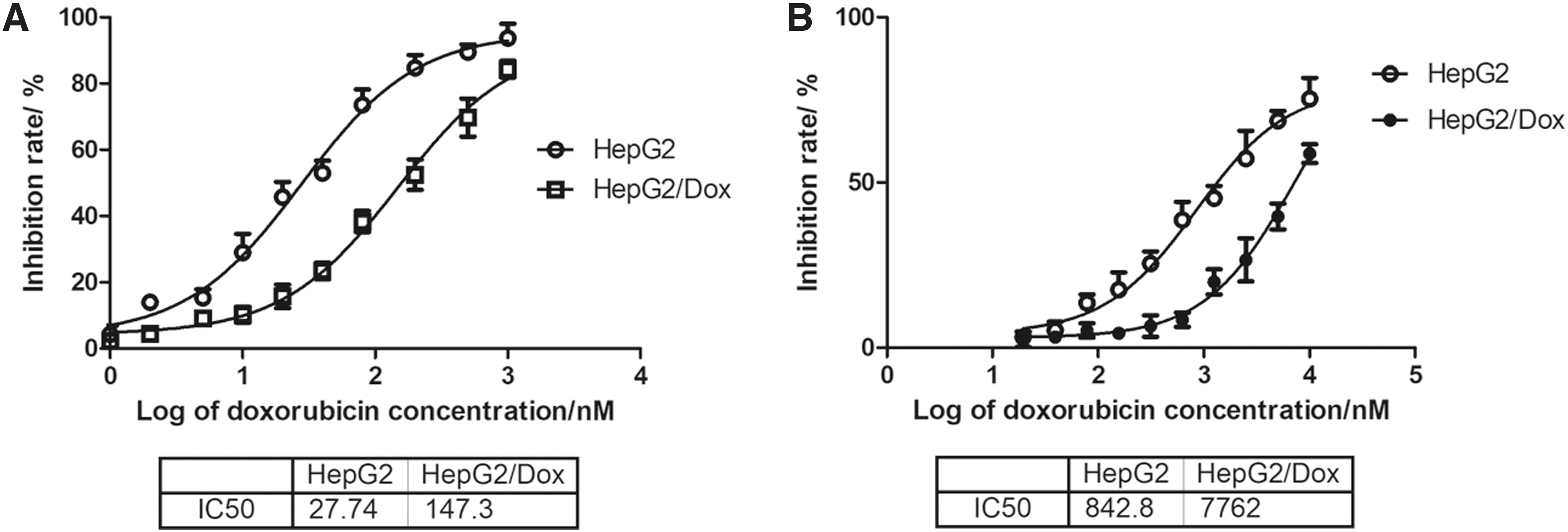
IC50 value is calculated in miR-183-transfected HepG2 and HepG2/Dox cells. HepG2 cells and HepG2/Dox cells were transfected with miR-381 human miRNA expression plasmid and treated with increasing concentrations of doxorubicin
IC50 Values for Doxorubicin and Cisplatin in HepG2 Cells and HepG2/Dox Cells
miR-381/β-catenin regulates the expression of ABCB1 in liver cancer cells
To further explore the molecular mechanism of ABCB1 in chemotherapeutic drug-resistant cells, miR381 human miRNA expression plasmid and control plasmid express microRNA precursors from CMV promoter (pCMVMIR) vector were transfected into HepG2/Dox cells. As shown in Figure 5A, the results showed that the expression of β-catenin and ABCB1 was decreased in miR-381-transfected HepG2/Dox cells compared with that in control vector-transfected HepG2/Dox cells. Conversely, miR-381 anti-miRNA oligonucleotides (AMO) and pCMV-MIR vectors were transfected into HepG2/Dox cells, and the results showed that the expression levels of β-catenin and ABCB1 were upregulated in miR-381 AMO-transfected HepG2/Dox cells, compared with the control vector group (Fig. 5B).
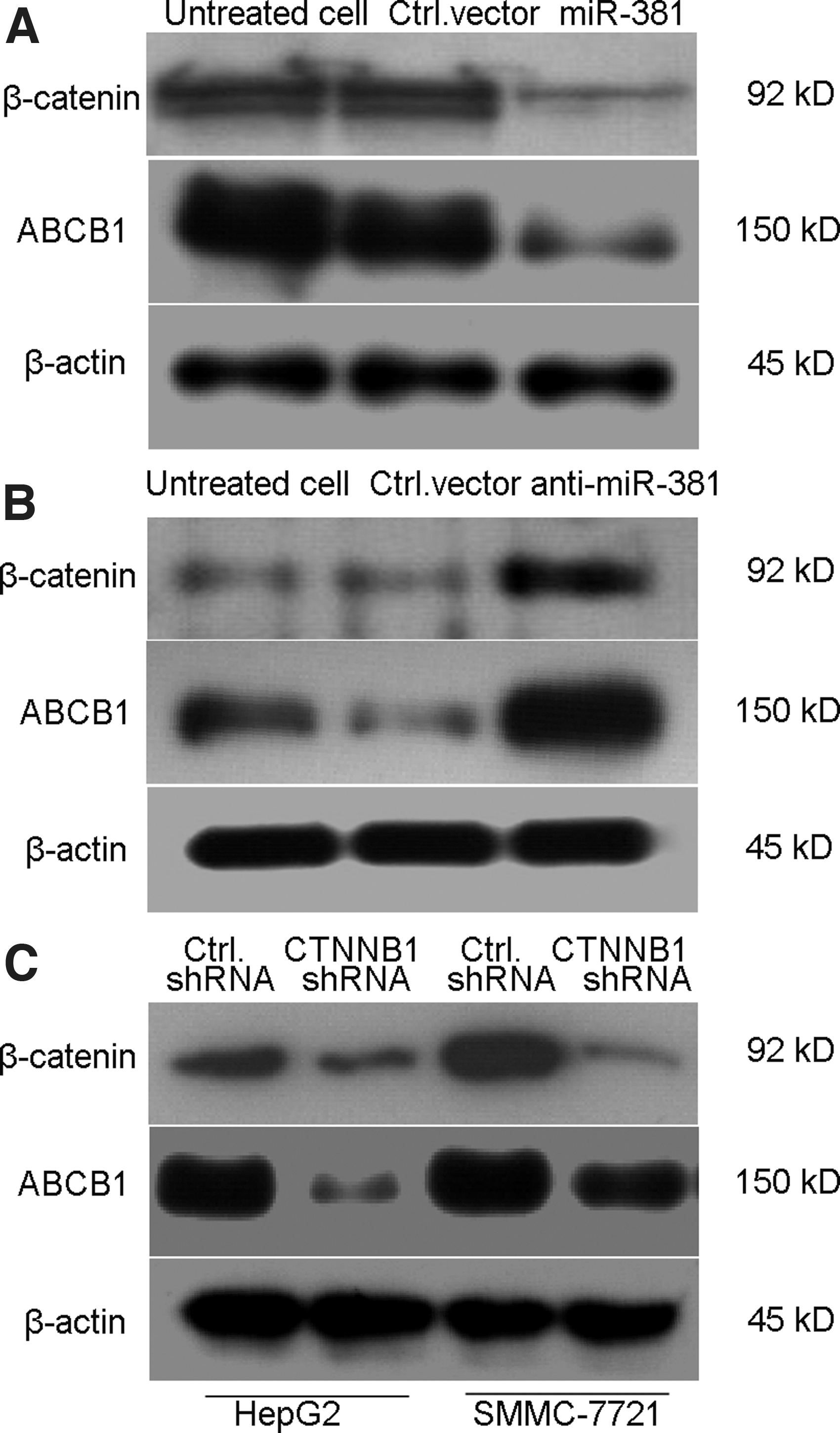
The expression of ABCB1 in liver cancer cells was regulated by miR-381/β-catenin.
Next, to confirm whether the expression of ABCB1 is regulated and mediated by the β-catenin-related pathway, we first knocked down the expression of β-catenin in HepG2 and SMMC-7721 cells. The expression of ABCB1 was tested by Western blotting analysis. As shown in Figure 5C, the level of ABCB1 decreased in CTNNB1 shRNA-transfected HepG2 cells and SMMC-7721 cells. All data revealed that miR-381 regulated the expression of ABCB1 and reversed the chemotherapeutic drug resistance, mainly regulated by the expression of the β-catenin-related pathway in liver cancer cells.
CTNNB1 and ABCB1 have a positive correlation in liver cancer patients
We analyzed data from TCGA (
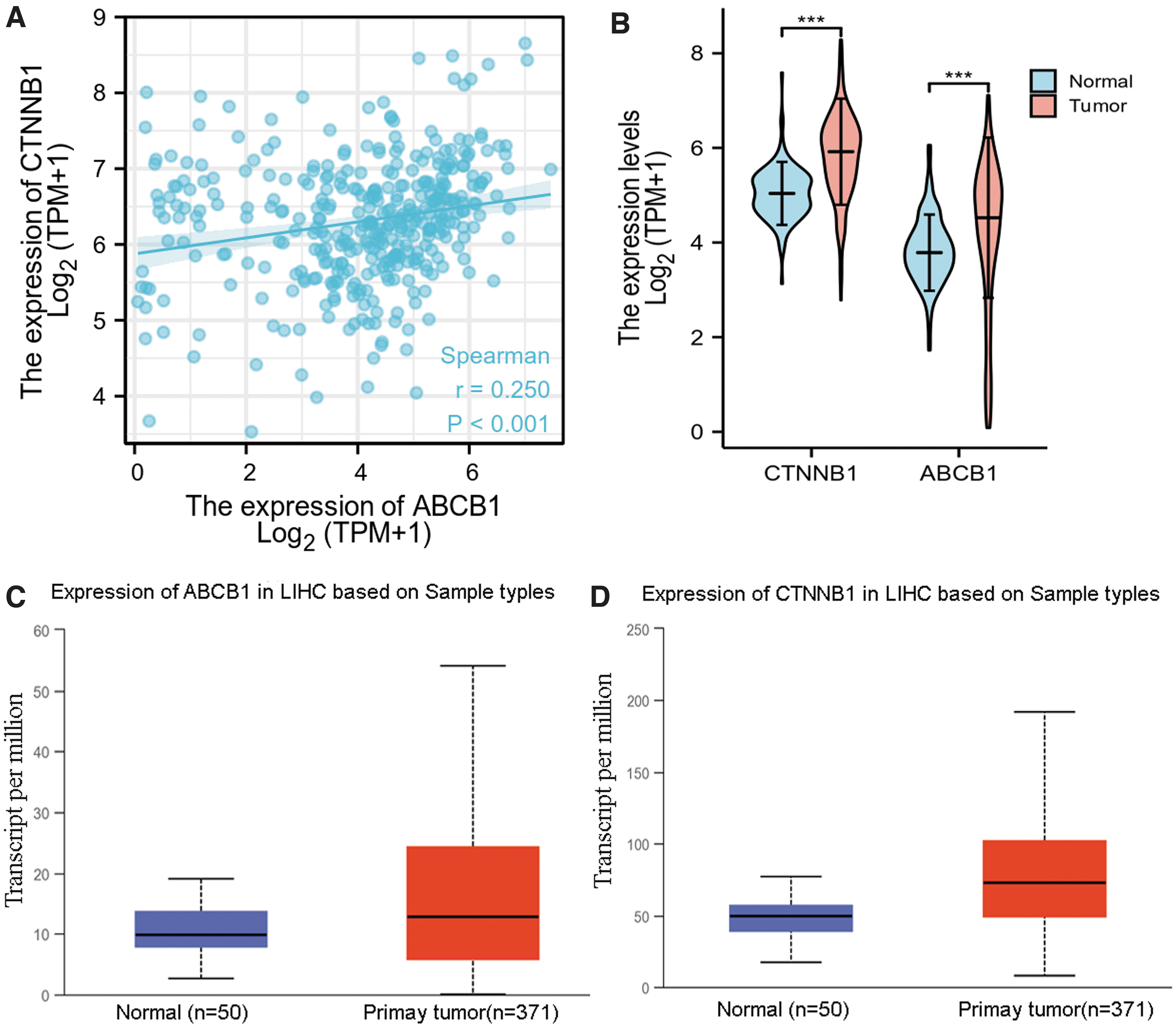
Higher expression of CTNNB1 and ABCB1 in LIHC patients was associated with shorter survival probability compared with lower expression of CTNNB1 and ABCB1 in patents.
The Statistical Description of CTNNB1 and ABCB1 in LIHC Patients
IQR, interquartile range; LIHC, liver hepatocellular carcinoma; SD, standard deviation; SE, standard error.
Increased expression of CTNNB1 and ABCB1 is found in liver cancer patients
Next, we analyzed the RNAseq data from the TCGA LIHC (
Relationship between ABCB1, CTNNB1, and clinicopathological characteristics of liver cancer patients
The association between the expression of ABCB1 and CTNNB1 and clinicopathological characteristics, including gender, age, race, weight, height, BMI, tumor status, pathologic stage, T stage, N stage, M stage, histologic grade, residual tumor, adjacent hepatic tissue inflammation, AFP concentration, albumin concentration, prothrombin time, Child-Pugh grade, Ishak fibrosis score, vascular invasion, overall survival (OS) event, disease-specific survival (DSS) event, and progression-free interval (PFI) event status of LIHC patients, was investigated using the RNAseq data in level 3 HTSeq-FPKM format in TCGA LIHC project. As shown in Table 3, 187 patients were classified as having high ABCB1 expression, and 187 patients were classified as having low ABCB1 expression. As shown in Table 3, the expression of ABCB1 was significantly correlated with gender (p < 0.001), age (p < 0.001), weight (p = 0.010), height (p < 0.001), residual tumor (p = 0.026), and AFP concentration (p < 0.001) in LIHC patients. There was no significant correlation between ABCB1 expression and other clinicopathological factors, including race (p = 0.298), BMI (p = 0.312), tumor status (p = 0.987), T stage (p = 0.298), N stage (p = 0.624), M stage (p = 0.123), pathologic stage (p = 0.098), histologic grade (p = 0.199), adjacent hepatic tissue inflammation (p = 0.723), albumin concentration (p = 0.736), prothrombin time (p = 0.097), Child-Pugh grade (p = 0.070), Ishak fibrosis score (p = 0.204), vascular invasion (p = 0.462), OS events (p = 0.447), DSS events (p = 0.479), and PFI events (p = 1.000). Furthermore, the data in Table 4 showed that the level of CTNNB1 in LIHC patients was significantly correlated with AFP concentration (p = 0.02) and Child-Pugh grade (p = 0.005) and there was no significant correlation with gender (p = 0.825), age (p = 0.232), race (p = 0.170), weight (p = 0.505), height (p = 0.957), BMI (p = 0.528), tumor status (p = 0.336), pathologic stage (p = 0.135), T stage (p = 0.503), N stage (p = 0.623), M stage (p = 0.622), histologic grade (p = 0.583), residual tumor (p = 0.218), adjacent hepatic tissue inflammation (p = 0.980), albumin concentration (p = 1.000), prothrombin time (p = 0.658), Ishak fibrosis score (p = 0.888), vascular invasion (p = 0.978), OS event (p = 0.447), DSS event (p = 1.000), and PFI event (p = 0.836).
Analysis of CTNNB1 Expression and Baseline Data
Analysis of ABCB1 Expression and Baseline Data
DSS, disease-specific survival; PFI, progression-free interval; OS, overall survival.
The diagnostic and prognostic value of ABCB1 and CTNNB1 expression in LIHC patients
Next, we tested whether ABCB1 and CTNNB1 could be used as independent prognostic markers in LIHC patients. Univariate Cox regression analysis was based on the TCGA dataset. Univariate analysis revealed that pathologic stage, T stage, M stage, and tumor status were significantly associated with OS. There were no statistically significant factors associated with OS in LIHC patients, including age, sex, race, weight, height, N stage, histologic grade, residual tumor, adjacent hepatic tissue inflammation, Ishak fibrosis score, vascular invasion, prothrombin time, AFP concentration, albumin concentration, and the expression of ABCB1 and CTNNB1.
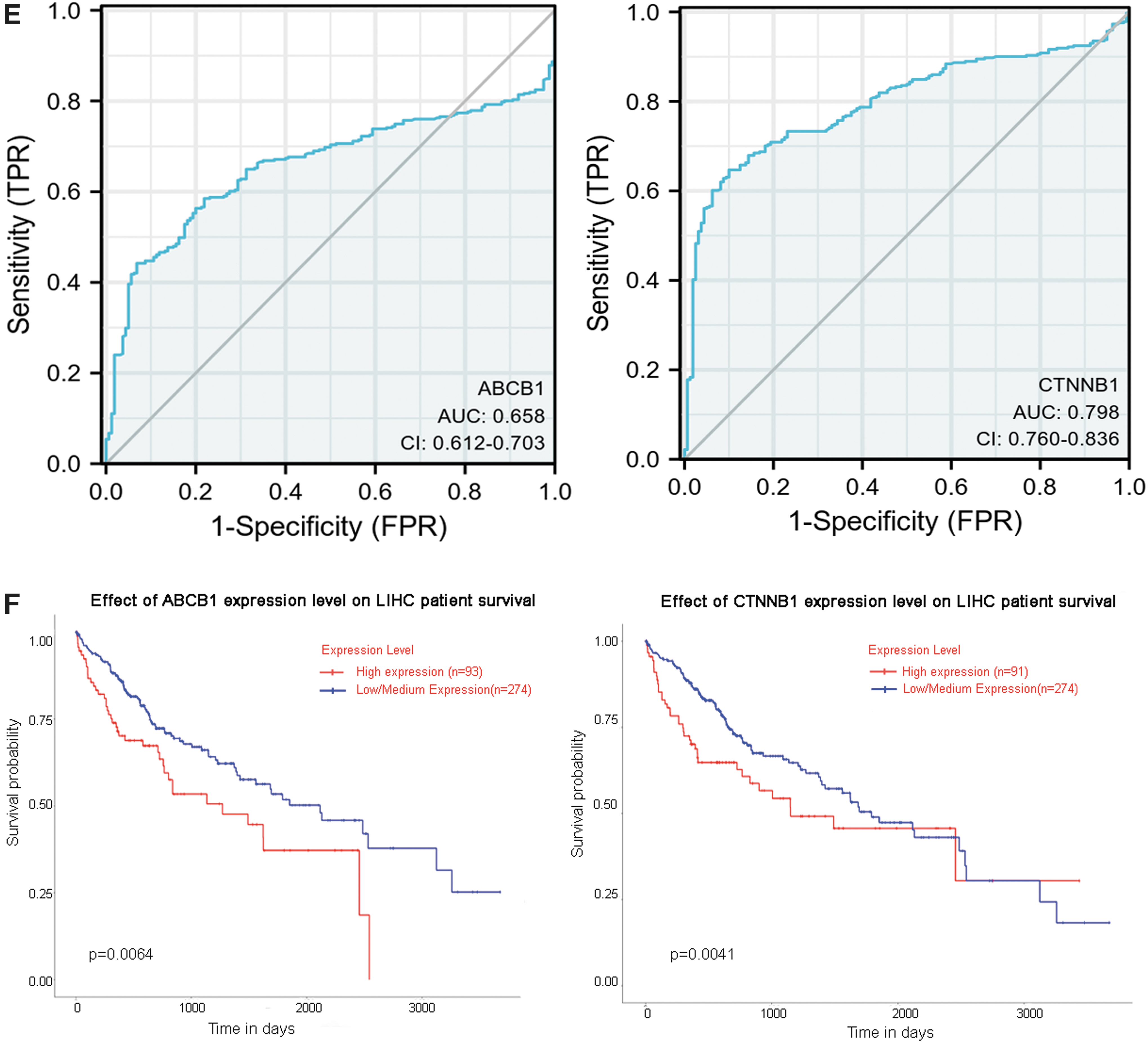
We conducted ROC curve analysis of ABCB1 and CTNNB2 expression data to evaluate the diagnostic value of these genes. The area of CTNNB1 was 0.798, which revealed that in predicting normal and tumor outcomes, the predictive ability of the variable CTNNB1 had a moderate accuracy (AUC = 0.798, CI = 0.760–0.836). In addition, the area of ABCB1 was 0.658, suggesting that the predictive ability of the variable ABCB1 had a lower accuracy (AUC = 0.658, CI = 0.612–0.703) (Fig. 6E). All data suggested that the expression of ABCB1 or CTNNB1 was not an independent diagnostic factor in LIHC patients (Table 5).
Univariate Analysis in LIHC Patients from TCGA Database
HR, hazard ratio.
High expression of ABCB1 and CTNNB1 indicates poor prognosis of liver cancer patients
The relationship between ABCB1 and CTNNB1 expression and clinical characteristics of LIHC patients in the GEPIA database. As shown in Figure 6F, high expression of ABCB1 was associated with a worse survival probability in LIHC patients (p = 0.0064). Similarly, higher levels of CTNNB1 were also associated with poor survival probability in LIHC patients (p = 0.041). All data revealed that higher levels of ABCB1 and CTNNB1 indicated poor prognosis in patients with liver cancer.
Discussion
The incidence of HCC has gradually increased worldwide. MDR is the biggest obstacle in clinical liver cancer treatment (Ramos-Penafiel et al., 2018). Thus, a better understanding of chemotherapeutic drug resistance mechanisms is crucial to improve the early diagnosis of liver cancer, develop novel targets to overcome drug resistance in liver cancer, improve the therapeutic effect of liver cancer, delay tumor recurrence and metastasis, and improve the prognosis of patients (Luo et al., 2020). ABCB1 is the first ABC transporter subtype to be discovered. Under normal circumstances, ABCB1 has a cytoprotective function, which can transport harmful toxins produced by human metabolism out of the cell. However, in chemotherapeutic drug-treated patients, ABCB1 normally acts as an efflux pump that transports chemotherapeutic agents out of cells and contributes to chemotherapeutic drug resistance (Yuan et al., 2017). In this study, we constructed doxorubicin-resistant HepG2/Dox cells with paired doxorubicin-sensitive HepG2 cells. The Western blotting analysis showed that ABCB1 expression was significantly increased in HepG2/Dox cells compared to HepG2 cells.
To determine how ABCB1 expression was regulated in doxorubicin-resistant HepG2 cells, we first predicted the possible miRNAs using TargetScan online tools. The results showed that the position 501–530 of ABCB1 3′ UTR might be the putative miRNA targeting site with hsa-miR-381. The dual-luciferase activity assay results showed that ABCB1 was directly targeted by hsa-miR-381 and negatively regulated by has-miR-381. In HepG2/Dox cells, miR-381 exerted antitumor effects and reversed chemotherapeutic drug resistance. This was consistent with several studies by Qiao et al. (2019), who have found that miR-381 functions as a tumor suppressor in pancreatic cancer; miR-381 and miR-489 were reported to inhibit the proliferation of gastric cancers by targeting CUL4B through Wnt/beta-catenin signaling (Fang et al., 2019). In addition, miR381 was found to regulate cancer progression in colorectal cancer (Han et al., 2021), osteosarcoma cancer (Yang et al., 2020), and ovarian cancer (Yang et al., 2020).
A miRNA can bind to several target genes at the 3′ UTR of mRNA, and multiple miRNAs can also target a single mRNA. Next, we found that miR-381 was also directly targeted at position 807–835 of CTNNB1 3′ UTR and decreased the luciferase activity of CTNNB1. This suggests that miR-381 negatively regulates the Wnt/β-catenin signaling pathway by directly targeting CTNNB1 in doxorubicin-resistant HepG2 cells. The Wnt/β-catenin signaling pathway is involved in chemotherapeutic drug resistance in various human cancers. For example, β-catenin activation is associated with castration resistance in prostate cancer (Patel et al., 2020). Inhibition of the Wnt/β-catenin pathway is a new therapeutic method for drug-resistant pancreatic cancer (Ryu et al., 2021). Moreover, β-catenin has been reported to be involved in the chemosensitivity of drug-resistant breast cancer cells (Alshaer et al., 2019; Alkaraki et al., 2020), prostate cancer (Khurana and Sikka, 2019), gastric cancer (Ryu et al., 2019), and cisplatin-resistant human cervical carcinoma (Hou et al., 2020). In this study, our results showed that miR-381 could directly target the 3′ UTR of targeted genes, including CTNNB1 and ABCB1, and reverse the chemotherapeutic drug resistance of HepG2/Dox cells.
Furthermore, the diagnostic and prognostic values of CTNNB1 and ABCB1 were evaluated in patients with LIHC. The expression of ABCB1 was significantly correlated with gender (p < 0.001), age (p < 0.001), weight (p = 0.010), height (p < 0.001), residual tumor (p = 0.026), and AFP concentration (p < 0.001) in LIHC patients, and the level of CTNNB1 in LIHC patients was significantly correlated with AFP concentration (p = 0.02) and Child-Pugh grade (p = 0.005). ROC curve analysis showed that the predictive ability of the variable CTNNB1 had a moderate accuracy (AUC = 0.798 and CI = 0.760–0.836) and the variable ABCB1 had a lower accuracy in predicting LIHC (AUC = 0.658 and CI = 0.612–0.703). Interestingly, both ABCB1 (p = 0.0064) and CTNNB1 (p = 0.041) were associated with poor survival probability in LIHC patients, suggesting that higher levels of ABCB1 and CTNNB1 indicated poor prognosis in LIHC patients.
Thus, in this study, all data showed that overexpression of hsa-miR-381 partially reversed the MDR of HepG2 cells by directly targeting and negatively regulating the expression of CTTNB1 and ABCB1. Moreover, a high expression of ABCB1 or CTNNB1 indicated poor prognosis in liver cancer patients.
Footnotes
Disclosure Statement
No competing financial interests exist.
Funding Information
The work was supported by Luzhou Science and Technology Bureau-Southwest Medical University Joint Project 2015LZCYD-S01 (2/15) and Sichuan Provincial Department of Science and Technology Project 2020ZHFP0058.