
Abstract
One of the main health issues in the modern world is cancer, with breast cancer (BC) as one of the most common types of malignancies. Different environmental and genetic risk factors are involved in the development of BC. One of the primary genes implicated in cancer development is the p53 gene, which is also known as the “gatekeeper” gene. p53 is involved in cancer development by interacting with numerous pathways and signaling factors, including microRNAs (miRNAs). miRNAs are small noncoding RNA molecules that regulate gene expression by binding to the 3′ untranslated region of target mRNAs, resulting in their translational inhibition or degradation. If the p53 gene is mutated or degraded, it can contribute to the risk of BC by disrupting the expression of miRNAs. Similarly, the disruption of miRNAs causes the negative regulation of p53. Therefore, the p53/miRNA axis is a crucial pathway in the progression or prevention of BC, and understanding the regulation and function of this pathway may contribute to the development of new therapeutic strategies to help treat BC.
Introduction
Breast cancer (BC) is currently among the most common and malignant cancers. This condition has emerged as a major concern in today’s society, especially among women, since it leads to financial burden and loss of lives. According to the global statistics report in 2020, about 2.26 million new BC cases and 685,000 deaths from this cancer were recorded. While it is not possible to entirely prevent certain factors that contribute to BC, in some cases, health interventions such as changing one’s lifestyle and eating habits, as well as environmental factors and minimizing radiation exposure, can reduce the incidence of cancer (Khodabandeh et al., 2022, Sung et al., 2021, Xu et al., 2023). BC is a form of cancer that often metastasizes to other organs such as the brain, liver, lungs, and bones, making it difficult to cure. However, early detection can significantly improve the prognosis and survival rate of patients (DeSantis et al., 2016). Mammography is a commonly used screening method that has been proven to effectively reduce the mortality rate of patients. Other screening approaches, such as magnetic resonance imaging (MRI), have also been examined and implemented over the past decade due to their high sensitivity (Alabousi et al., 2021, Sun et al., 2017). There are several risk factors associated with BC, including sex, age, exposure to estrogen, family history, gene mutations, and unhealthy lifestyle choices (Sargazi et al., 2023). BC predominantly affects women, with the number of cases being 100 times higher in women than in men. Despite the increasing incidence of BC worldwide, the mortality rate has decreased due to widespread early screening and advanced medical therapies. Biological treatments have been developed in recent years and proven to be beneficial for patients with BC (Kashyap et al., 2022, Youn and Han, 2020). The p53 protein is a transcription factor that is frequently mutated in human cancers and is involved in regulating a variety of cellular processes, such as cell cycle, apoptosis, angiogenesis, metabolism, cell senescence, and DNA repair mechanisms. It is estimated that around 30% of BC tumors exhibit mutations in the TP53 gene. Recent research has shown that the frequency, spectrum, and timing of these mutations may vary depending on the molecular subtype of the disease (Klusmann et al., 2016, Xiao et al., 2020). The frequency of p53 mutation in BC is lower than in other solid cancers and is associated with a poor prognosis. Patients with p53-mutated BC are resistant to some chemotherapy drugs and are associated with more invasive power and worse overall survival (Dajti et al., 2024, Gasco et al., 2002).
Noncoding RNAs (ncRNAs) are RNA molecules transcribed from the nonprotein-coding regions of the genome. Although once considered “junk DNA,” a growing body of evidence has demonstrated the critical role of ncRNAs in various biological processes, including development, differentiation, stress response, and disease pathogenesis. The different types of ncRNAs, such as microRNAs (miRNAs), long noncoding RNAs (lncRNAs), PIWI-interacting RNAs (piRNAs), and circular RNAs, have been found to efficiently regulate gene expression and modulate each other’s expression levels. In cancer, these ncRNAs have been reported to function as either oncogenes or tumor suppressors, depending on the type of cancer and the cellular context. They play a significant role in cancer initiation, progression, and metastasis and are being studied as potential diagnostic and therapeutic targets for cancer treatment (Ashrafizadeh et al., 2024, Cui et al., 2019, Parfenyev et al., 2021). The tumor suppressor p53 protein and miRNAs both play a key role in regulating gene expression and are involved in the development and progression of cancer. According to previous studies, p53 can directly regulate the expression of certain miRNAs, and conversely, these miRNAs can regulate the expression of p53 and its downstream targets (Chen et al., 2021) (Fig. 1). p53 regulates the function of miRNAs in two steps: In the first stage, which is the transcription stage, p53 binds to the promoters of miRNA genes and controls their transcription, and in the other stage, p53 regulates the maturation of miRNAs by affecting the RNA-induced silencing complex (RISC). Research has shown that miRNAs that are regulated by p53 not only modulate the p53 pathway but also regulate other pathways that are involved in various diseases (Liao et al., 2014). Therefore, in this review, we decided to examine the interaction between p53 and miRNAs in BC. However, more studies are needed to fully understand the mechanisms of p53 and miRNA involvement in BC and how to target them as an important strategy in the treatment of BC.
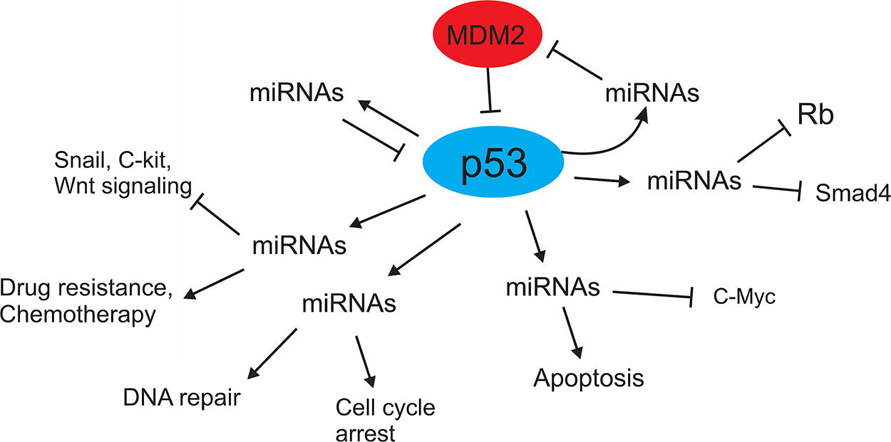
Correlation of p53 with miRNAs. This figure indicates that p53 regulates many important functions of the cell, including DNA repair, signaling pathways, angiogenesis, apoptosis, MDM2, and cell cycle arrest through miRNAs. Also, some miRNAs that affect p53 through some pathways have been shown. MDM2, mouse double minute 2 homolog; RB, retinoblastoma; SMAD4, mothers against decapentaplegic homolog 4.
Breast Cancer
After skin cancer, BC is the most common cancer among women, affecting more than 1 in 10 women worldwide, and its prevalence varies among different populations (Yedjou et al., 2019). According to the literature, the incidence of BC is the same among Black and White women. However, the incidence rate of BC in Black women under the age of 45 is higher than that in White women, whereas the incidence rate of this cancer is higher in White women over the age of 60 (Copeland et al., 2013). BC is divided into different categories based on various criteria, including etiology, response to treatment, molecular characteristics, and clinical manifestations. One of the classifications of BC is according to the expression of biomarkers such as progesterone receptor, estrogen receptor, and human epidermal growth factor receptor 2 (Beilankouhi et al., Fakhrioliaei et al., 2024, Momenimovahed and Salehiniya, 2019). BC is a complex disease with various risk factors involved in its development. Some of these factors include age, blood type, age of menarche, age of menopause, abortion, pregnancy characteristics, genetic factors, family history of BC, lactation, and lifestyle factors (Beilankouhi et al., 2024, Kim et al., 2015, Maghsoodi et al., 2023, Momenimovahed and Salehiniya, 2019). Research indicates that individuals with blood type AB and rhesus (Rh) negative have a lower risk of developing BC, whereas those with blood type A and Rh positive have a higher risk of developing BC (Meo et al., 2017, Saxena et al., 2015). It has been demonstrated that menstruation at a younger age increases the risk of BC by about two times (Thakur et al., 2017). Individuals over 50 years old have a higher risk of developing BC (Laamiri et al., 2015). Moreover, a high incidence of abortion is associated with an increased risk of BC (Bhadoria et al., 2013). A functional study revealed that women who give birth to their first child before the gestational age of 8 months show a higher incidence of BC (Innes and Byers, 2004). In addition, lactation has been shown to be a protective factor against BC and reduces its incidence (Laamiri et al., 2015). Lifestyle factors, such as obesity, alcohol consumption, inactivity, smoking, diet, and sleeping patterns, are also related to BC (Kerlikowske et al., 2017, Khodabandeh et al., 2022, Lu et al., 2017, Miller et al., 2018, Sargazi et al., 2023, Tong et al., 2014). Mutations in genes associated with BC including TP53, CHEK2, Notch, PTEN, CDH1, PI3K/AKT/mTOR, Wnt/β-catenin, STK11, PALB2, and miRNA genes increase the risk of BC (Allison, 2012, Colditz et al., 2012, Fattahi et al., 2020, Mohammadi et al., 2024). One of the most common mutations in BC is the p53 mutation, which occurs in ∼30% of BC cases. Based on gene expression patterns, BC is divided into four groups: HER-2 positive, luminal like (luminal-A or luminal-B), basal like, and normal like (Marvalim et al., 2023). miRNAs play a role in the regulation of posttranscriptional cellular processes such as migration, apoptosis, proliferation, and angiogenesis and potentially contribute to the regulation of BC. They are also regarded as biomarkers in the prognosis, diagnosis, and progression of BC (Yang and Liu, 2020). If BC is detected in the early stages, it can be successfully treated. Common methods for BC diagnosis are mammography, MRI, and ultrasound scan (Berg et al., 2004, Tang et al., 2024). Mammography has a higher error rate due to false positive and negative results and has more disadvantages due to high ionizing radiation (de Gonzalez and Darby, 2004, Marmot et al., 2013). Ultrasound screening is another method of BC diagnosis, which involves utilizing echoes directed toward the breast. Ultrasound is not very beneficial since it is not able to distinguish between benign and malignant tumors (Kwon and Lee, 2016). Compared with mammography and ultrasound, MRI is more sensitive in detecting small and aggressive abnormalities and is effective in detecting dense cancers. However, due to the location of the breast, MRI is prone to errors in BC diagnosis and is relatively expensive (Aldhaeebi et al., 2019, Orel and Schnall, 2001). Another method that does not have the disadvantages and limitations of the abovementioned methods is microwave-based imaging, which is favored due to its high sensitivity, safe radiation, low cost, and ease of use (Aldhaeebi et al., 2020).
p53
p53 is one of the most significant tumor suppressors, which is also known as a guardian gene. The primary function of p53 is involvement in apoptosis, DNA repair, and cell cycle, as well as the regulation of metabolic pathways leading to cell senescence (Engeland, 2022, Lane, 1992). TP53 is one of the most commonly mutated genes in different cancers. It is estimated that, on average, about half of all cancers have mutations in the TP53 gene. It has been shown that apart from genetic inactivation, in some cancers, p53 function is lost as a result of other factors, such as the overexpression of MDM2 or viral oncoproteins. One of the target proteins of p53 is the p21 protein. p53 binds to the gene encoding p21 CDKN1A, thus activating it. In turn, p21 contributes to the inhibition of cyclin-CDK combinations that cause the hyperphosphorylation of the retinoblastoma protein RB. (El-Deiry et al., 1994, Xia et al., 2023). The p53-p21-RB signaling regulates critical cellular functions including cell division. For example, the p53-p21-RB axis controls the mitogen-activated protein (MAP) kinase pathway, which includes the MAP kinase MAP3K5/ASK-1 (Engeland, 2022). Wild-type (WT) p53 is involved in DNA replication during the S phase of the cell cycle and helps to safeguard the genome by improving the processivity of replication forks. Missense mutations in p53, which are the most common types of mutation, typically occur within the DNA-binding domain of the protein (Stiewe and Haran, 2018, Xiao et al., 2020). Some of the functional roles of p53 include DNA repair, cell cycle regulation, metastasis, and senescence. In the presence of DNA damage, p53 is activated and regulates the cell cycle in G1-S and G2-M phase restriction points, which are checkpoints that are important in the cell division phase. In addition, activated p53 causes cell cycle arrest through the regulation of p21, 14-3-3σ, Cdc25c, and GADD45. Both intrinsic and extrinsic apoptotic pathways are regulated by p53, which induces apoptosis (Chen, 2016, Marvalim et al., 2023, Mijit et al., 2020, Williams and Schumacher, 2016). The drug and chemotherapy resistance of different cancer types is mostly related to cancer stem cells (CSCs), and mutated p53 plays a fundamental role in the maintenance of CSCs. Mutant p53 prevents the suppression of CD44, c-KIT, NANOG, and OCT4, which are markers of CSCs, induced by WT p53 (Borrero and El-Deiry, 2021, Levine and Oren, 2009, Lu and El-Deiry, 2009, Olivos and Mayo, 2016, Zhu et al., 2020). In addition to the effects of chemotherapy agents, genotypic changes also play an important role in the fate of cancer cells after exposure to these agents. In patients with BC, mutated p53 is involved in resistance to adverse drug reactions (Lin et al., 2019).
miRNAs
Today, with the increase of functional studies, it is clear that ncRNAs, which were previously assumed to be insignificant, have crucial roles in regulating cellular activities. ncRNAs serve various functions in cellular activities including transcription, replication, gene silencing, regulation of gene expression, RNA modification, and gene stability (Chen et al., 2023, Panwar et al., 2014). ncRNAs are divided into two groups based on their size and structural or regulatory features. One group consists of ncRNAs with <200 nucleotides, which include miRNAs, piRNAs, and small nucleolar RNAs, and the other group comprises ncRNAs with more than 200 nucleotides, which are known as lncRNAs (Feng et al., 2018). miRNAs are a type of small noncoding RNA with a length of about 19–24 nucleotides, which were first discovered in 1993 and are involved in the regulation of gene expression after transcription (Huang et al., 2007, Lee et al., 1993, Mamalo et al., 2023, Mohammadi et al., 2024, Zhou et al., 2022). miRNAs play their role either by suppressing mRNAs or by degrading them. In recent years, various studies have been conducted on the role of miRNAs in different cellular processes, including proliferation, apoptosis, differentiation, and metabolism, as well as their involvement in the initiation, spread, and progression of cancer (Garzon et al., 2009, Tan et al., 2018). Changes in miRNAs involved in necroptosis and apoptosis can lead to pathogenicity and carcinogenesis by influencing physiological conditions. A growing number of studies show that miRNAs originate from specific tissues and have the capability to detect tissues that have caused diseases of unknown origin (Li et al., 2012, Rosenfeld et al., 2008). miRNA biosynthesis includes nuclear and cytoplasmic stages. Initially, the miRNA precursor that is transcribed by RNA polymerase II is cleaved in the nucleus by Drosha (which binds to the UG motif) and the DGCR8 dimer (which interacts with the UGU motif). Then, the pre-miRNA is transferred to the cytoplasm by exportin5, where it is broken down again by Dicer and produces the miRNA-miRNA* duplex. This duplex is subsequently combined with the Argonaute protein; thus, the miRNA-induced suppressor (miRISC) is formed, and the miRNA* that is the passenger strand is degraded. However, the strand that is selected as the guide strand is preserved to form the silencing complex (Gregory et al., 2004, He et al., 2020, Hill and Tran, 2021, Nguyen et al., 2015). miRNA biogenesis is regulated by various processes including posttranslational modifications, such as phosphorylation, sumoylation, and ubiquitylation, as well as cellular signaling pathways. In addition, according to previous studies, the biosynthesis of miRNAs can be regulated by lncRNAs and RNA-binding proteins. In general, it can be claimed that the biosynthesis of miRNAs is meticulously and consistently regulated, and disruption in miRNA biosynthesis is related to numerous diseases, including cancer (Treiber et al., 2019). mRNAs are usually affected by different miRNAs, and each miRNA usually targets several mRNAs. However, the type of miRNAs is diverse, since they can act as oncomiRs, which play a significant role in tumor growth, or apoptomiRs, which are involved in apoptosis [67]. Changes in miRNAs can affect cell signaling processes and regulate complex genetic networks. In addition to diagnostic applications, miRNAs can be used as therapeutic targets for specific manipulation. The significance of miRNA application lies in their ability to regulate different processes, and a single miRNA can even regulate the entire cellular pathway. For example, miR-34a-5p is known to be central to the regulation of the T-cell network. In contrast, a pathway can be regulated by several miRNAs, which leads to the creation of a complex and potent network. Although using miRNAs for therapeutic purposes is promising, many challenges still need to be addressed, such as targeting tissues and cell types, identifying methods for administration and tissue targeting, ensuring stability within the body, and achieving the desired intracellular effects. Consequently, only a limited number of miRNAs have been used as drugs (Diener et al., 2022, Gebert and MacRae, 2019, Hart et al., 2019). The methods used to detect miRNAs include traditional approaches and new technological methods. Traditional approaches include northern blotting, quantitative polymerase chain reaction, and microarray analysis. New technological methods with improved sensitivity and specificity for miRNA detection use signal amplification strategies, including hybridization chain reaction, rolling circle amplification, isothermal exponential amplification, nanoparticle-based amplification, and a combination of these amplification strategies (Akmal et al., 2017, Niu et al., 2015, Schwarzkopf and Pierce, 2016, Torrente-Rodríguez et al., 2016).
miRNAs in BC
Dysregulation of cell proliferation is one of the symptoms of BC. In healthy cells, the capacity for division is limited and is determined by the optimal compaction capacity. Numerous studies have shown that miRNAs play a regulatory role in cell proliferation and progression of the BC cell cycle through interactions with the protein kinase family and their inhibitors, the cyclin family, and other growth stimulators and suppressors (Nahta et al., 2015, Zhou et al., 2022). The role of miRNAs in the process of cancer is supported by studies that have gradually shifted from theoretical to experimental studies. The first study on the relationship between miRNAs and BC in terms of clinical pathology was conducted in 2005 (Iorio et al., 2005). It has been shown that LZTFL1, which is a target of miR-21, prevents the metastasis and proliferation of BC cells by reducing the level of miR-21, and the expression of LZTFL1 is increased through epithelial–mesenchymal transition (EMT). In summary, the miR-21/LZTFL1 axis in the mammary gland increases the nuclear translocation of β-catenin, which is involved in the EMT process (Wang et al., 2019). According to the literature, most of the miRNA genes are found in the fragile region or in the regions of the genome that are related to cancer. Interference of miRNAs with many cancer types including lung, breast, stomach, prostate, thyroid, and colon cancer has been documented (Iorio et al., 2005) .Investigating the relationship between a specific type of miRNA and cancer is complex since one type of miRNA may act as a tumor suppressor in certain cancers, whereas the same miRNA may function as an oncogene in another type of cancer. For example, miR-29 acts as a tumor suppressor in lung cancer and as an oncogene in BC (Fabbri et al., 2007, Gebeshuber et al., 2009, Zaman et al., 2012). Similarly, miR-23b induces apoptosis and decreases invasiveness in renal cell carcinoma cell lines, but it induces proliferation and invasion in bladder cancer cells. miRNAs have great potential in regulating gene replication. It is estimated that about 60% of mRNAs are controlled by miRNAs, and an miRNA can participate in different pathways and have different roles in cell growth, proliferation, and survival, which depend on the gene expression pattern and cell type (Reddy, 2015). One of the most common methods to evaluate the expression level of a large number of miRNAs in cancer cells is miRNA microarray analysis. Previous studies have indicated that miRNA profiles in cancer cells are significantly different from normal cells of the same tissue. A study on 10 normal breast tissue samples and 76 BC samples revealed that the levels of miR-125b, miR-21, miR-145, and miR-155 were significantly different between the two samples, and the analysis of these miRNAs could potentially be used for cancer diagnosis (Iorio et al., 2005). A study by Zhu et al. showed that the overexpression of miR-143 suppresses MAP3K7, extracellular signal-regulated kinase ERK5, and cyclin D1 and decreases BC cell viability, whereas the suppression of miR-143 increases the survival of BC cells (Zhou et al., 2017). Another study showed that the upregulation of miR-455 by inhibiting cyclin D1 associated with Cdc2 and CDK14 inhibits the proliferation of BC cells while upregulating tumor suppressor p21 (Wang et al., 2017). Therefore, based on these findings, it is pivotal to shed light on the role of miRNAs in BC, which may prevent BC or cause the progression of BC. MiRNAs could also play an essential role in the prognosis and treatment of BC.
Correlation of p53 and miRNAs in cancers
The p53 protein, which is known as the most fundamental tumor suppressor protein, is located inside the cell. The p53 pathway detects cellular stress, DNA damage, and mitogenic misinduction. By integrating these signals, p53 inhibits angiogenesis and induces apoptosis or causes DNA repair. Studies have indicated that p53 suppresses genes that cause cell cycle progression or decrease apoptosis (He et al., 2007). The p53 gene, which is known as the guard gene, maintains genomic stability and prevents genomic mutations. According to the literature, mutations in the p53 gene occur in many tumors, which proves that defects in this gene are linked to multiple cancers, including BC (Xu et al., 2017). p53 is negatively regulated by two important antagonists, MDM2 and MDMX. This regulation can be affected by some p53 target miRNAs. MDM2, which is a ubiquitin ligase and causes the degradation of p53, suppresses its transcription through two miRNAs, namely miR-605 and miR-145, by binding to p53 mRNA (Xiao et al., 2011, Zhang et al., 2013). miR-29 transcription is induced by p53 upon DNA damage, and this miRNA represses Ppm1d phosphatase, which phosphorylates and destabilizes p53, thus upregulating it (Ugalde et al., 2011). In a study conducted by Liu et al., it was shown that miR-151a-3p promotes the growth, proliferation, and migration of nasopharyngeal carcinoma cells by inhibiting the expression of p53 and the downstream pathways of p53 (Liu et al., 2019). Ma et al. reported that miR-16 suppresses the expression of p53, and the level of miR-16 is regulated by p53, indicating the relationship between miR-16 and p53. In contrast, p53 simultaneously regulates the expression of survivin, CDK6, and cyclin D1 through miR-16 (Ma et al., 2013). In one study, PC-3 prostate cancer cells were transfected with WT p53 and mutant p53, and analyzing the results revealed that, after WT p53 transfection, the expression of miR-145 was upregulated and the number of apoptotic cells increased; however, with mutant p53 transfection, the expression of miR-145 was not changed (Suh et al., 2011). miR-125b inhibits p53 expression through binding to the 3′ untranslated region of p53 mRNA and prevents apoptosis in human lung fibroblast cells and human neuroblastoma cells (Le et al., 2009). When DNA is damaged, the expression of p53 increases, which induces the expression of miR-335. The increased expression of miR-335 suppresses the expression of Rb1 mRNA, which is a communication link between tumor suppressors and p53 (Scarola et al., 2010). A study on the relationship between miRNAs and p53 showed that miR-143 and miR-145 can negatively affect the expression of MDM2. In contrast, increasing the expression of p53 induces the expression of miR-143 and miR-145 genes, which in turn raises the level of p53 through an adverse effect on MDM2. These findings indicate that the reduction of miR-143 and miR-145 expression decreases the effect of these miRNAs on MDM2, which contributes to the development of epithelial cancer by positively regulating cell proliferation and negatively regulating apoptosis (Zhang et al., 2013). According to previous studies, p53 regulates EMT through the transcriptional activation of miR-200c. The expression of ZEB1, KLF4, and BMI1 is induced by increasing the expression of miR-200c, and the expression of E-cadherin, which was decreased by p53 knockdown, is increased (Chang et al., 2011) (Fig. 1).
Correlation of p53 and miRNAs in BC
As we mentioned in the previous section regarding the relationship between p53 and miRNA in all types of cancers, in this section we will also examine their relationship in BC. One of the important factors known as BC suppressor is FBXO11, the defect of which is associated with the progression and metastasis of BC. FBXO11 inhibits the proliferation of BC cells by increasing the sensitivity of cancer cells to chemotherapy drugs and by inhibiting EMT, which is a factor for metastasis. Interestingly, FBXO11 increases the invasiveness of non-EMT BC cells. It also helps tumor progression by adversely affecting the p53/p21 axis (Bagger et al., 2018, Fan et al., 2019, Tekcham et al., 2020). Xue et al. found that the level of miR-621 is inversely related to the expression of FBXO11 in patients with BC. Moreover, the expression of FBXO11 is associated with p53, inducing p53 dendylation while suppressing its expression. Overall, this study showed that miR-621 increases the sensitivity of BC cells to PTX/CBP chemotherapy drugs by preventing the suppression of p53 by FBXO11. As a result, miR-621 can be used as a therapeutic target and biomarker in the follow-up and treatment of patients with BC (Xue et al., 2016). mir-205, which is one of the transcriptional targets of p53, reduces cell proliferation and combats triple negative BC through E2F1 and LAMC1 target genes (Piovan et al., 2012). A study by Wang et al. on miR-214 in BC tissues revealed that the expression of miR-214 increases in BC cells, which promotes the invasion of BC cells by repressing p53. In contrast, by enhancing the expression of p53, the effects of miR-214 expression are nullified (Wang et al., 2015). A study conducted by Gu et al. aimed to establish a relationship between miR-34 and p53 in TNBC and MCF-7 cell lines. Their findings indicated that miR-34 leads to apoptosis in these cells through p53. However, SIRT1, which is an NAD+-dependent protein deacetylase, is another target of miR-34 in the apoptosis induced by the p53/miR-34 axis. miR34 reduces p53 deacetylation in BC cells by inhibiting SIRT1. Deacetylated p53 causes apoptosis and suppression of cell cycle in the G1/S phase. However, p53/miR-34a and SIRT1 induce a positive feedback axis in the stimulation of p53-mediated apoptosis (Imani et al., 2018, Rokavec et al., 2014). Another study related to the relationship between miRNAs and p53 in BC showed that p53 binds to the miR-30a promoter and induces the transcription of both miRNA strands. Both miR-30a-5p and -3p have the ability to target ZEB2, which is a transcription factor involved in drug resistance, tumor cell migration, and EMT. In this regard, p53 inhibits ZEB2 expression through miR-30a. Therefore, this study established a fundamental relationship between p53, EMT, and miR-30a (di Gennaro et al., 2018). In a study conducted by Sharma et al. on the relationship between miR-191-5p and p53 in MCF7 cells, it was revealed that the overexpression of miR-191-5p causes the downregulation of SOX4. Since SOX4 has a direct relationship with p53, the reduction of SOX4 leads to a decrease in p53. Moreover, the sensitivity of MCF7 cells to doxorubicin, which is an anticancer drug, was investigated in this study, and it was observed that miR-191-5p promoted apoptosis by decreasing the IC50 of doxorubicin (Sharma et al., 2017). Similar studies in this field have shown that the expression of miR-590-3p is reduced in BC cells. However, after transfecting miR-590-3p into BC cells, it was found that it prompted p53 acetylation and increased its expression, leading to the induction of p21 and BAX expression and ultimately resulting in apoptosis and the suppression of BC cell survival (Abdolvahabi et al., 2019). Curcumin, which is used in cancer treatment, induces apoptosis and inhibits MCF-7 cell growth through the miR-19/PTEN/AKT/p53 axis (Li et al., 2014) (Fig. 2). Taken together, these results revealed that miRNAs in association with p53 can be used for various purposes, including prognosis, diagnosis, and treatment of BC.
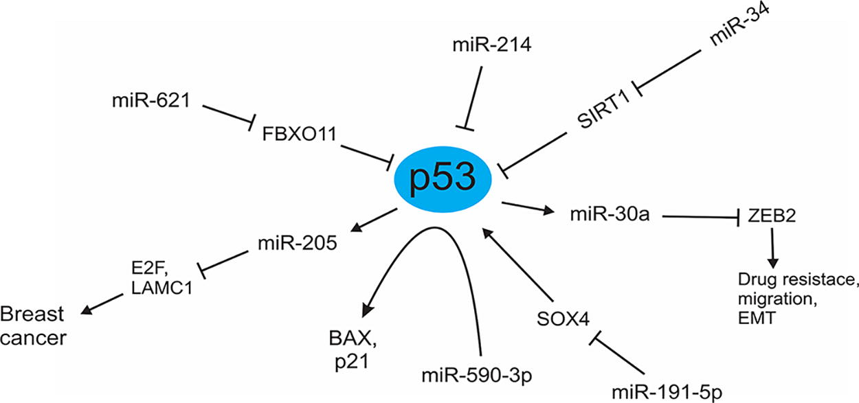
Interaction of p53 with miRNAs in BC. In this figure, the relationship between miRNAs and p53 is shown. As you can see in this figure, some miRNAs affect p53 directly and some through mediators and signaling pathways, or vice versa. For example, miR-214 directly inhibits p53 and promotes the invasion of BC cells. miR-34 activates p53 by inhibiting the SIRT1 pathway that inhibits p53. miR-191-5p inhibits p53 through the SOX4 pathway. On the other hand, p53 through miR-30a inhibits the ZEB2 pathway, which plays a role in drug resistance and migration of BC cells. Also, p53 through miR-205 with targeting E2F1 and LAMC1 genes, reduces cell proliferation and combats triple negative BC. BC, breast cancer; ZEB2, zinc finger E-box binding homeobox 2; SOX4, SRY-related HMG box transcription factor 4; LAMC1, laminin subunit gamma 1; E2F1, E2 promoter binding factor 1; miRNA, microRNA; SIRT1, sirtuin 1.
Conclusion
As previously mentioned, p53 is one of the most crucial regulatory factors in various cell pathways and activities, including apoptosis, cell cycle, angiogenesis, and oxidative stress, and acts as a gatekeeper or guardian gene. miRNAs are one of the components that p53 interacts with, and these molecules mutually regulate one another. Through miRNAs, p53 controls numerous critical cell activities, including angiogenesis and apoptosis. In contrast, miRNAs also regulate many cell activities through p53, which were outlined in this review. p53 regulates many cellular activities including the cell cycle by regulating genes related to the cell cycle. In this regard, it has been shown that p53 regulates these activities indirectly through miRNAs. In contrast, some miRNAs target p53 by targeting p53 directly and some target p53 regulators including MDM2 and MDM4. In contrast, it has been shown that p53 mutants that have “gain-of-function” activities are involved in tumorigenesis by regulating the expression of various miRNAs. Therefore, disruption in the function of each of these factors causes disruption in the other, leading to the development of various diseases, such as BC. Nevertheless, more studies are needed to clarify the relationship between miRNAs and p53 in BC to provide further insight into the additional mechanisms through which these two factors regulate each other and how this regulation occurs.
Footnotes
Acknowledgment
The authors would like to thank Kermanshah University of Medical Sciences for supporting this research.
Authors’ Contributions
R.S., S.M., and A.H. were involved in writing the article. H.K. and N.A. were involved in drawing figures and P.M. participated in the study design. P.B., A.H., A.K., and R.A.H.A.Z. were involved in data collection.
Ethical Approval
This article does not contain any studies on human participants or animals performed by any of the authors.
Data Availability Statement
Not applicable.
Author Disclosure Statement
The authors declare no potential conflict of interest with respect to research, authorship, and/or publication of this article.
Funding Information
The authors declare that the study had no funding.