




Abstract
The increasing use of silver nanoparticles (AgNPs) in aquaculture has raised concerns regarding their potential toxic effects on fish health, particularly on vital organs, such as the liver and kidneys. Licorice (Glycyrrhiza glabra) root, known for its medicinal and antioxidant properties, has gained attention as a natural agent capable of mitigating such toxicity. Furthermore, licorice extract can be used in the eco-friendly green synthesis of AgNPs, acting as both a reducing and stabilizing agent, as confirmed by characterization techniques including X-ray diffraction, Fourier-transform infrared spectroscopy, and transmission electron microscopy. This study aimed to evaluate the protective effects of dietary licorice root powder against AgNP-induced histopathological and physiological damage in common carp (Cyprinus carpio). A total of 150 fish were randomly assigned to seven dietary treatment groups for 56 days, including a control group, three groups receiving increasing doses of AgNPs (2.5, 5, and 7.5 mg/kg feed), and three groups receiving corresponding combinations of same amount of AgNPs with licorice root powder (2.5, 5, and 7.5 g/kg feed). Histopathological evaluation revealed that AgNPs alone induced severe liver and kidney damage, including hydropic degeneration, necrosis, and inflammatory infiltration. In contrast, fish receiving licorice-supplemented diets showed significantly reduced tissue lesions, indicating hepatoprotective and nephroprotective effects. In conclusion, licorice root powder effectively mitigated AgNP-induced toxicity and improved organ health in common carp. The combination of licorice and AgNPs offers a promising alternative to antibiotics in aquaculture, enhancing sustainability and fish welfare. Further studies are recommended to investigate the underlying molecular mechanisms and optimize application strategies in fish diets and to investigate another model of animal.
Introduction
Nanotechnology has gained significant attention in recent years due to its wide-ranging applications in fields such as medicine, agriculture, food production, and environmental science (Zaheer ud Din et al., 2023). Among its most notable developments are nanoparticles (NPs), particularly silver nanoparticles (AgNPs), which are known for their strong antimicrobial properties. However, the increasing use of AgNPs in aquaculture has raised concerns about their potential toxicity to aquatic organisms, particularly fish (Kakakhel et al., 2025).
Aquaculture plays a critical role in ensuring global food security and supporting economic development. Nevertheless, its rapid expansion is hindered by the frequent occurrence of infectious diseases, often exacerbated by intensive rearing conditions such as high stocking densities and limited water circulation. Conventional disease control strategies—including the use of antibiotics, antifungals, and antiparasitics, while initially effective—are associated with several drawbacks such as the development of antimicrobial resistance, environmental contamination, and harmful effects on nontarget organisms.
In response to these challenges, researchers are increasingly exploring natural alternatives to synthetic chemotherapeutics. Medicinal plants, which are rich in bioactive compounds with antioxidant, immunostimulant, and antimicrobial properties, represent a sustainable approach to enhancing fish health and performance. One such plant is licorice (Glycyrrhiza glabra), which contains beneficial phytochemicals including flavonoids, polysaccharides, and glycyrrhizin, and has demonstrated immune-enhancing and hepatoprotective effects in aquatic species.
Beyond its dietary applications, licorice root extract has also been utilized in the green synthesis of AgNPs. This eco-friendly approach employs plant-derived phytochemicals—such as hydroxyl, carboxyl, and amino groups—as natural reducing and stabilizing agents, eliminating the need for hazardous chemical reagents. The resulting AgNPs are biocompatible, stable, and effective against a wide range of pathogens.
The growing demand for sustainable and effective alternatives to conventional chemotherapeutic agents in aquaculture has driven increased interest in natural bioactive compounds and NPs. Among these, AgNPs are widely recognized for their potent antimicrobial activity; however, their application is not without risks. AgNPs can bioaccumulate in critical fish organs such as the liver, gills, and intestines, leading to oxidative stress, enzymatic alterations, and tissue damage (Lee et al., 2012; Naguib et al., 2020).
Despite their benefits, AgNPs can accumulate in fish tissues (e.g., liver, gills, intestines), inducing oxidative stress, inflammation, and histopathological damage (Kakakhel et al., 2021; Liaqat et al., 2021). However, the coadministration of licorice extract may offer protective effects against such NP-induced toxicity. Previous studies have shown that licorice can mitigate liver damage caused by toxicants like carbon tetrachloride in common carp (Malekinejad et al., 2010).
Materials and Methods
All procedures involving animals in this study were reviewed and approved by the Ethics & Research Registration Committee of the College of Veterinary Medicine, University of Sulaimaniyah. The research project was granted approval under Approval No. VMUS.EC. Doc 6-2024 in December 2024. All experimental protocols complied with institutional guidelines for the care and use of animals.
Preparation of G. glabra extract and green synthesis of AgNPs
Licorice roots (
To prepare the extract, the licorice root powder was subjected to solvent extraction using 80% methanol. The mixture was incubated in a shaker incubator (Lab Tech, EN61009) at room temperature (∼25°C) for 4 days. After incubation, the solution was filtered using standard filter paper, and the solvent was removed by rotary evaporation followed by thermal concentration using a thermocirculator (Lab Tech, D06121806). The resulting concentrated extract was stored at +4°C. The extraction ratio was optimized through preliminary trials to ensure adequate recovery of bioactive compounds for downstream applications.
The methanolic extract of licorice was then used in the green synthesis of AgNPs via a coprecipitation method. The synthesis was carried out at the Nanotechnology Laboratory, University of Raparin. For the reaction, 2.20 g of silver nitrate (AgNO3; CDH, catalog no. 640575) was dissolved in 50 mL of double-distilled water, and 1.00 g of sodium hydroxide (NaOH; Scharlau, UN 1823) was dissolved in 25 mL of the licorice extract. Both solutions were stirred separately at 70°C for 30 min. The NaOH–licorice solution was then added dropwise to the AgNO3 solution under constant stirring at 70°C. A visible color change from colorless to dark brown confirmed the formation of AgNPs.
The resulting suspension was centrifuged at 5,000 rpm for 30 min to collect the synthesized NPs. The pellet was then dried in an oven at 70°C for 24 h to remove any residual solvent, yielding 1.22 g of AgNP powder. The powder was stored at +3°C prior to characterization. A 0.05 mg sample was sent to the Photon Lab Center in Baghdad for structural and chemical analysis using X-ray diffraction (XRD), Fourier-transform infrared (FTIR) spectroscopy, and scanning electron microscopy, conducted with the X’Pert HighScore system (PANalytical).
Experimental animals
Experimental design and fish husbandry
The experiment was conducted at the Fish Disease Laboratory, College of Veterinary Medicine, University of Sulaimani, Iraq. A total of 150 common carp (Cyprinus carpio) were used in this study. The fish were obtained from a commercial fish farm located in TaqTaq, Sulaimaniyah Province. Their body weights ranged from 65 to 95 g, with an average of 81.73 g.
Before the start of the feeding trial, the fish were acclimated to laboratory conditions for 27 days and fed a commercial pelleted diet. The experimental feeding period lasted for 9 weeks. The chemical composition of the basal diet was as follows: crude protein 30.46%, crude fat 2.1%, crude fiber 4.8%, and dry matter 87.4%, as per the recommendations of the National Research Council.
Experimental system
Twenty-one plastic tanks (70 L) were used in this trial, representing seven groups with three replicates each. Each tank is provided with proper continuous aeration by Chinese air compressors, Hailea ACO-318 and 11 small aquarium air pumps, Luckiness 828 (power: 5-watt, air flow: 3.5 L/min), and stocked with five fish. The tanks (replicates) were randomly allocated to minimize differences among treatments.
Experimental design
The experiment uses a completely randomized design consisting of seven groups and three replicates with five fish per replicate. Treatments are as follows: Negative control (G1) group: Standard diet without any licorice and nano silver. Group 2 (G2): Adding 2.5 mg NPs/kg diet. Group 3 (G3): Adding the 5 mg NPs/kg diet. Group 4 (G4): Adding the 7.5 mg NPs/kg diet. Group 5 (G5): Adding the 2.5 g of licorice roots powder/kg diet with 2.5 mg AgNPs. Group 6 (G6): Adding the 5 g of licorice roots powder/kg diet with 5 mg AgNPs. Group 7 (G7): Adding the 7.5 g of licorice roots powder/kg diet with 7.5 mg AgNPs.
Diet formulation: Diet preparation and feeding protocol
The dietary ingredients and chemical composition of the fish diets were formulated based on the guidelines of the NRC (2011). The experimental diets were prepared using standard commercial ingredients obtained from local markets in Sulaimani city and were supplemented with licorice (G. glabra) root powder and AgNPs.
Seven different diets were formulated to include various levels of licorice root powder and AgNPs, both individually and in combination, according to the experimental design. The ingredients were thoroughly mixed, and the pellets were prepared using a Kenwood multiprocessor. The resulting pellets were air-dried at room temperature for 4 days and subsequently ground into fine particles.
During the first week of the trial, fish were fed at a rate of 3% of their body weight per day, divided into two feedings at 09:00 a.m. and 02:00 p.m. Fish were weighed biweekly in each tank, and feeding rates were adjusted based on the updated body weights. The feeding trial continued for a total duration of 9 weeks.
Histopathological examination
At the end of the feeding period, three fish were randomly selected from each group, anesthetized using clove powder for a few minutes, and then sacrificed for organ collection. The liver, intestine, gills, spleen, and kidney were carefully removed to prevent damage and preserved in a 10% buffered formalin solution. Then samples were routinely processed for histological analysis, including dehydration in graded ethanol and clearing in xylene, after that, the tissue samples were impregnated and embedded in paraffin wax. Thin sections (4 μm) were obtained using a rotary microtome and subsequently stained with hematoxylin and eosin. The tissue sections were examined and photographed (
Histopathological assessment
Following capturing each tissue section in different groups, the score lesions for liver, kidney were recorded as seen in (Table 1).
Score Assessment of the Kidney and the Liver’s Histological Features and Evaluation Score of Histopathological Characteristics in the Kidneys
Statistical analysis
All data generated will be subjected to one-way analysis of variance (ANOVA), using the General Linear Model procedure of XLSTAT 2016 Version.02.28451. Differences between treatment means will be compared by Duncan’s multiple range test and test for significance at p < 0.05.
Ethical considerations
Ensure that the experiment will be conducted under ethical guidelines for animal research and that the welfare of fish is prioritized with that of the College of Veterinary Medicine/University of Sulaimaniyah.
Results and Discussion
Characterization and optimization of AgNPs
FTIR spectroscopy analysis
The FTIR spectral analysis revealed seven distinct absorption peaks corresponding to functional groups involved in the synthesis and stabilization of AgNPs:
Interpretation
The presence of these functional groups confirms the involvement of bioactive compounds in licorice extract as both reducing and stabilizing agents in the synthesis of AgNPs. Hydroxyl (O–H) and carbonyl (C=O) groups facilitate the reduction of silver ions (Ag+) to elemental silver (Ag0), while other groups such as C–O and C=C serve as capping agents that stabilize the NPs and prevent agglomeration.
Figure 1 and Table 2 present the

Spectra of silver nanoparticles.
Peak Number of Silver Nanoparticle
The spectrum confirms the successful involvement of functional groups in AgNP stabilization and synthesis.
XRD analysis
XRD analysis identified sharp peaks at specific 2θ values: 38.338°, 44.394°, 64.794°, and 77.740°, corresponding to interplanar d-spacings typical of a face-centered cubic (FCC) crystalline structure. The most intense diffraction peak at
Graphical interpretation (Fig. 2)
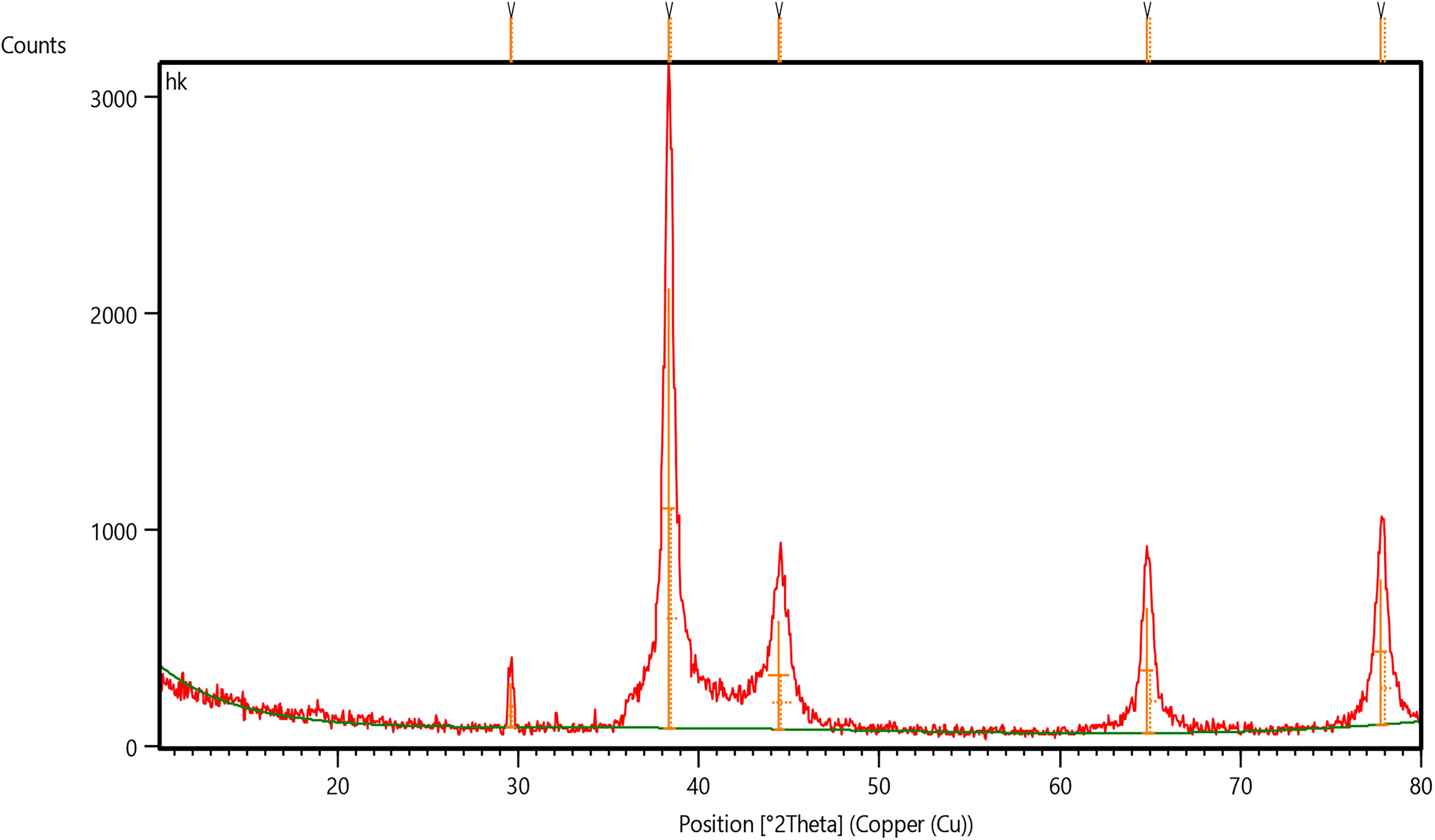
The X-ray diffraction pattern of silver nanoparticles, along with a corresponding table summarizing key peak parameters.
Table 3 provides detailed information:
Peak List of Silver Nanoparticle
FWHM, Full width at half maximum.
Synthesis optimization
The combined FTIR and XRD results confirm the successful green synthesis of crystalline AgNPs using licorice extract. Biomolecules in the extract functioned effectively as natural reducing and stabilizing agents. The FTIR analysis identified key functional group interactions, while the XRD pattern verified the structural purity and crystallinity, indicating optimal synthesis conditions.
Characterizing AgNPs synthesized via green methods is essential for understanding their properties and potential applications. These NPs possess notable antimicrobial and catalytic properties, strongly influenced by their size, morphology, and dispersion.
Transmission electron microscopy (TEM) analysis
TEM analysis was used to evaluate the morphology and size distribution of the synthesized AgNPs. As shown in Figure 3 (panels a, b, and c), the NPs appeared predominantly spherical, with limited aggregation and some forming small clusters. The particle sizes ranged from 20 to 100 nm, with an average size of approximately 30 nm. The relatively uniform size distribution reflects effective stabilization by bioactive compounds in the licorice extract. Minor aggregation was observed, likely due to drying artifacts during sample preparation.
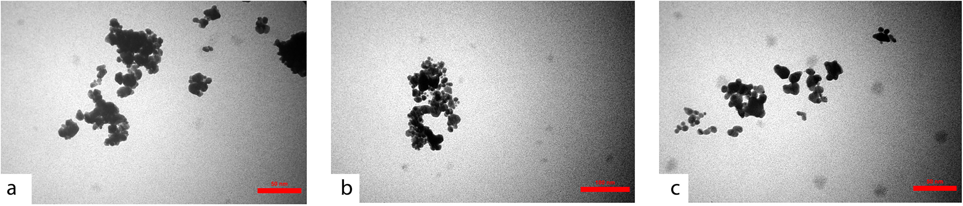
TEM images showing the morphology and size distribution of silver nanoparticles at different scales. TEM, transmission electron microscopy.
Optimization of NP synthesis
Optimal synthesis conditions were identified to enhance the stability and uniformity of AgNPs produced using G. glabra (licorice) extract. A slightly alkaline pH environment was found to improve NP stability and reduce aggregation. Additionally, an elevated reaction temperature (∼70°C) accelerated reaction kinetics, facilitating uniform particle formation. Higher concentrations of bioactive compounds in the licorice extract enhanced capping efficiency, thereby contributing to increased colloidal stability.
A
The use of licorice extract as both a reducing and capping agent demonstrated an efficient green synthesis method for AgNPs. Characterization results support this approach: FTIR analysis confirmed the presence of functional groups responsible for reduction and stabilization of silver ions. XRD patterns verified the crystalline nature and high purity of the synthesized NPs. TEM images showed well-dispersed, spherical NPs with uniform size and minimal aggregation.
Collectively, these findings highlight the effectiveness of using G. glabra extract in the green synthesis of AgNPs, suggesting strong potential for applications in antimicrobial, catalytic, and other nanotechnology-related fields.
Histopathological
Histopathological of liver
Regarding the liver section, in the control negative group, the liver section presented the normal architecture of the central vein, rows, or plates of hepatocytes that were separated by tortuous sinusoidal capillaries without any evidence of congestion and inflammation (Fig. 4a).

Microscopic section of liver tissue from common carp. Group 1
The microscopic of the liver in fish of G2 revealed ballooning degeneration of hepatocytes characterized by clear distended vacuoles with eccentrically located nuclei, congestion of the central vein and pancreas vasculature, and the liver macrophage (Kupffer cell) increased in number with mild infiltration of chronic inflammatory cells (Fig. 4b, c).
In comparison to G3, the lesion progressed that showed hepatic necrosis characterized by swollen, eosinophilic hepatocytes with pyknotic and karyolysis nuclear features, also congestion, Kupffer cell proliferation, and a mild inflammatory reaction seen in the liver parenchyma and pancreas (Fig. 4d, f).
In G4, besides congestion of vasculature, hydropic degeneration of hepatocytes with hyperplasia of Kupffer cells, the inflammatory response progressed to moderate to extensive infiltration of pus exudate (Fig. 4g, i).
In comparison, the fish that were treated with Licorice attenuated the effect of AgNPs and reduced the lesion in the liver and pancreas; in G5, the liver section revealed moderate hepatic ballooning, and mild congestion with only a minimum inflammatory response (Fig. 4k, l).
While in G6, there was no inflammatory cell infiltration (Fig. 4m, n).
The G7 only mild alteration recorded such as hydropic degeneration, congestion without any inflammatory reaction (Fig. 4o, p).
At the end Score assessment of the liver histological features is shown in Figure 6.
Histopathological of kidney
The kidney section in the control negative group presented intact normal features of glomeruli that consist of glomerular tuft capillary, Bowman’s capsule, juxta glomerular apparatus, also proximal and distal convoluted tubules with a huge number of hematopoietic cells that were distributed among interstitial tissue (Fig. 5a, b).
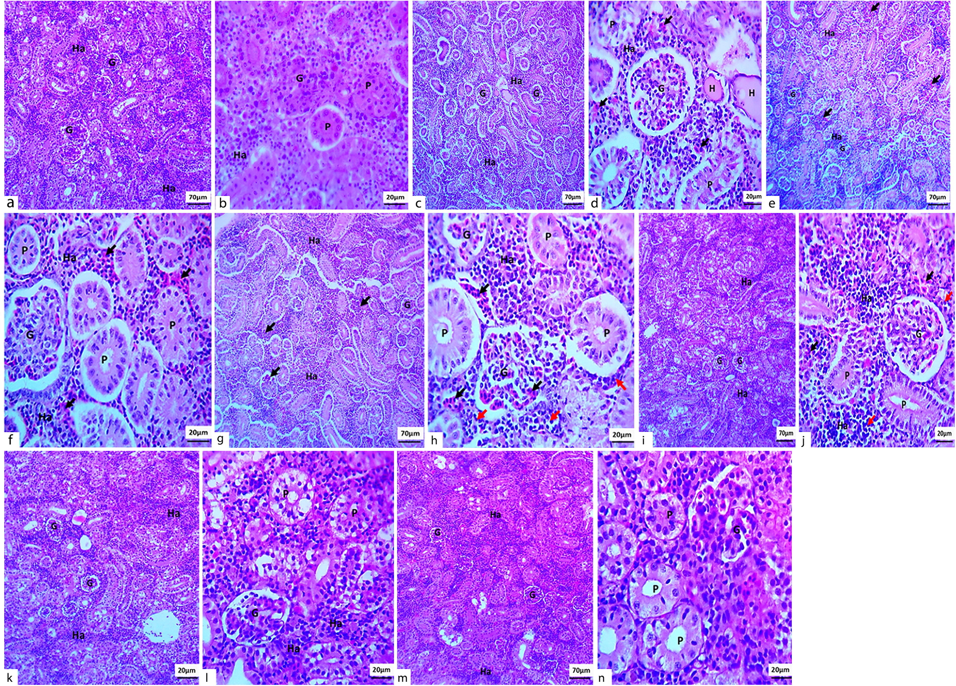
Light microscopic sections of kidney tissue in common carp.

Effect of licorice and silver nanoparticles on hepatic histopathology in common carp.
In comparison to the control negative group, the NP-induced toxicity by alteration of the histological structure of the kidney and in G2 revealed; mild swelling of glomeruli and widening of bowman’s space, moderate swelling of the lining epithelium of renal tubules, more specifically in proximal convoluted tubules, and free RBC or hemorrhage were seen in interstitial that mixed with mild chronic inflammatory cells and hematopoietic cells (Fig. 5c, d).
In G3, the glomeruli were still mildly swollen, and the renal tubules were swollen moderately, with moderate interstitial hemorrhage and mild chronic inflammatory reaction (Fig. 5e, f).
In comparison to G4, the lesion degree increased and showed swollen and congestion of glomeruli with focal atrophy of glomerular segments, severe tubular swollen, and severe interstitial hemorrhage with marked chronic inflammatory response (Fig. 5g, h).
Whereas, adding licorice alleviated the damaging effect of NPs and showed in G5; only mild swelling of glomeruli and collecting tubules were seen with infiltration of chronic inflammatory cells with few RBC in the interstitial tissue in combination with hematopoietic cells (Fig. 5i, j).
Versus: In the fish’s kidney in G6 (Fig. 5k, l), the lesion to mild swelling of glomeruli and renal tubules with a minimum inflammatory reaction, that mixed with hematopoietic cells.
In comparison to the G7, which restored the kidney to normal histological structures (Fig. 5m, n).
At the end, score assessment of the kidney histopathological features is shown in Figure 7.
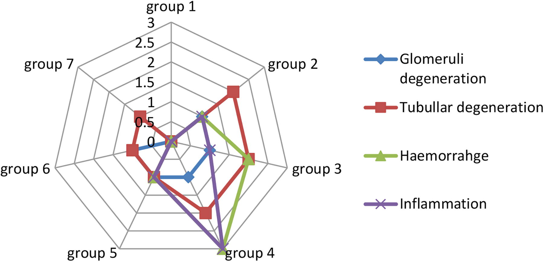
Histopathological kidney lesion scores in common carp (Cyprinus carpio) exposed to licorice-silver nanoparticle treatments.
Discussion
The findings of this study highlight the critical role of licorice (G. glabra) extract in the green synthesis and stabilization of AgNPs. Using FTIR spectroscopy, XRD, TEM, and histopathological analyses, significant insights were obtained into the structural, chemical, and biological properties of AgNPs synthesized with natural reducing and capping agents. These analyses also demonstrated a reduction in toxicity when AgNPs were used at appropriate concentrations during treatment.
FTIR spectral analysis revealed the presence of functional groups essential for reducing and stabilizing AgNPs. Peaks corresponding to hydroxyl (O–H) and carbonyl (C=O) groups confirmed the active role of biomolecules in reducing silver ions (Ag+) to elemental silver (Ag0). Additionally, C–O and C=C functional groups served as stabilizing agents, capping the NPs and preventing aggregation. These findings underscore the dual function of licorice extract as both a reducing and stabilizing agent, consistent with eco-friendly synthesis approaches (Ali et al., 2013; Ahmed et al., 2016). Glycyrrhizin, a major bioactive compound in licorice, was identified as a key component responsible for reducing Ag+ to Ag0 and enhancing NP stability (Feng et al., 2022). The green biosynthesis of metal NPs using plant extracts supports applications in antibacterial therapy, cancer treatment, and environmental remediation (El-Seedi et al., 2024; Kandav and Sharma, 2024).
XRD analysis confirmed the crystalline nature of the synthesized AgNPs, with sharp peaks at 2θ values corresponding to the (111), (200), (220), and (311) planes of a FCC structure (Kakakhel et al., 2021). The most intense peak at 38.338°, representing the (111) plane, indicated high crystallinity and purity. These structural features are essential for the antimicrobial and catalytic potential of AgNPs (Krishnaraj et al., 2010; Philip, 2010). The average crystallite size was approximately 20.26 nm, indicating a clean, cost-effective, and environmentally sustainable synthesis method (Ali et al., 2023). In comparison, Schiff base ligand-capped AgNPs sized between 5 and 10 nm have demonstrated multifunctional applications as catalysts, sensors, antioxidants, and antimicrobial agents (Suneetha and Manjari, 2022). XRD results also linked AgNPs to antibacterial efficacy, especially against multidrug-resistant bacteria, by inhibiting biofilm formation and reducing cytotoxicity (Xie, 2024).
TEM imaging showed that the synthesized AgNPs were predominantly spherical with an average size of approximately 30 nm and minimal aggregation. The uniform particle size distribution was attributed to effective capping by bioactive compounds in licorice extract. Minor aggregation observed was likely due to sample drying during preparation, consistent with previous reports (Nabikhan et al., 2010; Sharma et al., 2009). Optimization studies revealed that slightly alkaline pH conditions enhanced NP stability, while an elevated temperature (∼70°C) accelerated reaction kinetics and promoted uniform NP formation. Higher concentrations of licorice-derived biomolecules improved capping efficiency, enhancing stability and minimizing aggregation. An optimized reaction time of 30 min was effective in reducing particle size and aggregation (Iravani, 2011; Khalil et al., 2014).
Liver tissue from AgNP-exposed common carp (C. carpio) exhibited hepatocellular vacuolation, sinusoidal dilatation, and inflammatory cell infiltration, with lesion severity increasing in a dose-dependent manner. Notably, fish exposed to 10 µg/L AgNPs displayed nearly normal liver architecture, supporting the hypothesis that low-dose exposure elicits a milder hepatic response. Intestinal tissues showed villi shortening and fusion, goblet cell hyperplasia, and infiltration of the lamina propria. Gill tissues revealed epithelial hyperplasia, lamellar fusion, and necrosis—conditions exacerbated at higher AgNP concentrations. These findings suggest that lower concentrations may elicit a subdued response or permit partial compensation through antioxidant and immune mechanisms. Subepidermal edema and inflammatory cell infiltration were observed in high-dose AgNP-treated groups (Mabrouk et al., 2021).
In another study, freshwater common carp (C. carpio) were exposed to AgNPs synthesized using animal blood serum to assess toxicity, mortality, bioaccumulation, and histological changes at concentrations of 0.03, 0.06, and 0.09 mg/L. Minimal behavioral changes were observed even at the highest concentration, although significant bioaccumulation occurred in the liver, intestine, gills, and muscle, in that order. This bioaccumulation corresponded with histopathological damage, particularly in gill and intestinal tissues, at the highest exposure level (0.09 mg/L) (Kakakhel et al., 2021).
Histopathological evaluation of liver and kidney tissues revealed that licorice extract conferred protection against AgNP-induced toxicity. Fish treated with AgNPs alone exhibited severe changes, including congestion, hydropic degeneration, necrosis, and inflammation. In contrast, those co-treated with licorice extract showed significantly reduced tissue damage. Licorice mitigated inflammatory responses, minimized cellular degeneration, and promoted tissue regeneration, attributed to its antioxidant and anti-inflammatory properties (Rajeshkumar and Malarkodi, 2014; Durán et al., 2011). These findings align with previous reports describing necrosis, nuclear degeneration, and other NP-induced lesions in liver and kidney tissues (Shobana et al., 2018; Wu and Zhou, 2013). Furthermore, licorice extract demonstrated hepatoprotective activity, improving liver histology and biochemical markers in (C. carpio) (Malekinejad et al., 2010).
However, some studies reported no significant histopathological changes in fish exposed to AgNPs, with liver and kidney structures remaining intact (Clark et al., 2021). This variability may be attributed to differences in NP concentration, exposure duration, or the use of protective agents such as licorice extract. In the current study, exposure to AgNPs caused structural alterations in the gills of C. carpio, including epithelial lifting, lamellar fusion, clubbing, subepithelial edema, and necrosis—features indicative of impaired respiratory and osmoregulatory functions. A decrease in carbohydrate content was also observed, as evidenced by weak periodic acid–Schiff (PAS) staining, suggesting metabolic stress and impaired mucopolysaccharide synthesis. While partial recovery was noted after a depuration period, residual structural abnormalities persisted, including lamellar curvature and hyperplasia. In muscle tissues, histopathological findings included fiber splitting, degeneration, necrosis, and inflammatory infiltration (Sayed et al., 2020).
Overall, this study validates the efficacy of licorice extract as a natural reducing and stabilizing agent in the green synthesis of AgNPs. Comprehensive characterization confirmed the structural integrity, stability, and biocompatibility of the synthesized NPs, emphasizing the potential of eco-friendly synthesis strategies in producing biologically safe nanomaterials for diverse applications (Raveendran et al., 2003; Singh et al., 2016).
Green synthesis of AgNPs by using glycyrrhizin. The eco-friendly synthesis of AgNPs, a major bioactive compound found in licorice (G. glabra), has been demonstrated. The synthesized AgNPs exhibited significant antibacterial activity against Escherichia coli and Staphylococcus aureus and showed lower toxicity to human kidney epithelial cells compared to chemically synthesized AgNPs (Feng et al., 2022)
The acute toxicity and physiological effects of biosynthesized AgNPs in Nile tilapia, specifically biogenic AgNPs synthesized from Aspergillus tubingensis, have been evaluated. The findings indicate that while these AgNPs are less toxic than silver nitrate, they still induce physiological and morphological changes in the fish, such as alterations in gill structure and swimming behavior (Ribeiro et al., 2025).
In Shelke (2023), the chronic toxicity of AgNPs on zebrafish (Danio rerio) was investigated, focusing on liver, gill, and muscle tissues. The study reported decreased levels of reduced glutathione and total protein, alongside increased lipid peroxidation and lactate dehydrogenase activity, indicating oxidative stress and tissue damage.
Younas et al. (2022) examined how harmful chemically and green-synthesized AgNPs were to silver carp (Hypophthalmichthys molitrix). Compared to their chemically produced counterparts, green-synthesized AgNPs showed reduced toxicity and caused fewer abnormalities in hematology and histopathology.
The effects of prolonged exposure to elevated levels of AgNPs on common carp (C. carpio) were investigated in a (Kakakhel et al., 2021) study. The study illustrated the possible dangers of extended AgNP exposure in aquatic environments and showed how AgNPs bioaccumulate in different organs, resulting in histological changes in intestinal and gill tissues.
Conclusion
Licorice root powder demonstrated a significant protective effect against AgNP-induced histopathological alterations in the liver and kidneys of common carp. Its incorporation into the diet not only mitigated tissue damage but also improved growth performance and immune response, highlighting its potential as a natural additive for sustainable aquaculture. The combined use of licorice and green-synthesized AgNPs presents a promising strategy to enhance fish health while reducing dependence on conventional antibiotics. This eco-friendly approach supports the development of safer and more sustainable aquaculture practices.
To fully harness the benefits of this combination, future studies should focus on elucidating the molecular and cellular mechanisms underlying the protective effects of licorice against NP toxicity; evaluating long-term safety and efficacy under commercial aquaculture conditions; optimizing dosages and delivery methods for different fish species and life stages; and investigating the effects on other physiological systems, such as the immune and reproductive systems.
Footnotes
Authors’ Contributions
H.K.F.: Sample collection, laboratory analysis, and draft manuscript preparation. N.M.A.: Study design, methodology development, supervision of sample collection and laboratory work, and manuscript revision.
Author Disclosure Statement
All authors declare that they have no known financial or personal conflicts of interest that could have appeared to influence the work reported in this article. No Interests to disclose.
Funding Information
No funding was received for this article.