




Abstract
Background and Purpose:
Retrograde ureteroscopic marsupialization is a pure natural orifice translumenal endoscopic surgery (NOTES). We retrospectively examined the feasibility and safety of this technique to manage symptomatic simple renal cysts.
Patients and Methods:
Sixteen patients with simple renal cysts were selected and treated by incising the cyst wall to drain into the collecting system through retrograde ureteroscopy. A retrospective observational study was performed to evaluate the patient's symptomatic and radiologic results after ureteroscopic marsupialization. Symptomatic success based on pain relief was evaluated using a visual analog pain scale preoperatively and postoperatively. Radiologic success was defined as no recurrence of the cyst or a reduction in cyst size by at least half.
Results:
There were no intraoperative or postoperative complications observed. The mean operative time was 35 minutes (range 20–50 min). The mean hospital stay was 3.4 days (range 2–5 d). Of the 16 patients, one patient was lost at follow-up. The symptoms based on pain had resolved in 13 (83%) cases but remained in 2 cases at a mean follow-up of 24.2 months (range 6–36 mos). The average visual analog pain scale decreased from 6.7 (range 4–9) to 1.1 (range 0–5) at the sixth month. The mean size of all cysts decreased from 6.8 cm (range 4–10 cm) to 1.3 cm (range 0–5 cm). Radiographic success was achieved in 93% (14/15) of patients. Cytology and cyst wall pathology reports revealed no evidence of malignancy.
Conclusions:
Retrograde ureteroscopic marsupialization is a complete transurethral NOTES marsupialization. With appropriate patient selection, the minimally invasive retrograde ureteroscopic marsupialization is feasible, safe, and effective. It can be preferred to more invasive laparoscopic or open surgical approaches.
Introduction
Currently available minimally invasive therapeutic options include percutaneous aspiration with or without sclerotherapy, 1 percutaneous marsupialization, 4 ureteroscopic marsupialization, 5 –7 and laparoscopic marsupialization by transperitoneal or retroperitoneal access 8 ; more recently, single-port laparoscopic surgery has become available. 9 Among these minimally invasive intervention options for symptomatic renal cysts, percutaneous aspiration with sclerotherapy has been commonly performed; however, this approach was associated with a high recurrence rate. 10 –12 Percutaneous marsupialization has also been described as another minimally invasive approach. This technique, however, is more suitable for a large single cyst at the lateral margin of the kidney. The long-term results have also been disappointing, with a recurrence rate up to 30% and residual cysts in 20% of patients. 13 Laparoscopy is an effective method to manage simple renal cysts, but it is technically difficult, necessitating a long learning curve for the surgeon, expensive, and needs extensive retroperitoneal dissection. 1,14,15
Ureteroscopic marsupialization was first reported in 19915 as a complete transurethral natural orifice translumenal endoscopic surgery (NOTES) 16 that was significantly less invasive than percutaneous nephroscopy or laparoscopy. 4,14 Unfortunately, no large case series studies have reported on the success of this technique in the management of simple renal cysts. To investigate the feasibility and safety of this technique, we retrospectively examined the results of retrograde ureteroscopic marsupialization in the management of symptomatic simple renal cysts using 2 years of follow-up data.
Patients and Methods
Our retrospective study included the outcomes of 16 patients who were selected to undergo ureteroscopic marsupialization to manage symptomatic renal cysts between October 2006 and July 2009. This study cohort was composed of seven men and nine women, with a mean age of 49.8 years (range 36–71 y). All 16 patients presented with appropriate and similar symptoms and complained of varying degrees of flank pain. Five among them experienced several episodes of renal colic.
All patients were evaluated before treatment by history taking, ultrasonography, and gross clinical, imaging, and laboratory examinations. Preoperative imaging examinations for all 16 patients demonstrated extrinsic compression protruding into the collecting system, with the cyst wall directly adjacent to the collecting system. The presence of Bosniak I cysts 17 was diagnosed with CT on all patients. The laboratory examinations revealed that two patients had microscopic hematuria, one patient had azotemia (ie, an increased serum creatinine level >1.4 mg/dL), and no patients had proteinuria or pyuria. Of those 16 patients, we found that 1 had a concomitant contralateral ureteral stone (Fig. 1), two had a history of previous renal surgery, three had mild hydronephrosis, and four had hypertension. The mean duration of symptoms was 3 months (range 1–24 mos).
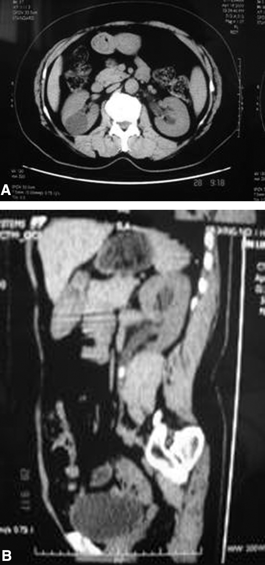
CT scan demonstrated a right 4-cm posterior cyst located in the interpolar area adjacent to the collecting system
Of all cysts in our patient cohort, seven were located on the left side and nine on the right. The cysts were peripelvic in nine patients, and cysts in seven patients were peripheral (four in the upper pole, three in the interpolar area). Our study excluded complex cysts and patients with autosomal dominant polycystic kidney disease, infectious renal cysts, cystic renal-cell carcinoma, pregnancy, solitary kidney, or coagulopathy. All patients provided written informed consent after receiving details about the procedure and its possible complications, and the study was carried out according to the protocol approved by the local ethics committee.
Patients were placed in a lithotomy position under regional or general anesthesia. Prophylactic intravenous antibiotics were given before endoscopic manipulation. Retrograde ureteroscopy marsupialization was performed with a 8/9.8F Storz semirigid ureteroscope and/or a 7.5F flexible ureteroscope. Macroscopic inspection of the bladder and the cyst's ipsilateral ureter showed no urothelial abnormalities in any of the cases. Inside the ureteropelvic region, the cyst wall was generally found to protrude into the collecting system and was noted to be obstructing the urinary collecting system.
The tip of a 3F to 5F open-end ureteral catheter with a thin inner steel core was cut to make the open end double, and then the thin steel core was inserted into the cyst under direct vision through a ureteroscope, as confirmed by ultrasonography. After that, the double open-end ureteral catheter was inserted into the cyst under inner steel core guidance. Then the steel core was removed; a clear or straw-colored cyst fluid was aspirated through the catheter by a syringe. An aliquot of the fluid sample was sent for bacteriologic and cytologic examinations.
After fluid protein examination confirmed the cyst diagnosis, direct ureteroscopic vision was used to guide a 3F needle electrode or laser fiber to incise the cyst wall at the extrinsic compression locale without any obvious vessel pulsation. In some cases, a portion of the cyst wall was first harvested by ureteroscopic biopsy forceps for histopathologic examinations. The full-thickness open incision was made to approximately 2 to 4 cm to communicate with the collecting system. The margin of incision was completely coagulated, and the cyst interior was completely inspected to avoid the misdiagnosis of cystic renal-cell carcinoma. A 5F double pigtail ureteral stent was positioned with the proximal end coiled in the cyst cavity and left in place for 4 weeks.
After surgery, all patients were followed up in the outpatient clinic at regular intervals. We compared preoperative and postoperative symptoms based on pain using a visual analog scale to determine success of the surgery in relieving symptomatology. All patients underwent repeated imaging examinations to demonstrate the communication between the unroofed cyst cavity and the collecting system after operation. The size of the cysts before the treatment and during follow-up was measured by ultrasonography. Radiologic success was defined as no recurrence of the cyst or reduction of cyst size by at least one-half after surgery. The patients were followed at 1, 3, 6, 12, 24, and 36 months postoperatively for any evidence of adverse events after treatment.
Results
Retrograde ureteroscopic marsupialization was successful in all patients without conversion to open surgery and with no inadvertent injury to the collecting system. There were neither intraoperative nor postoperative complications. During the surgery, the blood loss was minimal for all patients, and the mean operative time was 35 minutes (range 20–50 min). The mean hospital stay was 3.4 days (range 2–5 d). For incision of the cyst wall, 10 patients were treated with a 8/9.8F Storz semirigid ureteroscope with a 3F needle electrode, and six patients were treated with a 7.5F flexible ureteroscope with a 360-μm laser fiber. All cytology and pathology findings revealed benign renal cysts.
One patient was lost at follow-up, but complete data were available for the remaining 15 patents. At a mean of 24.2 months (range 6–36 mos) follow-up, symptomatic success based on pain relief was achieved in 87% (13/15) of patients, but pain remained in two patients. Severity of pain decreased from 6.7 (range 4–9) to 1.1 (range 0–5) at the sixth month. Radiographic success was achieved in 93% (14/15) of patients. The mean size of cysts had decreased from 6.8 cm (range 4–10 cm) to 1.3 cm (range 0–5 cm) at the end of follow-up.
For those two patients who failed to attain symptomatic relief, one had reported moderate pain before surgery but no significant relief after the surgery, despite the cyst being shown as resolved by ultrasonography imaging. Thus, the pain for that patient was thought to be caused by another unknown etiology, and the patient was given additional clinical outpatient care and continuous follow-up. Pain for the second patient became worse at the ninth month after the surgery, and a recurrent 5-cm peripelvic renal cyst was diagnosed by ultrasonography. That patient was recommended for resurgery, and reureteroscopic marsupialization was performed, which yielded a finding of the incision autoclosure. We speculated that the autoclosure was probably caused by the previous surgery having been inefficient in incising the cyst wall. That patient has been followed to date and reports good pain relief; radiographic success has been determined.
During follow-up, three of four patients with hypertension had well-controlled blood pressure that needed no medication after the surgery, while the fourth patient showed no evidence of improvement. The patient who presented with a concomitant ureteral stone repeated ultrasonography at the sixth month during which the cyst and the stone were removed; further signs of recurrence have been absent. The patient with azotemia showed the serum creatinine level drop to the normal range after surgery. For the three patients with hydronephrosis, the problem disappeared in two, while the third patient persisted with mild caliectasis. After the surgery, four patients complained of mild lower urinary tract symptoms, microscopic hematuria, and slight discomfort in the lumbar area. Those symptoms were cleared when the double pigtail ureteral stent was removed 1 month later. The comprehensive perioperative and follow-up data are presented in Table 1.
Discussion
Simple renal cysts are a common disease of the renal parenchyma. The etiology is unknown, and no genetic factor has been found that is associated with the condition, but tubular obstruction and ischemia from obstruction may be involved. 18 Fortunately, most patients are asymptomatic and need no treatment unless noticeable symptoms or complications develop.
With the recent advances in instrumentation and therapeutic techniques, minimally invasive surgical procedures have gradually replaced open surgery for managing symptomatic renal cysts. The introduction of percutaneous aspiration with or without sclerosis, 1 percutaneous marsupialization, 4 ureteroscopic marsupialization, 5 –7 and laparoscopic marsupialization 9 has made open surgery unnecessary in most cases.
Percutaneous aspiration with sclerotherapy has been commonly performed and succeeded more often when used for cysts that are separated from the collecting system. 19 This approach, however, is associated with a high recurrence rate, and it is also more dangerous for the cyst that is proximal to the collecting system and renal hilar. 10 –12 In addition, it has been shown to be less effective for peripelvic cysts. 20 Therefore, percutaneous aspiration with sclerotherapy is relatively contraindicated for peripelvic cysts, because extravasation or inadvertent instillation of the sclerosing agent into the retroperitoneum may induce severe perirenal inflammation and secondary ureteropelvic junction obstruction. 21
Since the advent of laparoscopy in the 1990s, laparoscopic management of renal cysts has been verified as safe and effective. 22 Laparoscopy duplicates all the steps of the open procedure, combined with the advantages of a minimally invasive approach. 9,23 Previous renal surgery, obesity, and peripelvic cysts have made the laparoscopic access more difficult, however. Previous renal surgery necessitated dissection to be more tedious and time consuming because of adhesions around the retroperitoneal area. Furthermore, obese patients need more fat to be dissected from the retroperitoneum to increase the working space. Laparoscopic decortication of peripelvic cysts necessitates advanced surgical skills because of the complexity of the cysts and the proximity to the renal hilar and the collecting system; as such, this is often associated with a greater estimated blood loss, longer operative time, and extended hospital stay. 20,24
Laparoscopy has the risk of injuring the adjacent organs and vascular structures during operation. Shiraishi and associates 25 also reported that the recurrence rate for this technique was up to 19%, as determined by analysis of follow-up data covering 67.2 months. The high level technical skill, expense, and the physician's lengthy learning curve remain the principal drawbacks of this technique.
Percutaneous marsupialization has also been described as another minimally invasive technique. Percutaneous marsupialization can be performed either directly, by creating a percutaneous access to the cyst, or indirectly, by creating a nephrostomy tract through a nearby calix and approaching the cyst from inside the collecting system. 13,26 Direct percutaneous access, in theory, is similar to the method used by open surgery or laparoscopy, in that these methods all use fulguration at the base of the cyst and incise the dome of the cyst. When using this technique, however, the working space, visibility, and management of bleeding are worse than that of laparoscopy. In addition, the direct percutaneous access is more suitable for large lateral margin posterior cysts. In fact, the indirect percutaneous access is similar to retrograde ureteroscopic marsupialization, where the cyst wall is incised to create communication between the cyst and the collecting system. As the percutaneous incision proceeds, however, the collapsing cyst impairs visibility, and this may lead to difficulty in management of the bleeding, inefficient incision, or inadvertent injury of the collecting system. The long-term results of percutaneous marsupialization are also disappointing; a recurrence rate up to 30% and residual cysts in 20% of patients have been observed. 13
Ureteroscopic marsupialization was first reported in 19915 and was a pure NOTES 16 marsupialization; the advantages of this new technique included that it needed no incision, minimized patient discomfort and hospital stays, had a short convalescence period, and afforded aesthetic benefits. The development of semirigid, fiberoptic, and actively deflectable flexible ureteroscopes has allowed urologists to gain access to all areas of the collecting system. 27
Simple renal cysts are usually asymptomatic. Symptoms such as pain and obstruction are probably a consequence of cyst enlargement and secondary compression of the collecting system, especially for peripelvic cysts that are in proximity to hilar vessels and the collecting system. 20,28 Relief of obstruction is the primary goal for the management of symptomatic renal cysts. 5 We used the pure transurethral surgery to incise the cyst wall into the collecting system to relieve the collecting system compression, and achieved satisfactory results.
Compared with antegrade percutaneous nephroscopy, retrograde ureteroscopy does not necessitate percutaneous nephrolithotomy and change of position, and reduces the risk of puncturing injury. In addition, it incises the cyst wall at an extrinsic compression place without vessel pulsation under direct vision and avoids the blind incision of percutaneous access. During the resection, the ureteroscopy remains outside of the cyst and allows an overview of the outer surface of the cyst; as such, it is very convenient to control bleeding and avoid inadvertent injury.
The introduction of the flexible ureteroscope and holmium laser has resulted in remarkable improvement in the outcomes of ureteroscopy for simple renal cysts in terms of feasibility and safety. We used the flexible ureteroscope and holmium laser in six patients. The fragility and high cost of this equipment are major drawbacks that restrict routine use. 29 Usually the semirigid ureteroscope can reach the lesion and incise the cyst wall successfully with good cost-effective results, particularly in female patients. We chose the flexible ureteroscope for tall male patients whose cysts were located in the upper pole. In these patients, the longer distance between the urethra and the cyst is more difficult for the semirigid ureteroscope to reach.
Our satisfactory results were related to careful selection of patients for the procedure. In the selected patients, all symptomatic simple renal cysts were adjacent to the collecting system. All patients' preoperative imaging examinations showed extrinsic compression protruding into the collecting system. We excluded cysts that were separated from the collecting system, because this type of cyst is difficult to create an adequate communication between the cyst and collecting system and injury to the renal vessels is more likely.
In some conditions, however, ureteroscopic marsupialization offers a favorable minimally invasive alternative for the management of symptomatic simple renal cysts, particularly in patients who have peripelvic renal cysts, previous history of surgery, obesity, or are undergoing simultaneous ureteroscopic management of other ureteral lesions. For peripelvic renal cysts that are unamenable to laparoscopy and contraindicated for aspiration with sclerosis, simultaneous ureteroscopic management of other ureteral lesions can avoid a staged treatment. Ureteroscopy not only avoids the risk of sclerotherapy damage to the collecting system, but also avoids resurgery difficulties in terms of surgical access because of absence of normal surgical landmarks and adhesive fibrous tissue around the kidney.
According to our experience, it is very important to place a double pigtail ureteral stent with the proximal end coiled into the unroofed cyst cavity for 4 weeks postsurgery. This procedure has many advantages. First, a double pigtail ureteral stent can continuously drain cyst fluid, collapse the cyst cavity so that the cyst walls can adhere to each other and obliterate the cyst cavity. Second, a biomaterial stent can produce a certain degree of foreign body reaction, facilitate the cyst wall inflammatory adhesion and fibrosis, and damage the cystic epithelium. Third, a ureteral stent can prevent the autoclosure of the incision, forming a permanent communication between the unroofed cyst cavity and the collecting system with long-term drainage. At the same time, a double pigtail ureteral stent is also a good internal drainage facility for inadvertent laceration of the collecting system and urine leakage.
Similarly, the size and direction of the incision are also very important in the operation. A too large incision may increase risks of injuring the collecting system, while a too small incision may not be suitable for efficient drainage. In our practice, a 2 to 4 cm long incision at the extrinsic compression locale, in a longitudinal direction, and without the vessel pulsation under direct ureteroscopic vision is recommended. To achieve adequate drainage and prevent urinary stasis, we do not recommend such surgery for cysts that are located in the lower pole. The interior of the cyst was not fulgurated in our operation, because it was adjacent to the collecting system and major renal vessels. In addition, we performed electroresection and electrocautery of the incision at a low energy setting. We not only reduced the possibility of cyst recurrence in theory, but also improved the safety of operation in practice.
Concerns may exist that the communication with the collecting system and the cystic epithelium secretion may lead to proteinuria or hypoproteinemia. Terada and colleagues 30 have reported that the average cyst size increase per year was 1.6 mm and average rate of cyst enlargement was about 3.9% per year. Thus, the volume of the cystic epithelium secretion per day is minimal, less than what is needed to lead to proteinuria. During the follow-up period, we did not find any proteinuria or hypoproteinemia evidenced by urinalysis or blood biochemistry. It is possible that as the cyst began adhesion and the cystic epithelium cell degenerated, cellular secretion declined. In addition, it remains unknown whether urine itself is able to destroy cystic epithelium secretion.
Although ureteroscopic marsupialization is a pure transurethral NOTES marsupialization, it necessitates regional or general anesthesia, hospitalization, skillful surgery, and needs a second procedure to remove the stent. A tortuous, kinked, or angulated ureter makes the ureteroscopy procedure more difficult. Scarring of the renal pelvis, perforation of the ureter, stasis of urine, infection, and formation of stones could occur. The incidence of both intraoperative and postoperative complications is low and not serious, however, if proper selection of patients and adequate preoperative preparations are carried out.
We also noted some limitations in our study, such as a relatively small sample size. Ultrasonography was used to measure the cyst size in most cases during follow-up, because it is less expensive. Sometimes it is difficult to identify the collapsed unroofed cystic cavity and the caliectasis by ultrasonography imaging. These limitations will be considered in our following studies. In spite of this, the results in this study provide useful information in evaluation of the complete transurethral NOTES marsupialization for the management of symptomatic simple renal cysts.
Conclusions
Retrograde ureteroscopic marsupialization is a complete transurethral NOTES marsupialization. With appropriate selection of patients, retrograde ureteroscopic marsupialization is feasible, safe, effective, and minimally invasive. It is recommended for consideration before other more invasive laparoscopic or open surgical approaches.
Footnotes
Disclosure Statement
No competing financial interests exist.