




Abstract
Background and Purpose:
During laser lithotripsy, working instruments are often in close proximity to the distal fiber tip and may be damaged accidentally or even intentionally. The aim of this study was to compare the amount of damage to a standard guidewire and the nitinol wires of endourologic retrieval baskets that were affected by three different clinically available laser systems.
Materials and Methods:
The impact of pulsed laser irradiation on a standard hydrophilic guidewire and a retrieval basket were investigated. One infrared (IR) laser system (holmium:yttrium-aluminum-garnet [Ho:YAG]: λ = 2100 nm) and two laser systems emitting light in the visible (VIS) spectral range (frequency-doubled double-pulse neodymium:YAG [FREDDY]: λ = 532 nm/1064 nm and flashlamp pulsed dye [FLPD]: λ = 598 nm) were used. Experimental parameters were fiber core diameter, laser pulse energy, and distance between the fiber tip and the investigated tool. Damage was evaluated by microscopic investigation and by quantifying the damage size and magnitude by creating laser impact related damage factors.
Results:
After application of one single laser pulse, IR-laser related maximum damage to guidewires occurred, depending on the pulse energy and the fiber core diameter, either in contact mode or in a distance of maximum 2 mm. Maximum VIS-laser related damage occurred in a distance range of 2 to 3 mm. The nitinol wires of the extraction tools could be destroyed completely by IR laser irradiation at pulse energies EP > 1200 mJ, depending on the fiber core diameter used. VIS lasers were solely able to set visible damage to guidewires without any disruption of nitinol wires.
Conclusions:
Ho:YAG laser induced damage to endourologic tools is significantly higher compared with the impact of the FREDDY or the FLPD-laser. Because complete disruption of guidewires and stone extraction tools occurred, a safety clearance must be kept between the fiber tip and the endourologic tool during Ho:YAG stone disintegration. If disruption is intended, such as in the case of basket-retrieval problems, it can easily be performed with Ho:YAG irradiation.
Introduction
Accidental damage to endourologic instruments during laser lithotripsy, however, may occur when the laser fiber tip is in close proximity to incorporated tools during the laser-firing period. In particular, this scenario is frequently seen with the use of safety wires during ureteroscopy or with retrieval instruments used to prevent calculus retropulsation during lithotripsy. Deliberate damage of a retrieval instrument, in addition, is performed in cases of entrapped devices. Therefore, the aim of this study was to compare the amount of damage to guidewires and retrieval baskets by a variety of commonly used clinical laser systems.
Materials and Methods
Laser devices
Laser impact on endourologic tools of three different, clinically approved and commercially available laser systems was systematically investigated.
The flashlamp pulsed dye (FLPD) laser (Lithognost, Basel Lasertech GmbH, Germany) emits light pulses in the wavelength λ = 594 ± 3 nm. The power output at the end of the bare fiber (core diameter 300 μm) could vary between Ep = 60 mJ/pulse to 150 mJ/pulse at repetition rates of 1 to 10 Hz and pulse duration of tP = 2.5 μs. This system includes an active stone recognition system to prevent laser tissue interaction if the fiber is placed in front of soft tissue instead of calculi. 6 –9 For the experiments, a 300 μm fiber was used, EP was 120 mJ/pulse and 150 mJ/pulse and the repetition rate was set to 3 Hz.
The frequency-doubled double-pulse neodymium:yttrium-aluminum-garnet (FREDDY, U100plus, WOM World of Medicine AG, Germany) is a short-pulse solid-state laser emitting both wavelengths at λ = 532 nm und λ = 1064 nm. 10 –14 The green 532 nm pulse is clinically intended to induce the formation of plasma between the fiber and the stone, while the infrared (IR) energy is deployed to pump the plasma amplifying bubble formation. The energy of a green pulse is not supposed to be high enough for any soft tissue interaction in front of the fiber tip. Instead, it penetrates into the soft tissue and is absorbed without major thermal effects. The IR light, which also penetrates into the soft tissue, causes only a slight increase of temperature. Therefore, this technique is supposed to provide “passive” safety for soft tissues. The pulse-duration of the laser system is fixed to tp = 1.2 μs, while the energy per pulse can be changed between EP = 120 mJ/pulse or 160 mJ/pulse as single or double pulse. The repetition rate can vary between 1 to 20 Hz. 4,10,11,13 In the experiments, the pulse frequency was set to 3 Hz and the 273 μm fiber was used exclusively.
The holmium:yttrium-aluminum-garnet (Ho:YAG) laser device (Auriga, StarMedTec GmbH, Germany), emits light of the wavelength λ = 2100 nm with a pulse duration tp in the range of 250 μs to 300 μs at repetition rates between 3 and 12 Hz. The impact of the following energy settings used for clinical Ho:YAG laser lithotripsy were studied: Ep = 300, 500 and 800 mJ/pulse with the 230 μm fiber; Ep = 300, 500, 800, 1200, 1600 mJ/pulse with the 365 μm fiber; and Ep = 300, 500, 800, 1200, 1600, 2000 mJ/pulse with the 600 μm fiber. The pulse frequency for every single experiment was set to 3 Hz.
Endourologic tools
For investigations of laser-induced damages to endourologic tools, guidewires (0.038 inch hydrophilic wire, Terumo® Europe N.V., Belgium) and retrieval baskets (2.2F tipples nitinol basket Sur-Catch N,™ GYRUS ACMI, USA) were used as targets.
Experimental set-up
Experimental laser energy application was performed in an “underwater” laboratory setup, thereby mimicking the clinical situation. The optical fiber was introduced through an 8F semirigid ureterorenoscope (Karl Storz, Germany). The optical axis of the fiber was directed toward the target tool at an angle of 90 degrees. The distance between the fiber tip and the target surface resembled the clinical situation and was set at 0 mm, 1 mm, 2 mm, 3 mm, and 4 mm, respectively. All components were fixed and adjusted using micropositioning devices to reproduce comparable geometril conditions (angel adjustment, distance between fiber tip and tool). Before each experiment, the angle was measured using a fine goniometer. For direct vision and documentation, the endoscope was coupled to a three-chip endoscope camera using a camera controller (Tricam SL Pal, Karl Storz, Germany), and a video monitor (Model PVM-2053MD, Sony, Japan).
The impact of one single laser pulse at the overall adjustable repetition rate of 3 Hz was investigated to achieve reproducible, comparable experimental conditions. After each single experiment, the wire was moved along its longitudinal axis by 10 mm to conduct the next laser firing experiment on an untreated area.
Evaluation
Each experiment was performed five times. Visual analysis of the induced damage was performed under a microscope (magnification ranging between 10x and 60x). Size calibration measurements were performed with photographs using 0.1 mm voxels.
Damage to guidewires
The current study aimed to classify the amount of damage on the endourologic tools using different parameters: The area of damage (which had been evaluated before), and the visually detectable damage relative to the diameter as a second parameter. The relative damage of the guidewire (C) was classified into no visible damage (0), thermal damage of coating (1), thermal damage of coating, visible wire spots (2), destruction of coating, visible wire spots (3) and destruction of the coating, severe damage to wire spots (4), as visibly demonstrated in Table 1. The damage area A was measured, therefore, as mm2 and determined using the picture processing software Adobe Photoshop 7.0 (Adobe Systems Incorporated, USA).
A damage factor, D, subsequently was calculated as D = C
Damage to retrieval baskets
Laser-induced ablation to the nitinol wires of the retrieval tools was evaluated by calculating the percentage of the remaining wire diameter after application of a single laser pulse according to the following formula:
Grade 1 damage represents laser-induced defects of 25% disruption, leaving 75% of the wire intact; grade 2, 50% disrupted and 50% intact; and grade 3, 75% disrupted and 25% intact, respectively. Grade 4 describes complete disruption of the wire Table 2 and Figure 1.

Ablation rate Q: Laser induced impact on nitinol wires of a 2.2F stone basket.
Results
Damage to guidewires
The results of the Ho:YAG laser-induced damages to the guidewires with respect to fiber size are shown in Figures 2, 3, and 4. The laser-induced impact depended on the fiber core diameter, the applied energy per pulse, and the distance between the fiber tip and the guidewire surface. For all three Ho:YAG fibers tested, the damage factor D was at a maximum at a distance of 1 to 2 mm. This was because the damage class C was at a peak in full contact manner (0 mm), while the damage area A increased at distances of 1 to 2 mm, depending on the divergence of the laser beam.
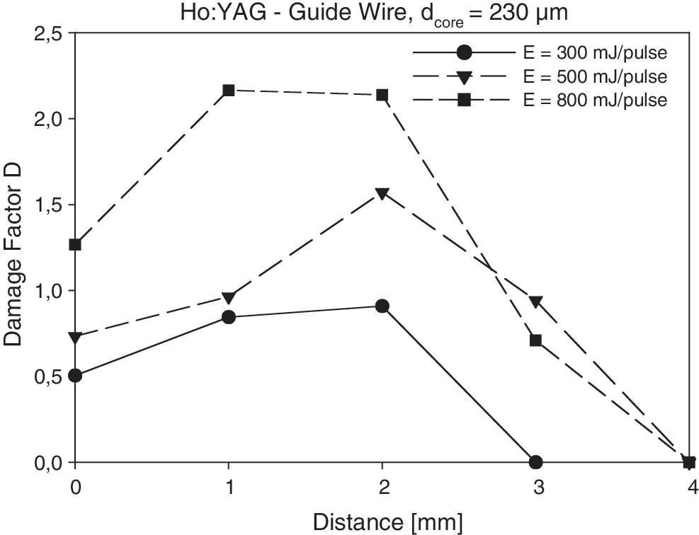
Ho:YAG laser (230 μm fiber): Damage factor D plotted as a function of the distance between the laser fiber tip and the guidewire. Ho:YAG = holmium:yttrium-aluminum-garnet.

Ho:YAG laser (365 μm fiber): Damage factor D plotted as a function of the distance between the laser fiber tip and the guidewire.
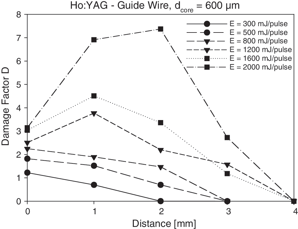
Ho:YAG laser (600 μm fiber): Damage factor D plotted as a function of the distance between the laser fiber tip and the guidewire.
Extending the distance over 2 mm resulted in a decrease of factor D based on the fact that the transmitted laser energy consequently reduced toward a Ho:YAG-related threshold value for detectable impact, irrespective of the fiber core diameter used. At distances of ≥ 4 mm, no more visible detectable damage occurred.
The distance-dependent increase of Ho:YAG laser impact in the range of 1 to 2 mm subsequently occurred with increasing magnitudes of energy per pulse with respect to the fiber used (eg, 230 μm at 500 mJ/pulse; 365 μm at 800 mJ/pulse; 600 μm at 1200 mJ/pulse). Damages induced by the Ho:YAG laser differed dramatically from the FREDDY and FLDP lasers. Here, the initial increase of the damage class D resulted from the extension of the damaged area because of the numeric aperture of the fiber while the visible damage sensation remained constant. As shown in Figure 5, the damage impact induced by the FLPD laser increased within a distance range of 2 mm to reach a saturation, which may decline with larger distances. The FREDDY laser showed a different distance behavior as shown in Figure 6. Because of the wavelength combination, the green portion penetrates efficiently through the water to induce a certain amount of damage to the wire. In the distance range of up to 4 mm, a slight increase of the damage factor D could be observed with FREDDY and FLDP, not exceeding a threshold of 2.
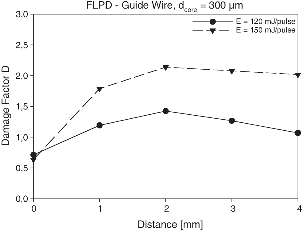
FLPD laser (300 μm fiber): Damage factor D plotted as a function of the distance between the laser fiber tip and the guidewire. FLPD = flashlamp pulsed dye.

FREDDY laser (273 μm fiber): Damage factor D plotted as a function of the distance between the laser fiber tip and the guidewire. FREDDY = frequency-doubled double-pulse neodymium: yttrium-aluminum-garnet.
Damage to stone baskets
Using the Ho:YAG laser, the damage to the nitinol wires of the retrieval baskets depended significantly on the applied energy per pulse and the fiber core diameter (Table 3) showing an equivalent distance dependency as evaluated in the guidewire experiments. Complete disruption of the nitinol wires could be achieved using the 365 μm fiber with a pulse energy of 1600 mJ and by means of the 600 μm fiber starting at EP = 1200 mJ/pulse. Contrary to the Ho:YAG laser, the impact of the other two investigated lasers did not exceed grade 2. Maximal energy per pulse resulted in partial disruption of the nitinol wires with a remaining diameter of 50% (grade 2) in the case of the FLPD laser and of 25% (grade 3) using the FREDDY laser (Tables 4 and 5). Even when applying multiple pulses with any of these laser systems, no disruption of the wires could be observed.
Ho:YAG = holmium:yttrium-aluminum-garnet.
FLDP = flashlamp pulsed dye.
FREDDY = frequency-doubled double-pulse neodymium:yttrium-aluminum-garnet.
Discussion
In laser-induced lithotripsy. the primary mechanisms that cause stone decomposition and fragmentation depend on the laser system selected and the parameters used. Direct absorption of laser pulse energy plays a major role in the Ho:YAG-induced destruction process. Ho:YAG irradiance (λ = 2100 nm) is highly absorbed by water. At smaller distances, parts of the laser pulse energy last for inducing a vapor channel (“Moses effect”), allowing direct laser energy transmission that results in increased temperature of the irradiated volume above a critical threshold, leading to thermal disruption. The magnitude of the generated shockwaves is low and therefore contributes little to the fragmentation process. 12,15,16
Photomechanical shockwave-induced fragmentation occurs during plasma expansion and on bubble collapse in Q-switched neodymium:YAG (FREDDY) and pulsed dye lasers (FLDP). 7,9,10 The magnitude of pressure waves generated at cavitation bubble collapse contributes significantly to the predominant fragmentation process that is described as laser-induced shockwave lithotripsy.
In contrast to the Ho:YAG laser devices, there is negligible water absorption of the visible irradiation (FLPD: λ = 595 nm, FREDDY: λ = 532 nm). Even the absorption of the IR fraction of the FREDDY device (λ = 1064nm) amounts to about 1/10 of the absorption at the 2100 nm Ho:YAG laser wavelength. The irradiation of the FREDDY and FLDP lasers penetrates the hydrous medium over longer distances. 6,8,10,11,13 Furthermore, calculi tend to absorb the complete laser energy and the fragmentation process from plasma formation, the generation of cavitation bubble collapses, shockwaves, and water jet takes place at the same time.
Laser material processing depends on the energy applied to the targeted material exceeding the ablation threshold energy. There are different explanations of how material decomposition may occur. One is direct absorption of light, resulting in temperature increase and subsequent melting of the material. Another is pulsed light energy-induced plasma formation with electrons being emitted by the cannonaded metal, creating an effective cavitation process for shockwave induction. When differentiating the predominant laser-induced damage process to endourologic tools, the wavelength of the laser should be taken into account. FREDDY and FLDP laser wavelengths are minimally absorbed by the surrounding water; their absorption coefficient is about a factor of 10,000 times lower compared with Ho:YAG lasers. Therefore, these lasers transmit energy without any dominant losses.
The Ho:YAG laser loses energy on its way through the water before being absorbed by the targeted surface. In direct contact with the material, the higher pulse energies and the longer pulse duration of the Ho:YAG laser affect a longer interaction time, resulting in the effective destruction of the target. At distances between fiber tip and tool surface more than 4 mm, the emitted pulse energy is completely absorbed by water, and direct laser light transmission is impossible.
Incidental thermal fracture of accidentally irradiated endourologic tools is a common phenomenon during Ho:YAG laserlithotripsy. 17 –19 Honeck and associates 19 have demonstrated that the time needed to disrupt wires may be reached during endourologic procedures. Nitinol baskets especially disrupt within seconds using standard clinical Ho:YAG laser settings. The authors reported an average time of transection for a 0.025-inch Terumo wire with energy settings of 800 mJ/pulse at 5 Hz using a 230 μm fiber to be 40 ± 1.7 seconds after direct laser application, with the disruption time decreasing to 34 ± 0.7 seconds when a 365 μm fiber was used.
Reeves and colleagues 20 demonstrated in their series of experiments of Ho:YAG impact on a 0.038-inch guidewire a pulse energy, cumulative energy, and distance dependency. The experiments presented here, though, aimed to compare the impact of laser light irradiation of three different clinically available laser types with typical settings used in clinics. Single laser pulses were applied with an overall adjustable repetition rate of 3 Hz. The impact of the first laser pulse was investigated to obtain reproducible, comparable conditions, showing the amount of impact of a single laser pulse.
Up to now, investigations of damage that was caused by laser systems on endourologic tools were limited to the evaluation of the damaged area and the time to complete disruption. Our measurements, however, do take into consideration that one single laser pulse may be sufficient to impair the function of the tool significantly. Therefore, we introduced for the first time the relative destruction of the tools as a second parameter. The determination of the damage factor D for the guidewire experiments and the ablation rate Q for experiments with the stone baskets allowed direct laser impact comparisons.
With respect to the damage of the guidewire and the nitinol wires of the retrieval basket, Ho:YAG laser-induced damages exceeded those of both the FREDDY and FLDP lasers, showing a significant distance dependency. It could be demonstrated that FREDDY and FLDP laser impact was not capable of compromising the functionality of the endourologic tools tested.
In a clinical setting, as in the case of an impacted stone basket, the holmium laser has proven to fracture the basket safely and rapidly. 21 –23 This scenario is hardly conceivable for FLPD or FREDDY lasers. The Ho:YAG-laser is currently the most efficient, versatile, and widely used laser lithotripsy device available, whereas the FLPD or the FREDDY lasers do not play an important role in everyday endourologic practice. 4,5 Based on the results of the current study, extreme care has to be taken when using the Ho:YAG-laser to avoid direct contact of applied laser energy with wires and baskets to prevent unintended disruption.
Conclusion
The current study demonstrated that the amount of laser-induced damage to endourologic tools and guidewires depends on fiber core diameter, pulse energy, and distance. The collateral damage induced by Ho:YAG lasers exceeds FREDDY- and FLPD-induced laser damage. Therefore, a safety distance, depending on laser systems, is necessary to prevent laser-induced disruption of endourologil tools. If disruption is intended, the Ho:YAG laser is the instrument of choice.
Footnotes
Disclosure Statement
No competing financial interests exist.