
Abstract
Background and Purpose:
With the rise in detection of small renal masses that are amenable to nephron-sparing surgical approaches, there has been an increasing need for renal tumor models that create discrete lesions suitable for training exercises. We aim to investigate a handful of commonly used compounds, subjectively evaluating their ease of implementation and imaging characteristics.
Materials and Methods:
After an initial ex vivo study, we selected five compounds for an in vivo porcine investigation. These compounds included metagel with barium, Smooth-Cast 320, Silfome with and without barium, and Kromopan. The compounds were injected under laparoscopic guidance with the aim of creating discrete renal tumors. The kidneys were then imaged under ultrasonography and CT. The animals were euthanized, and nephrectomy was performed. Handling characteristics were noted.
Results:
All compounds were relatively easy to inject. Most of the compounds were susceptible to some degree of subcapsular spread. Kromopan had a high propensity for infiltration of the collecting system. On imaging, metagel was clearly distinguishable from normal renal parenchyma on both CT and ultrasonography. Silfome and Smooth-Cast were difficult to resolve on ultrasonography. Metagel was prone to rupture during surgical manipulation.
Conclusions:
No single compound provided the ideal combination of ease of implementation, resistance to extravasation, ease of resolution on imaging, and resistance to rupture. Therefore, compound selection should be dictated by the particular aims of a training simulation.
Introduction
As such, some interest has recently been directed toward the creation of renal mass models for in vivo use in minimally invasive training laboratories. Scott and associates 5 and Taylor and colleagues 6,7 have adapted an agarose-based model that was used previously in hepatic tumor modeling and described its use as a renal tumor model for radiofrequency ablation training. Despite its low cost and ease of use, their technique appears to be limited by the propensity for extravasation when attempting to create tumors beyond 1 cm. 6,8
In 2005, our institution explored the use of Smooth-Cast 320 (Smooth-On, Easton, PA), a dental impression molding, to create an artificial tumor in the porcine kidney, which was evaluated favorably by participants in a laparoscopic training course during which the model was used. 9 Most recently, Eun and coworkers 8 described two additional models that allow for the creation of larger tumor models. Kromopan hydrocolloid (Kromopan USA, Morton Grove, IL), also a dental impression product, was evaluated favorably as a mimic for solid renal masses. For cystic renal masses, a mixture of Metamucil, gelatin, and methylene blue was used, a compound the authors named “metagel.” 8 This compound is very similar to that used in previous hepatic tumor model studies. 10 Other compounds explored in the literature include a wide range of industrial caulks, sealants, and epoxies. 8
Recently, our laboratory has become interested in evaluating the use of Silfome (ArtMolds, Summit, NJ) in renal mass modeling. Silfome is a silicone-based casting agent used in animatronics and toys for its ability to realistically model flesh and soft tissue. We sought to explore this compound, as well as other previously described compounds, to evaluate their ease of use and imaging characteristics. While previous studies have evaluated the imaging characteristics of mimic compounds under ultrasonography, 5 –9 to our knowledge, no study to date has attempted to evaluate these lesions under CT. Therefore, CT imaging was a main focus of the present evaluation.
Materials and Methods
After obtaining approval from our Institutional Animal Care and Use Committee, an initial in vivo and ex vivo pilot study was performed to evaluate seven compounds under noncontrast CT imaging. These included metagel with and without barium, Elite H-D (Zhermack, Badia-Polestine, Italy), Silfome with and without gelatin and barium, Kromopan, and Smooth-Cast 320. Ease of handling was determined by injecting the compounds into discarded porcine nephrectomy specimens from another approved study. To evaluate imaging characteristics preliminarily on CT, the compounds were rolled into roughly spherical masses and imaged alongside additional discarded nephrectomy specimens. Based on these imaging findings, as well as the ease of use of the products, five compounds were selected for further study: Metagel with barium, Smooth-Cast, Silfome with barium and Silfome without barium, and Kromopan. The Elite H-D was not further investigated because of prohibitive difficulty with handling and administration.
Subsequently, two adult female domestic pigs (Oak Hill Genetics, Ewing, IL) were selected for our study. After sedation with a weight-appropriate dose of buprenorphine, the animals were placed under general endotracheal anesthesia.
The pigs were placed in a lateral decubitus position, and the abdomen was insufflated with carbon dioxide to a pressure of 15 mm Hg. The kidney was then identified, and tumor sites were selected, one each in the upper and lower poles of the kidney. A laparoscopic needle was then advanced percutaneously under direct vision into the renal parenchyma, using visual cues to ascertain depth of penetration. One of five compounds was then injected with the aim of creating an exophytic renal mass. These compounds included metagel with barium, Smooth-Cast, Silfome with barium, Silfome without barium, and Kromopan.
Once tumor placement had been accomplished on one side, the incisions were closed using nylon sutures, and the pig was then repositioned to the opposite side, where placement of upper and lower pole tumors was performed in the same fashion as described above. These incisions were also closed with nylon sutures.
The animals were then transported under anesthesia to our imaging facility, where abdominal CT scans were performed with and without intravenous contrast. Transabdominal ultrasonograpphic imaging was then performed to evaluate the in vivo appearance of the masses before the animals were humanely euthanized according to standard protocol. Flank incisions were then used to expose the kidneys, and direct imaging with ultrasonography was also performed. Radical nephrectomy was then performed through the flank incision, taking care to minimize direct handling of the tumors.
Results
Pilot study
The metagel, Silfome, and Smooth-Cast 320 were subjectively the easiest compounds to inject into the porcine kidney. For reasons that are not quite clear, Kromopan had a high tendency to extravasate into the collecting system, while the Elite H-D proved very difficult to inject because of its quick-setting properties.
Ex vivo CT imaging revealed that the metagel and Kromopan provided the most homogenous masses. The Hounsfield unit (HU) density of metagel ranged from 91 HU without barium, to 623 HU with barium. The Kromopan had a density of 260 HU.
Smooth-Cast 320 and Silfome demonstrated low attenuation, even with the addition of barium to the Silfome mixture, with densities of −500 to −523 HU. As such, they were not visible on default abdominal windows. In addition, once properly windowed, these compounds demonstrated considerable heterogeneity, with Silfome demonstrating a central area of low attenuation. The Elite H-D was visible on default windows, with a density of 360 HU, but also demonstrated significant heterogeneity.
Live animal study
Tumor sizes of up to 1.5 × 2.8 cm were created using the selected compounds. All studied compounds proved relatively easy to inject, and all grossly created discrete exophytic masses on instillation. Hemostasis appeared to be excellent before concluding the cases, and no animal suffered any intraoperative complication. On delayed imaging, however, it was noted that in one pig, a retroperitoneal hematoma had developed that appeared to arise from a lower pole injection site where metagel was administered.
Despite initially creating discrete masses, delayed imaging with CT demonstrated that most compounds were prone to delayed subcapsular spread. Once again, Kromopan was noted to extravasate readily into the collecting system.
HU densities were similar between the two subjects for Smoothcast (119 HU and 111 HU), and Silfome appeared to be consistent, regardless of the administration of contrast (−150 to −182 HU). Kromopan demonstrated a density of 249 HU.
The metagel showed considerable variation, ranging from 146 HU to 459 HU. The higher value, however, was associated with the lower pole injection that likely gave rise to the perinephric hematoma noted in that subject, and the presence of blood may have altered the imaging characteristics.
The administration of intravenous contrast did not aid in the identification of the masses or their extent. Conversely, it did not hinder the identification, nor were image characteristics different for the masses after the administration of contrast.
In vivo transabdominal ultrasonography proved highly challenging because of the overlying ribs, and despite attempts by multiple investigators, the results were highly variable, echoing the experience of Hidalgo and colleagues. 9 Metagel, however, consistently demonstrated a discrete mass with heterogeneous echotexture. Silfome was extraordinarily difficult to image, with large signal dropout and shadowing, likely because of the aeration of the compound. Smooth-Cast 320 was isoechoic, rendering it difficult to distinguish from normal renal parenchyma. Kromopan demonstrated a sharp hyperechoic interface with the renal parenchyma; however, acoustic shadowing made finding the deep margin extremely difficult.
Most all of the compounds remained stable on extraction, except for one metagel tumor, which ruptured during gentle manipulation; the other metagel tumor remained stable. The delayed spread of the masses that were noted on CT were also noted on gross specimens.
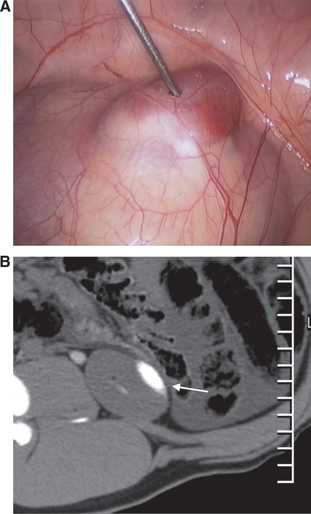
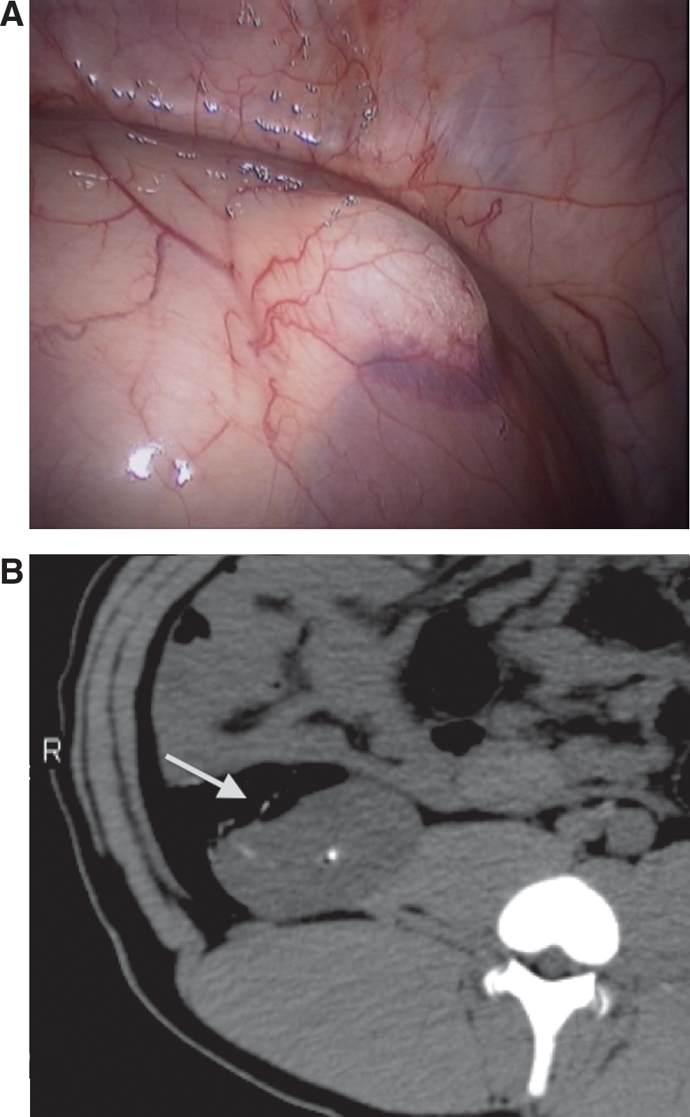
A comparison of the relative strengths and weaknesses of each compound can be found in Table 1. In addition, an overview of the gross appearance of the masses, along with their corresponding appearance on imaging, can be found in Figures 1 to 4.


Discussion
No one compound in our study proved ideal under all circumstances. Instead, our results suggest that the value of a particular compound depends on the context in which it will be used.
Some variability was noted in our study, especially between the ex-vivo and in-vivo CT imaging. Overall, there was a trend toward lower absolute values of attenuation, likely because of multiple factors, including absorption of X-rays from surrounding structures, differences in density between room and body temperature, as well as changes in the texture of the compounds from the process of injecting the compounds rather than rolling them manually into a sphere. The stark difference in the Smooth-Cast 320 density between ex-vivo and in-vivo images is unknown but is perhaps because of temperature differences.
In terms of creating reliable and stable lesions, Smooth-Cast 320 retained a discrete parenchymal lesion despite the tendency to infiltrate the collecting system. Because of its quick-setting properties, however, it must be used quickly. In addition, the compound appeared to mimic normal renal parenchyma quite closely, which makes identifying the interface between normal kidney and mass somewhat difficult on ultrasonography. Therefore, it would likely be inadequate for a simulation that evaluates endophytic lesions or one for which margin status is of primary importance.
Metagel also proved to be a very easy compound to manipulate, and carries with it an advantage of extremely low cost. While the substance was prone to a small degree of subcapsular spread, the masses nevertheless remained relatively discrete. In addition, the masses show up distinctly on abdominal windows on CT and are easily differentiated from normal renal parenchyma on ultrasonography. The metagel, however, was the only substance that ruptured during extraction of the kidneys during this experience, a property also noted by Eun and associates. 8
Silfome proved extremely easy to inject; however, it is prone to a larger degree of subcapsular spread than metagel. In addition, it is very difficult to resolve on CT, even with the addition of barium to the mixture, and nearly impossible to resolve on ultrasonography, likely because of its high degree of aeration.
Kromopan was difficult to inject, and it was prone to infiltration into the collecting system, for reasons that are not entirely clear and that could not be elucidated by evaluation of the gross specimen. In their experience, however, Eun and coworkers 8 noted that Kromopan masses were more stable than metagel during surgical simulations, a property that we also found, although our experience was small and not specifically designed to evaluate the durability of the masses.
Our study does have a few limitations. Because our aim was to compare a wide variety of compounds, we were unable to focus on exhaustively testing the properties of all of the materials. Indeed, while successful as a pilot study, our small sample sizes prohibit the present experience from being entirely conclusive. The utility of the models for surgical simulation of ablation and partial resection was not explicitly tested in our study, because this had been described elsewhere. 5 –9
Rather, our focus was primarily on the CT appearance of commonly used compounds. While it is not anticipated that CT will be regularly used in training situations, with the possible exception of percutaneous ablative techniques, our study nevertheless provides some indication of the stability of masses over time in that our images were obtained roughly 90 minutes after injection. Therefore, our results indicate that lesions that are designed for large training courses should not be created more than 1 hour ahead of time and are best installed at the start of the training simulation.
Conclusions
No single compound serves as an effective renal mass model for all situations. The needs of a particular study or training simulation should be weighed against the properties of each compound, and task-appropriate substances should be selected.
Footnotes
Acknowledgment
The authors wish to thank Nitin Das, Ahmed Tawfik, and Geneva Baca for their assistance with this investigation.
Disclosure Statement
Dr. Benway is a consultant for Viking Systems, Westborough, MA, and Dr. Bhayani is a consultant for Intuitive Surgical, Sunnyvale, CA. The other authors have no conflicts of interest to declare.