




Abstract
Purpose:
To investigate in vitro whether pH ranging between 6 and 9 has an effect on the degradation of stent fibers made of poly(
Materials and Methods:
The fibers were divided into three groups and immersed in sodium phosphate-buffered saline (Na-PBS) solution with three different pH values: 6, 7.4, and 9. The mechanical and thermal properties were studied, and scanning electron microscopy (SEM) images were taken at specific time points of hydrolysis.
Results:
The tensile testing showed that the strength of the fibers decreased through hydrolysis and was lost at 8 weeks in all groups. The Tm and Tg of the PLGA fibers did not indicate any significant differences between the different groups. In SEM images taken at 4 weeks, there were no significant differences between the fibers immersed in Na-PBS solutions of different pH values. However, at 8 weeks the surface of the fiber immersed in saline with a pH of 6 seemed coarser than that of those immersed in neutral (pH 7.4) or alkaline (pH 9) Na-PBS.
Conclusion:
The studied pH values did not influence the degradation behavior of the PLGA 80/20 fibers. Therefore, rabbits can be used as model animals for human biodegradable urological devices even though the pH of their urine is different.
Introduction
Studies investigating the degradation of poly(α-hydroxyacid)s in phosphate buffers of different pH values have found that the hydrolysis rates are similar. Studying the polymers in several other buffers has revealed that the degradation rate increases in strongly alkaline conditions and at higher ionic strengths. 2,3 Hurrel and Cameron 2 explained this phenomenon for poly(α-hydroxyacid)s as follows: The rate at which the degradation proceeds through a polymer depends on the rate at which acidic degradation products diffuse out, creating space for water in the structure. If the solution is made more alkaline or more concentrated, the degradation products are mainly neutralized, driving the hydrolysis reaction forward. Further, the solubility of the neutralized acids increases. These effects appear to dominate over autocatalysis caused by degradation products.
A braided bioabsorbable stent for urological applications has been previously developed by our research group to avoid the problems caused by biostable stents. 4 –8 The degradation of bioabsorbable stents has been previously studied in sodium phosphate-buffered saline (Na-PBS) with a pH of 7.4. 9,10 Animal testing of the stents has been carried out in the anterior urethra of New Zealand white rabbits 11 ; the pH of rabbit urine has been shown to be alkaline, normally ranging from 7.6 to 8.8, 12 but it can be strongly affected by the nutrition of the rabbits. On a low-alkali diet, the average pH of rabbit urine can decrease to 6.26 due to a dramatic reduction in the soluble bicarbonate excreted daily. In contrast, a high-alkali diet does not raise the pH of rabbit urine that remains below 9 as it does with a normal diet as well. 13 As the pH of the human urine is between 4.5 and 7.8, but commonly around 6, 14 it is questionable whether the rabbit model provides reliable results on the degradation of bioabsorbable stents.
The aim of the present study was to investigate in vitro whether pH ranging between 6 and 9 has an effect on the degradation of stent fibers made of poly(lactide-co-glycolide) and, therefore, whether a rabbit model can be used to simulate human conditions.
Materials and Methods
The polymer, a copolymer of
The monofilament fibers were melt-spun and oriented by drawing from the vacuum-dried PLGA. The die temperature of the single screw extruder (Gimac microextruder, Gimac, Castronno, Italy) was 273°C and the diameter of the die was 1.5 mm. The drawing process consisted of three separate stages where the extruded preform was drawn through ovens heated to 100°C to 130°C. The final diameter of the monofilament fiber was 0.4±0.02 mm, and the obtained draw ratio for the fiber was 5.1. The fibers were cut, packed to pouches, and gamma irradiated for sterility using a standard procedure of a 25 kGy dose. The temperature during irradiation was kept low with dry ice.
After sterilization, the fibers were divided into three groups and immersed at 37°C into Na-PBS solutions of three different pH values: 6, 7.4, and 9. The pH change was achieved by changing the concentration ratios (mmol/L) of the Na2HPO4 and NaH2PO4 * H2O 3.65:26.35, 24.52:5.48, and 29.95:0.05, respectively. The NaCl concentration was the same in all cases, namely 100.96 mmol/L. The pH of the buffered saline solutions was measured weekly at room temperature from three randomly selected samples of each set. At the same time, the solutions were changed weekly to maintain the pH at the desired level. For reference, saline solutions with no samples immersed were followed up for detecting the effects of the degradation of PLGA on the pH.
The tensile strength of the fibers was measured as manufactured and as gamma-irradiated at 0, 1, 2, 3, 4, 6, and 8 weeks' follow-up. Both ends of a fiber were placed between the hydraulic grips of the materials testing machine (Instron 4411 Materials Testing Machine, Instron plc, High Wycombe, United Kingdom) and pulled with a crosshead speed of 30 mm/minutes. Five parallel samples of each set were tested (n=5).
The thermal properties of the fibers were measured from the vacuum-dried fibers as manufactured and as gamma-irradiated at 0, 2, 4, 6, 8, and 12 weeks' follow-up using a differential scanning calorimeter (DSC, TA Instruments Q1000, New Castle) with indium calibration and a heating speed of 20°C/minutes. The melting temperatures (Tm) as maximum temperature and the glass transition temperatures (Tg) as onset value were measured each time.
In addition, the fibers were examined using scanning electron microscopy (SEM: JEOL T-100, Tokyo, Japan) at 0, 2, 4, and 8 weeks. The samples were dried and gold sputtered for examination.
Results
pH values
The solutions having a pH of 6 and 7.4 retained a steady pH level throughout the follow-up period of 12 weeks. The most alkaline solution (pH 9) showed changes over the 1-week intervals of solution replacement, starting at 5 to 6 weeks in vitro. At first the changes were relatively moderate, but from week 6 onward the pH changed from 9 to 7.8 to 8 within one week. The lowest pH value measured for that set was 7.81 at week 12.
Mechanical testing
At baseline, the tensile strength of the stent fibers was 377 MPa before and 340 MPa after gamma-irradiation. Figure 1 presents the tensile strength of the fibers in different pH environments during the hydrolysis. The decreasing trend of the strength was practically similar for all samples types. After 8 weeks the tensile strength was less than 15 MPa for all groups.
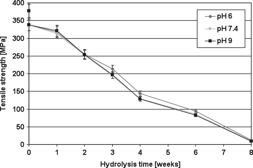
Tensile strength of stent fibers in different pH environments before sterilization, after sterilization, and during hydrolysis.
Thermal properties
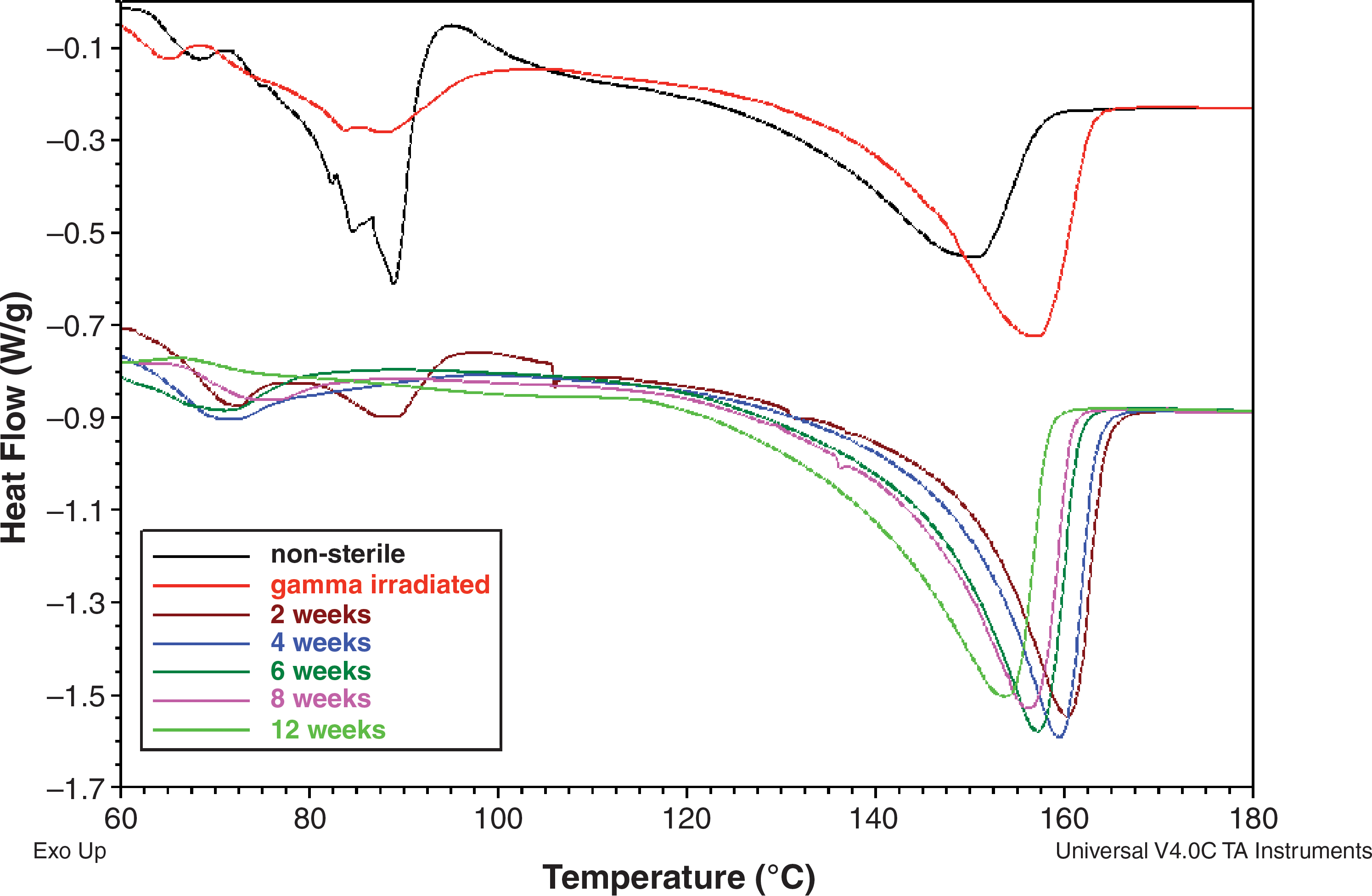
The initial 150°C melting point (Tm) increased during the gamma irradiation process to 155°C. During the hydrolysis period, Tm reached its peak value of approximately 160°C at 2 weeks, after which it steadily decreased. At 12 weeks, Tm had decreased back to 155°C.
The glass transition temperatures (Tg) of the PLGA samples were analyzed. Gamma irradiation influenced the Tg, decreasing it from 56.7°C to 55.9°C. During the hydrolysis, Tg decreased in all sets until week 6 when the Tg for the samples hydrolyzed in pH 7.4 and pH 9 was 49°C and for the pH 6 samples 46°C. The Tg of the pH 6 samples remained at almost the same level (48.2°C) until 12 weeks, but for the others, Tg started to elevate; for pH 7.4 samples Tg was 51°C and for pH 9 samples it was 53.2°C.
Figure 2 shows the DSC curves of the samples before hydrolysis and when immersed in a solution with a pH of 6 during the hydrolysis. Gamma irradiation smoothed the relaxation peaks at glass transition and cold crystallization areas (below 100°C), shifted the Tm to the lower temperature, and increased the melting enthalpy of the fibers. Figure 3 shows the comparison between the melting areas of the DSC curves of the samples hydrolyzed in different pH environments at 2, 6, and 12 weeks. It is clearly demonstrated that the behavior is similar for all three pH Na-PBSs.
Differential scanning calorimeter curves of poly(
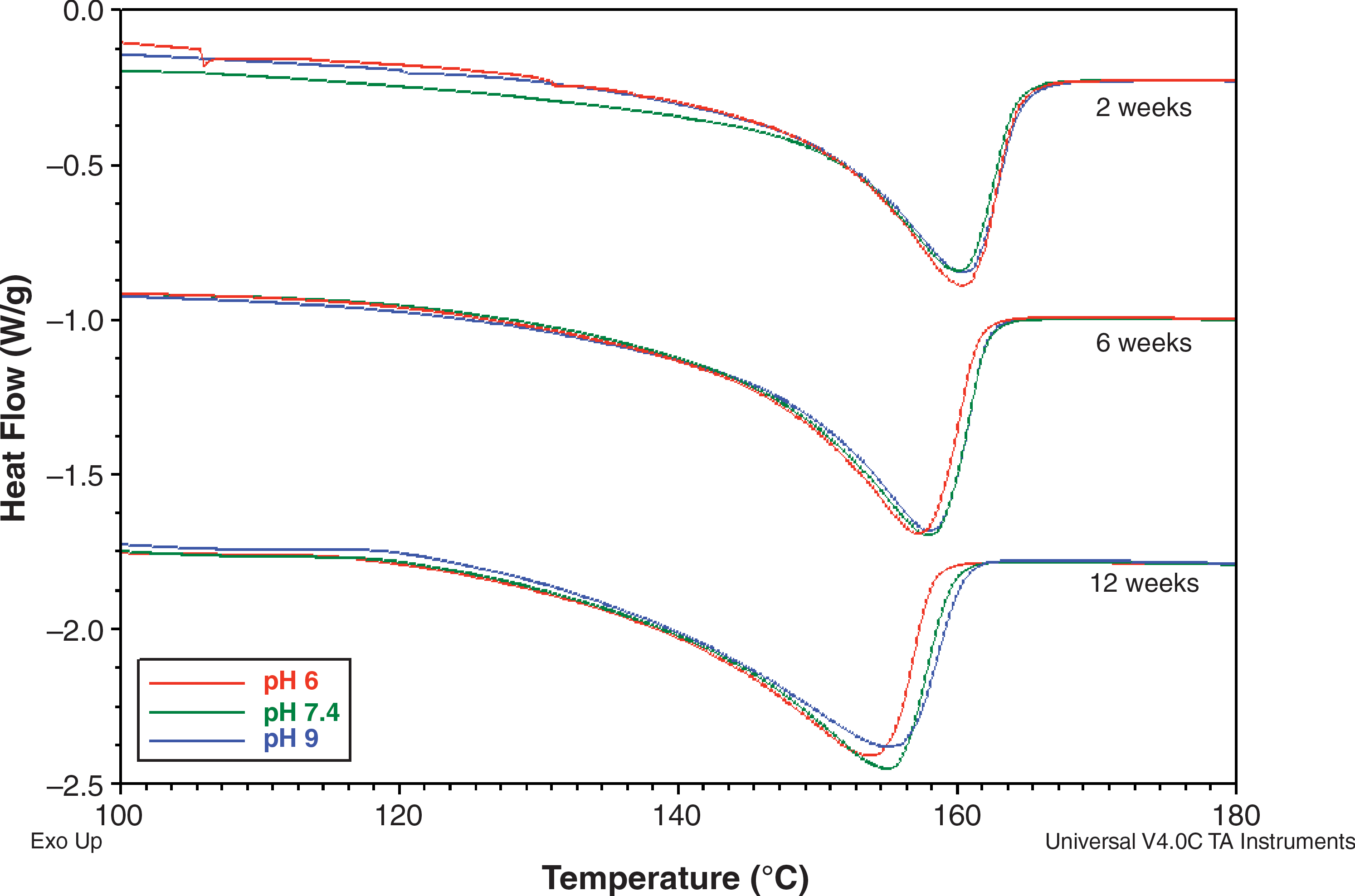
The melting peaks of PLGA samples after 2, 6, and 12 weeks of hydrolysis in different pH Na-PBSs.
SEM images
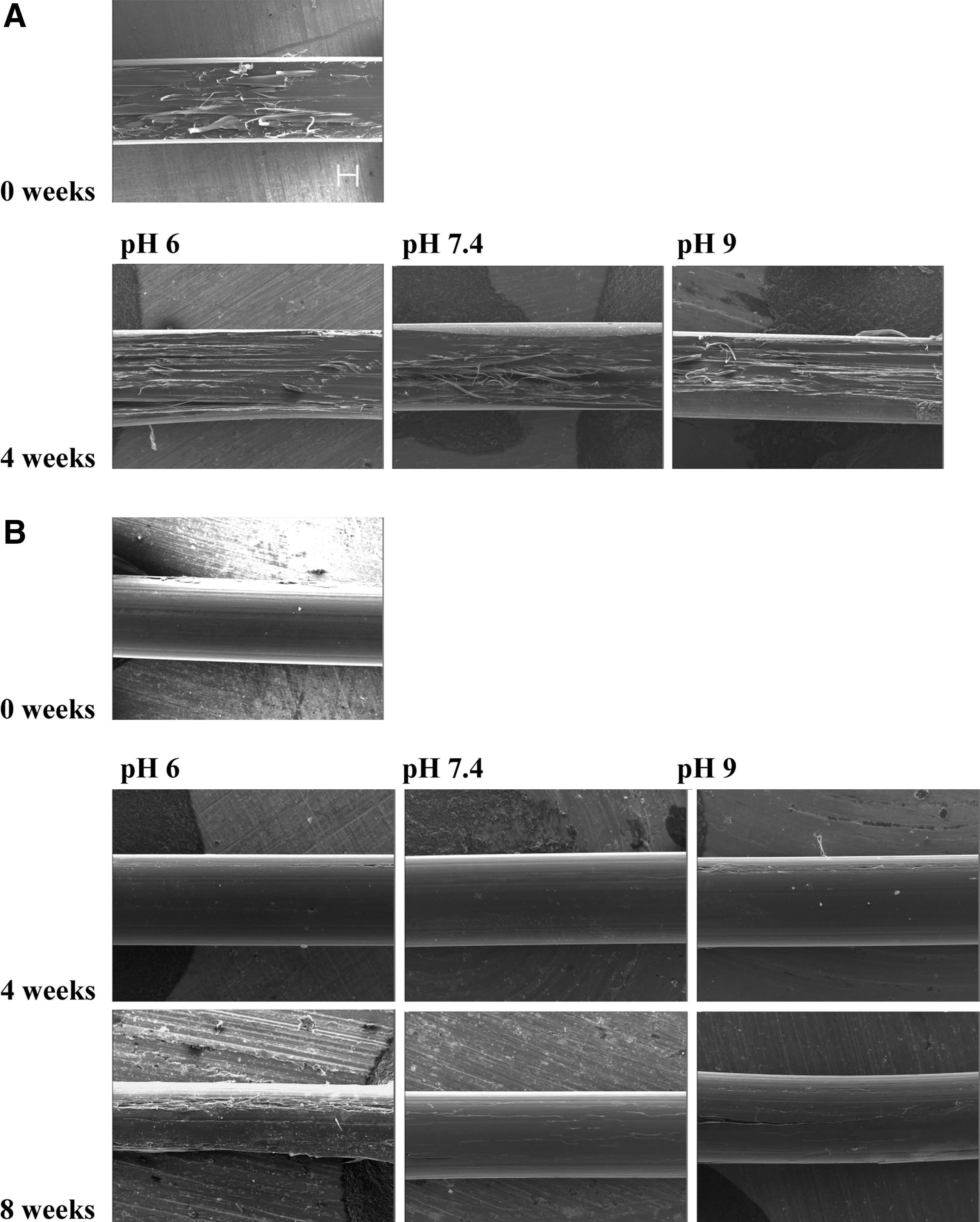
Figure 4 presents the SEM images of the PLGA fiber surfaces before hydrolysis (t=0) and at 4 weeks of hydrolysis (A), in addition to the cross sections of the fibers before hydrolysis and at 4 and 8 weeks (B). There were no significant differences between the fibers in different pH environments at 2 weeks (data not shown) and at 4 weeks. However, at 8 weeks the surface of the PLGA fiber immersed in Na-PBS with a pH of 6 showed clear signs of degradation as opposed to those immersed in a neutral (pH 7.4) or alkaline (pH 9) Na-PBS.
Discussion and Conclusions
The three different Na-PBS solutions were prepared so that the ion concentration of the phosphate was the same in the different Na-PBSs. This caused a problem of the reagent NaH2PO4 * H2O having so low a concentration in the saline of pH 9 that the buffering effect was not sufficient. Thus, the reference Na-PBS with a pH of 9 also had a tendency to decrease. Although the set with pH 9 did not maintain its original pH level, it remained in the same range as rabbit urine (pH 7.6–8.8) throughout the study. In all cases, the pH had dropped more in the saline with PLGA samples than in the reference saline. This was due to the acidic degradation products of PLGA, as documented earlier. 15
The present tensile strength results showed a loss of mechanical strength practically in 8 weeks. This behavior was expected since PLGA copolymers are known to degrade and lose their strength faster than the plain homopolymers. In the present study, we did not observe any significant difference in degradation time as measured by the loss of mechanical integrity at the studied pH values. Therefore, regarding the pH value rabbits can be used as model animals for human urological devices despite the difference between the pH values of rabbit and human urine.
Even though it was possible to perform the tensile test for the PLGA fibers after 8 weeks' hydrolysis and a moderate tensile strength was observed, the preparation of the dried fiber specimens for cross-sectional SEM studies was impossible at this time point due to the fragmented nature of the samples. A surface examination of the fibers at 8 weeks revealed only moderate changes (namely, slight cracking) for samples in a neutral or alkaline saline, but those hydrolyzed in a Na-PBS of pH 6 appeared to have deteriorated more and the fibers also appeared somewhat thinner. This may indicate a faster structural degradation of PLGA at pH 6 than at pH values of 7.4 or 9, but it did not critically influence the mechanical properties of the fibers.
The changes in thermal properties of the polylactides indicate the degradation of the polymer. The melting temperature decreases when the degradation is progressing and the mechanical properties of the fibers are decreasing. The measured melting points (Tm) increased with gamma irradiation and during the first 2 weeks of hydrolysis after which the tendency was sloping. As seen in Figure 2, the melting peaks had a tendency to broaden as the hydrolysis progressed, demonstrating a broader melting area and indirectly indicating the loss of molecular weight. 16 However, as shown in the presented figures, no big differences were observed in thermal property changes between the samples hydrolyzed at different pH values. However, at 12 weeks one can observe a slight change in the Tm of pH 6 samples that may confirm the observations of the earlier structural degradation from SEM images.
If the hydrolysis and the study of thermal properties were continued further after 12 weeks, it could be expected that more differences would appear between the properties of the separate sets. However, this has no relevance in practice since the PLGA stents do not show any radial force after 12 weeks of hydrolysis in vitro 17 and they may have already fragmented and disappeared from the body with urine.
As a conclusion, it can be said that there was no significant difference in the degradation rate between the three groups with different pH environments in the hydrolysis in vitro. Earlier studies—performed with a PLGA copolymer with DL-lactide instead of
Footnotes
Acknowledgment
The authors wish to thank Mrs. Raija Reinikainen for her technical assistance. The funding from the National Technology Agency of Finland (Tekes) NeoText project is greatly appreciated.
Disclosure Statement
No competing financial interests exist.