




Abstract
Purpose:
Conventional two-dimensional (2D) laparoscopy has been limited by the lack of depth perception and spatial orientation. This disadvantage may affect surgical performance, operative time, or morbidity. Depth perception, spatial resolution, and accuracy may be improved by three-dimensional (3D) vision. This study was designed to evaluate the effect of 3D imaging on the performance of novice and experienced laparoscopic surgeons executing standardized laparoscopic tasks in an ex-vivo setting.
Materials and Methods:
A 3D imaging system with a digital 3D full high definition (HD) and a 2D imaging with a conventional HDTV camera were used. A total of 10 laparoscopic experts and 10 novices were evaluated while performing standardized skill tasks in a pelvic trainer. Participants were divided into two groups working either with 2D or 3D imaging. The parameters measured were time until completion of each skill and total procedure time, number of losses of working material, number of missed grasps, and number of needed support. Statistical analysis was performed using the Wilcoxon test.
Results:
The results showed a significant difference in favor of the 3D system for the amount of missed grasps in the experts as well as the novice group (P<0.0001). A slightly significant difference was also seen for the loss of working materials (P=0.0381 and P=0.0693). The study is limited by its small sample size.
Conclusion:
The 3D imaging system significantly improves spatial orientation and depth perception during laparoscopy under ex-vivo conditions.
Introduction
Three dimensional (3D) imaging may be beneficial by eliminating the need for mental processing of the 2D image into a 3D image using motion parallax like changes in shades or position of instruments. 1 Studies evaluating the advantages of 3D systems have reported conflicting results. One point of criticism in these studies is that a steep learning curve for laparoscopy using 2D systems making any improvement by 3D vision unnecessary. 2 –5 Some authors also criticize the quality of the 3D systems. 2 –4 In part, this is a result of the lower resolution of first-generation 3D technology compared with standard 2D systems. In older 3D systems, the 3D effect was achieved by presenting two slightly different images to each eye in an alternating sequence. 3 In contrast to that, the modern 3D systems have two separate optical channels creating two unique images for the right and left eye. 1 Therefore, the imaging of current systems is similar to stereoscopic vision, in which the depth perception is achieved by different unique images received by each eye. 6
This study was designed to evaluate the effect of a modern 3D imaging system on efficiency and accuracy for laparoscopic skills. The study will allow an evaluation of the effects of 3D vision using a modern 3D camera system on both novice and experienced laparoscopic surgeons.
Materials and Methods
The 3D imaging system (Einstein system, Schölly Fiberoptics, Germany) consisted of a 10-mm outer diameter 30-degree stereoscopic endoscope, a digital 3D full high-definition (HD) camera, and a 32-inch 3D full HD monitor (Fig. 1). Camera and endoscope were fixed on a maneuverable robotic arm system (Schölly, Denzlingen, Germany). The operator had to wear special 3D glasses to be able to see the 3D image (Fig. 2).
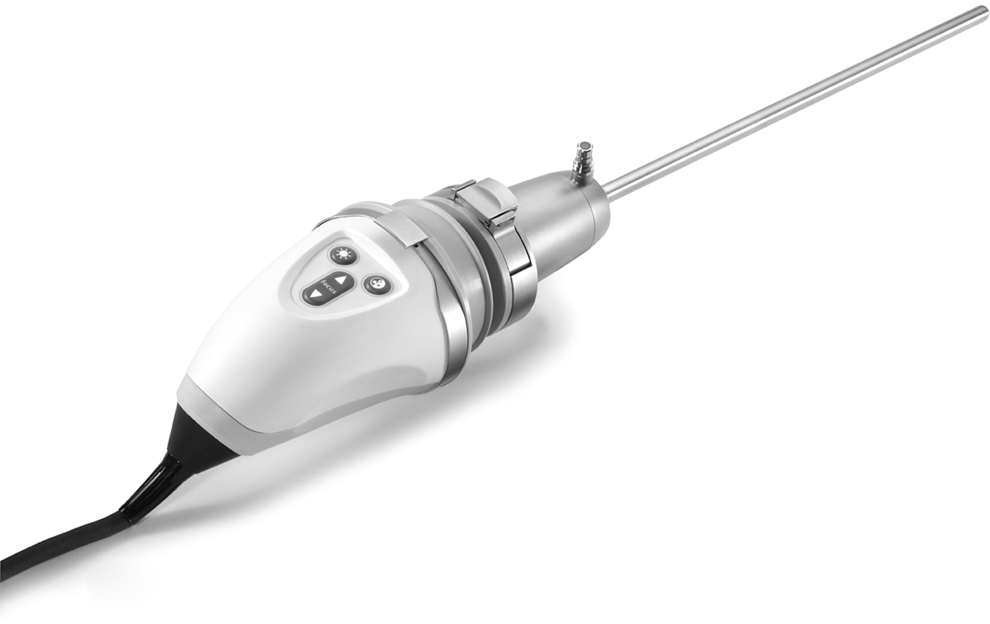
A 10-mm outer diameter stereoscopic endoscope with a digital three-dimensional full high-definition camera.

Three-dimensional (3D) glasses for the Einstein 3D imaging system.
The 2D imaging system used was a conventional high definition television camera with a 30-degree and 10-mm diameter laparoscope (Karl Storz, Tuttlingen, Germany). The images were also seen on a 32″ full HD 3D liquid crystal (LC) monitor. The camera was mounted on the robotic arm in the same way as the 3D camera.
A pelvic trainer incorporating two working ports and a camera port was used. The camera was positioned in a fixed angle, and working trocars were always placed in the same position for all participants. Participants were asked to perform a standardized list of laproscopic skill tasks. Skill 1 consisted of the placement of three plastic rings on three different cones performed with the dominant (D) hand and the nondominant (N) hand (Skill 1D and 1N). The parameters measured were time until completion, number of ring losses, number of missed grasps, and number of needed support. Time for skill completion was measured in seconds; each loss of a working material or a missed grasp was counted as an error. Each support needed by the participants through an experienced surgeon was counted as a needed support.
Skill 2 involved the grasping of three rings starting with the D hand, transferring it into the N hand and placing it on one of three cones. The same procedure was then performed starting with the N hand (Skill 2D and Skill 2N). The same parameters were evaluated as mentioned before. Skill 3 included the passage of a needle through a metal ring. Measured parameters were procedure time, needle loss, missed grasps, and numbers of support needed. Skill 4 consisted of cutting a preknotted suture. Measured parameters were procedure time, missed grasps, and number of supports needed. Skill 5 included the tying of one knot. Measured parameters were procedure time, needle loss, missed grasps, and numbers of support needed. Skill 5 was only performed by experienced laproscopic surgeons, because for novices without any laparoscopic experience, tying a knot without training in advance was considered a too difficult task.
A total of 10 experts and 10 novices were evaluated. All experts had performed at least 50 laparoscopic procedures by themselves. Novices had not performed any laparoscopic surgery so far. Participants were randomized into the conventional or 3D group. Five novices and five experts performed the tasks using a conventional laparoscopic system and five novices and five experts performed the same tasks with a modern 3D camera system. The 3D group did not have additional 2D vision. In addition, all experts in the 3D group were asked to judge the comfort and ease of handling of the 3D system.
Statistical analysis was performed using the Wilcoxon test. SAS software, release 9.2 (SAS Institute Inc, Cary, NC) was used for statistical analysis.
Results
A total of 10 novices and 10 experienced laparoscopic surgeons were enrolled in this study. The results for both groups are displayed in Table 1. The overall time for skill completion did not differ significantly in either group (experts P=0.3125, novices P=0.4735). Especially in the novice group, however, the standard deviation was very high (±687). In the novice group, we could see a slightly significant difference in favor of the 3D system in skill 2D and in the number of losses of working material (ring or needle, P=0.0381). A significant difference in favor of the 3D system was seen in the amount of missed grasps (P<0.0001). In the experts group, there was also a slightly significant difference in the number of losses of working material (P=0.0693) and a significant difference in the number of missed grasps (P<0.0001). None of the participants in the 3D groups had problems with achieving 3D vision. The 3D glasses were not regarded as an impairment during the procedure. The subjective impression of those participants working with the 3D image system was consistently positive regarding operating conditions and operability.
2D=two dimensional; 3D=three dimensional; D=dominant; N=nondominant.
Discussion
The introduction of laparoscopy has produced new concepts and benefits for the urology patient population, and its long-lasting impact in urologic surgery is an undeniable fact. Conventional 2D laparoscopy has been limited by the lack of depth perception and spatial orientation. This disadvantage may affect surgical performance, operative time, or morbidity. Depth perception, spatial resolution and accuracy may be improved by 3D vision. Especially complex tasks, such as intracorporeal suturing and precise dissection, need a high degree of skill and depth perception. To compensate for this lack, 2D imaging uses monocular references like motion parallax, relative position and size of anatomic structures, shading of light and dark, and tissue grading. 3,4,7,8 Experience in laparoscopy, though, improves the processing of monocular cues. 9 It can be assumed that these adaptions account for the increased mental fatigue and strain associated with 2D imaging as higher visual centers recalibrate to interpret perpetual input. 3,10
3D vision offers the advantage of improved depth perception and accuracy, and separate input from two unique images allows for neural summation on a cortical level. 11 –13 Previous studies comparing 2D and 3D imaging have been limited by the technology available at that time, and numerous studies showed no difference in performance. 2,12 The Einstein system, however, is a true 3D imaging system with two separate optical channels and a light source. The 3D images present directly on the 32" full HD 3D LC monitor. The image separation for each eye is provided by passive polarized glasses. The right and left eye receive unique images from the camera. Two imagers with true full HD resolution each are used. Falk and associates 7 were able to show using motion analysis that the 3D imaging system of the da Vinci robot, which is similar to the Einstein system, resulted in shorter performance times through increased peak velocity and acceleration and decreased rates of deceleration. Operators were able to perform more percise adjustments by using 3D vision when compared with 2D vision.
Surprisingly, we did not see a sigificant difference in the total procedure time in our statistical analysis. This might be because of a combination of different reasons. First of all, the study groups are rather small, with only 10 participants in the experts and novice groups. Regarding the total procedure times in the novice group, there is also a very high standard deviation of 687 seconds. These variations in combinations with the small study group might well explain the missing significance. The experts group, on the other hand, did not show a significant difference in total procedure time as well. This was not unexpected, because for experts, it can be assumed that laparoscopic skills are already so well trained that a significant difference in performing these skill tasks is unlikely. This might be different when performing actual surgery over a longer time interval of several hours.
Based on the small sample size, slightly significant results should also be regarded noteworthy. The loss of rings or needles was slightly significant in both groups. In combination with the highly significant reduction of missed grasps in both groups, this seems to illustrate the most prominent benefit of the 3D imaging system in terms of depth perception and spatial orientation. The subjective impression of the 3D participants supports this assumption, because all of them reported a much better spacial resolution and depth perception. This benefit might well be even more substantial under realistic conditions when complex and detailed skills such as intracorporeal suturing or difficult dissection are needed.
Although it is a subjective judgement, all experts who worked with the 3D system reported that is more comfortable than working with conventional 2D systems, which might well result in less mental fatigue and strain.
Conclusions
The 3D imaging system seems to improve especially spacial orientation and depth perception during laparoscopic procedures even for expert surgeons. More studies with larger sample size and evaluation under actual surgical conditions are needed to show a marked difference from conventional laparoscopy.
Footnotes
Disclosure Statement
Prof. Knoll is a member of the advisory board for Schölly Fiberoptics, Germany. For the remaining authors, no competing financial interests exist.