
Abstract
In the first two patients who underwent laparoscopic anatrophic nephrolithotomy, because of the large size of the stones, we attempted to optimize the exposure of the collecting system, making the renal incision slightly forward of the Brodel line, almost at the lateral edge of the kidney. With this approach, we were attempting full exposure of the calculi.
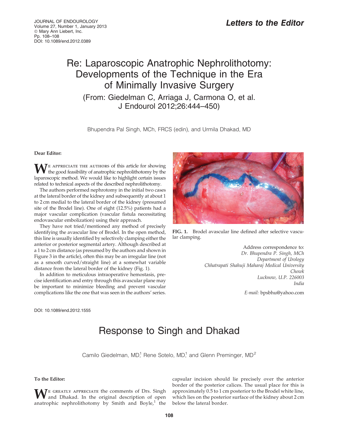
In the third and following cases, we decided to move the incision to the real line of Brodel to limit the bleeding and to obtain better visualization of the parenchyma and the collecting system.
We believe that in the development of the laparoscopic technique, dissection beyond the division of the renal artery to locate the posterior branch is unnecessary, because an optimal view of the Brodel line and unique clamping of the principal renal arterial trunk is enough to perform the nephrotomy incision. An example of this approach has been reported previously in a large number of cases performed by an open or laparoscopic technique, using only anatomic landmarks. 2 –4 We also considered that arterial clamping should be performed only once, to reduce the possibility of organ reperfusion syndrome. Therefore, clamping of the posterior branch of the renal artery is not recommended, because we believe that “double clamping,” with subsequent ischemia, to aid in identification of the Brodel line is not the preferred method.
Renal arteriovenous fistula may occur in any renal surgical procedure. There are many references describing arteriovenous fistulas after radical nephrectomy, 5,6 partial nephrectomy, 7 open anatrophic nephrolithotomy, 8,9 percutaneous nephrolithotomy, 10,11 and even after renal biopsy. 12,13 In our case, once we identified the complication, management with selective arterial embolization was successful. 7,10,11,13