
Abstract

Abstracts
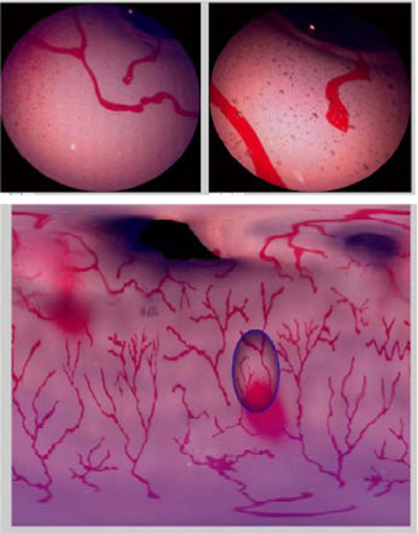
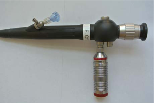
IN VIVO AND EX VIVO COMPARISON OF OPTICS AND PERFORMANCE OF A NOVEL DUAL CHANNEL FIBEROPTIC URETEROSCOPE
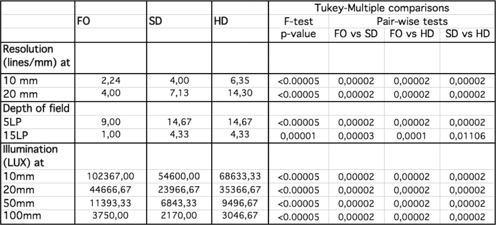
Resolution, Depth of field and Illumination
Resolution, Depth of field and Illumination

University of Chicago Medical Center, Urology
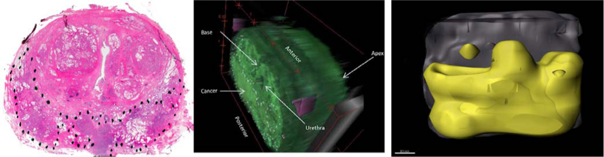
a) Whole mount pathological radical prostatectomy slide with outlined tumor, b) 3D reconstruction of scanned and stacked images, c) Volume rendering of 3D reconstructed smoothened tumor area.

A SINGLE CENTRE PROSPECTIVE 5 YEAR CLINICAL EXPERIENCE WITH ROBOTIC-ASSISTED TRANSPERINEAL PROSTATE BIOPSY IN PATIENTS WITH PREVIOUS NEGATIVE BIOPSIES
Yong Loo Lin School of Medicine, National University of Singapore
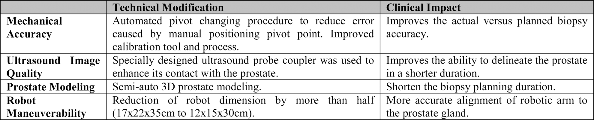
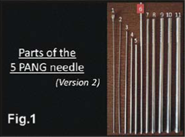
Summary of Modification Made to iSR'obot™ Mona Lisa
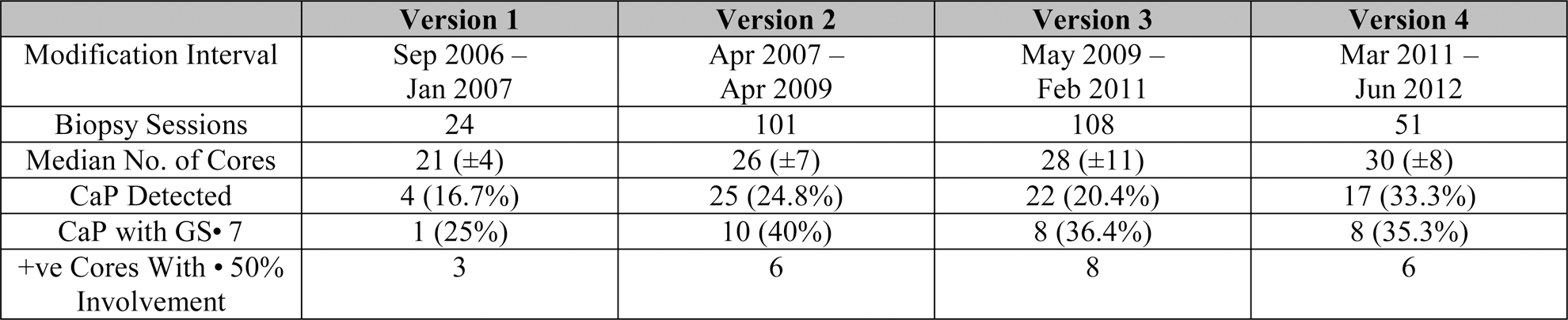
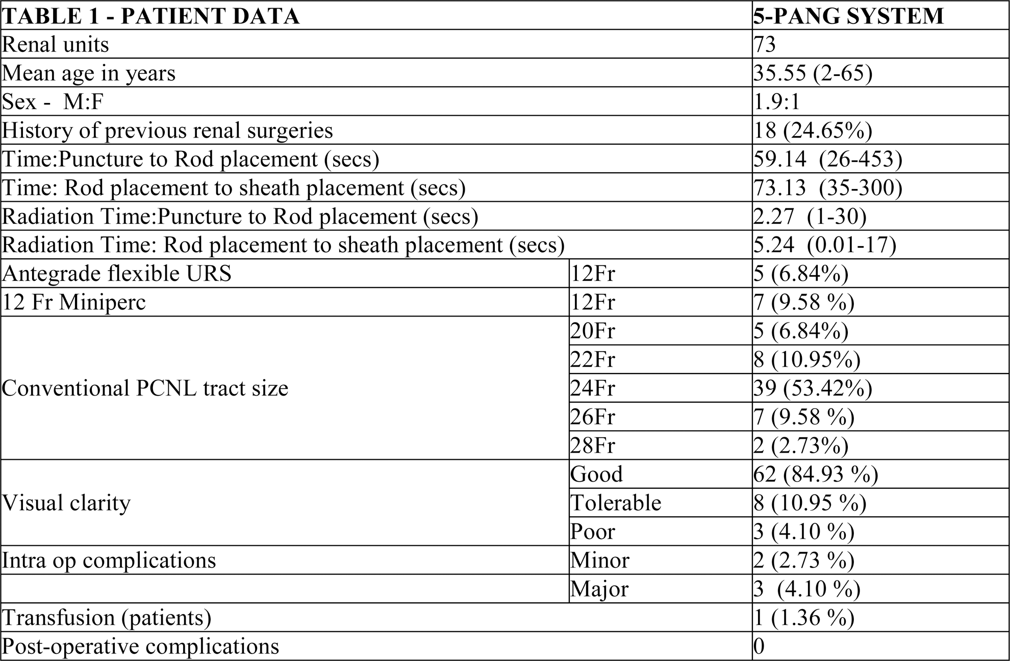
Patient Demographics and Clinical Outcome
University of California Irvine Biomedical Engineering

3-D volumetric rendering of the kidney hilum created in SolidWorks. Arteries are shown in blue, veins shown in red, and nerves are shown in light blue. The model was then used to precisely characterize the path of all nerves as well as distances from other anatomic structures.
Department of Urology, University of Florida College of Medicine, Gainesville, FL
INDENTER STUDY: HIGHER ELASTICITY OF PROSTATE IS ASSOCIATED WITH LOWER TRACT SYMPTOMS
Department of Urology, Urological Science Institute, Yonsei University College of Medicine
Reference: Ahn B, Lorenzo EI, Rha KH et al. Robotic palpation-based mechanical property mapping for diagnosis of prostate cancer. J Endourol 2011; 25: 851–7.
BEST PAPER AWARD
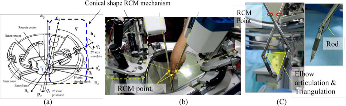
(a) Conceptual design and parameter definition of the conical shape RCM mechanism, (b) 1st prototype of cone actuator 4DoF, (c) surgical instruments 5DoF.
In vivo animal test for design out verification (Nephrectomy Rt, porcine model, 35 kg).
ICVS/3B's - PT Government Associate Laboratory, School of Health Sciences, University of Minho- Braga/Guimarães, Portugal
Department of Urology, University of Pittsburgh Medical Center, Pittsburgh, USA
TOP 10 ABSTRACT
Dept. of Radiology, Memorial Sloan-Kettering Cancer Center, New York, NY
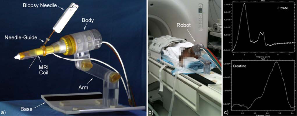
a) MRI-Safe Robot, b) Animal experiment, and c) MRSI graphs with built-in robot coil
Winter Haven Hospital & University of Florida

Direct visualization of ice ball formation during micro-cryoablation of peri-spermatic cord ilioinguinal and/or genitofemoral nerve branches.
PROSPECTIVE, RANDOMIZED COMPARISON BETWEEN MICROPERC AND RETROGRADE INTRA-RENAL SURGERY OF RENAL CALCULI LESS THAN 1.5 CM
NOVEL LAPAROSCOPIC DEFOGGING AND CLEANING DEVICE FOR ROBOT-ASSISTED LAPAROSCOPIC PROSTATECTOMY (RALP)
SouthWest Urology, LLC, Middleburg Heights, OH, USA;
TOP 10 ABSTRACT
Stony Brook University Medical Center, Stony Brook, NY
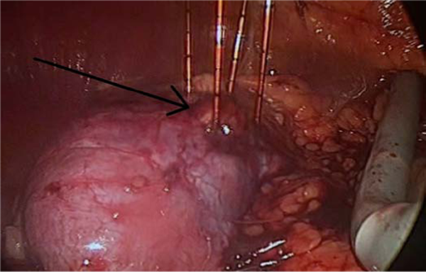
Laparoscopically-placed needles surrounding partially exophytic renal lesion.
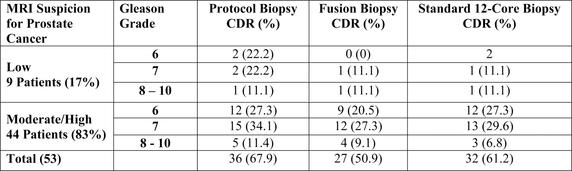
CRITICAL EVALUATION OF MRI-TARGETED TRUS-GUIDED TRANSPERINEAL FUSION BIOPSY FOR DETECTION OF PROSTATE CANCER
Department of Urology, University Hospital Heidelberg, Heidelberg, Germany
Department of Urology, University of California, Irvine
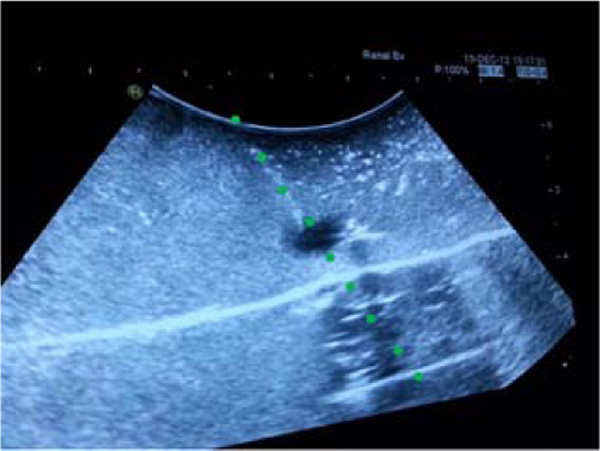
Echotip needle puncturing cyst at 30 degree angle.
TOP 10 ABSTRACT
First, needle insertion was done through the fixed needle-guide, to measure the natural lateral deflection of the needle. Then, the robot was used to tilt the needle-guide to prevent the deviation of the needle point from a straight path.

Uncontrolled curved (left) and controlled straight (right) needle paths. Please play the
Vanderbilt University, Department of Urologic Surgery
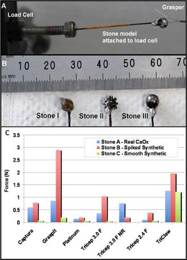
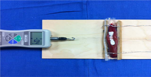
Grip strength set up. (B): Three stone types designed to replicate common shapes. (C) Average maximum grip strength measurements for each grasper per stone.
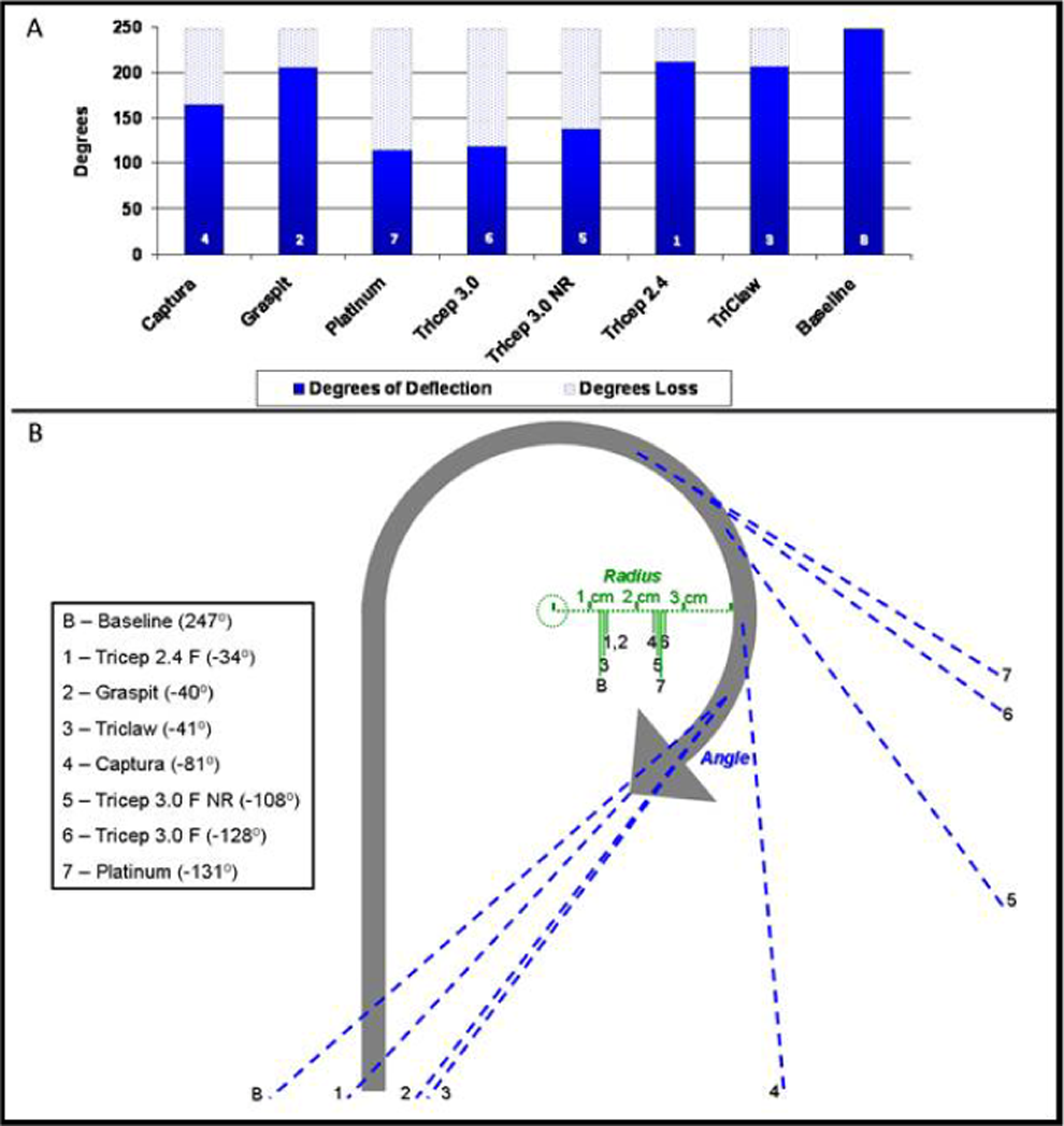
Total impact on the angle of ureteroscope deflection (A and B), and radii (B) varied among grasper devices.
Joint Bioengineering and Endourology, Developmental Surgical Laboratory, Division of Endourology, Laparoscopy, and Minimally-Invasive Surgery, Department of Urology, University of Miami Miller School of Medicine, Miami, FL, USA

Transverse view of the kidney that was ablated with 180 Watts for 2 minutes.
University of California, Irvine, Department of Urology, Orange, California, USA

Measurement anterior/lateral and medial/lateral position of the kidney.
GRIS, TU Darmstadt, Germany, 2MedCom GmbH, Darmstadt, Germany
[1] Han et.al: Geom. Evaluation of Systematic Transrectal Ultrasound Guided Prostate Biopsy, J. Urology, Vol. 188, 2404–2409, 12/ 2012
[2] Hadaschik et.al: A Novel Stereotactic Prostate Biopsy System Integrating Pre-Interventional Magnetic Resonance Imaging and Live Ultrasound Fusion, J. Urology, Vol. 186, 2214–2220, 12/ 2011
[3] Kuru et.al.: Phantom Study of a Novel Stereotactic Prostate Biopsy System Integrating Preinterventional Magnetic Resonance Imaging and Live Ultrasonography Fusion, JOURNAL OF ENDOUROLOGY, Volume 26, Number 7, 807–813, 7/2012
Muljibhai Patel Urological Hospital, Nadiad, Gujarat, India
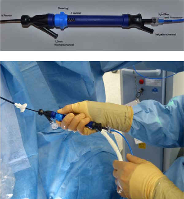
Prototype (Polydiagnost®) model for digital ureteroscopy.
In comparison, the mean apical prolapse in patients without a history of PFR was grade 3.2. These patients experienced 3 Clavien grade II complications. Mesh erosion did not occur in any of these patients. Grade I to III apical prolapse recurred in 14 patients, at 17.9%, 5.4% & 1.8%, respectively. None of these patients were symptomatic.
Demographic and Perioperative Data
Demographic and Perioperative Data
Post-Surgical Prolapse Recurrence
UMDNJ/New Jersey Medical School, Newark, New Jersey USA
1. Disposable cystoscope with Operating Sheath; 2. Bag of irrigation fluid with tubing; 3. Laptop/Tablet computer (Windows 7 or Windows 8); and 4. USB 2.0 Connecting Cable.
A NERVE COMPRESSION DEVICE AND ELECTROPHYSIOLOGICAL RECORDING OF NERVE RESPONSE TO PRESSURE COMPRESSIONS
Department of Biomedical Engineering, University of Minnesota
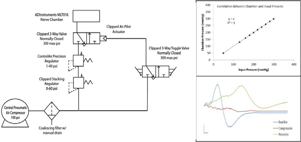
Test results and a diagram of the nerve compression system. a. Device diagram. b. correlation between input and chamber pressure. c. CAPs before, during and after the compression.
CHUM Section of Urology, Department of Surgery, Centre Hospitalier de l'Université de Montréal, QC, Canada.
Montefiore Medical Center, Albert Einstein College of Medicine

Sample H&E slide after laser ablation
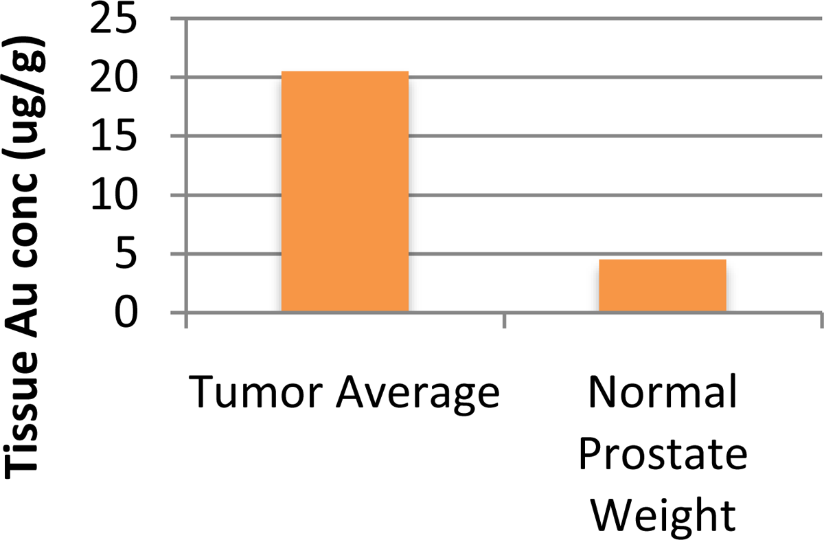
Nanoshells accumulation of gold.
TOP 10 ABSTRACT
EVALUATION OF ACCEPTABILITY OF PHYSICAL SIMULATION MODEL FOR TRAINING OF LAPAROSCOPIC PYELOPLASTY
Department of Urology, University of Minnesota, Minneapolis, MN, USA

Pyeloplasty model under room lighting showing (a) exterior and (b) interior and (c) under UV lighting conditions showing BLASTTM assessment lines.
Department of Urology, Singapore General Hospital

Grip strength set up. (B): Three stone types designed to replicate common shapes. (C) Average maximum grip strength measurements for each grasper per stone.
THE CHARACTERISTICS OF MICROCIRCULATION IN RENAL CELL CARINOMA BY CONTRAST-ENHANCED ULTRASOUND (CEUS)
A PROSPECTIVE COMPARISON BETWEEN NBI AND STANDARD WHITE LIGHT CYSTOSCOPY IN CASES OF NON-MUSCLE INVASIVE BLADDER CANCER
TOP 10 ABSTRACT
IN VITRO COMPARISON OF A NOVEL FACILITATED ULTRASOUND TECHNOLOGY VERSUS STANDARD TECHNIQUE APPROACH FOR PERCUTANEOUS RENAL BIOPSY
Department of Urology, University of California, Irvine
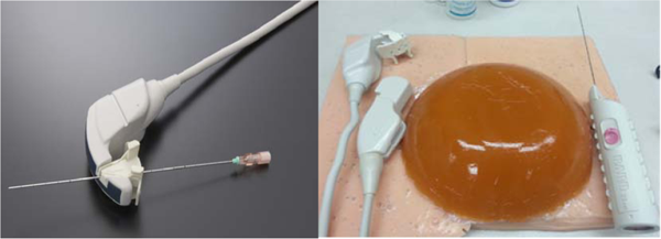
a) Close up of FUT probe with biopsy needle guide, courtesy of Hitachi Aloka©. b) Total impact on the angle of ureteroscope deflection (A and B), and radii (B) varied among grasper devices.
HISTOTRIPSY ACCURACY: CHARACTERIZATION WITH A NOVEL RED BLOOD CELL PHANTOM
HistoSonics, Inc.
Transverse section of prostate plug from pelvic phantom after treatment with histotripsy. Grid line spacing is 1 mm.
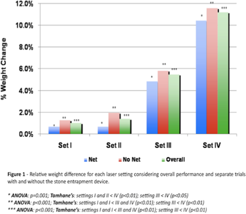
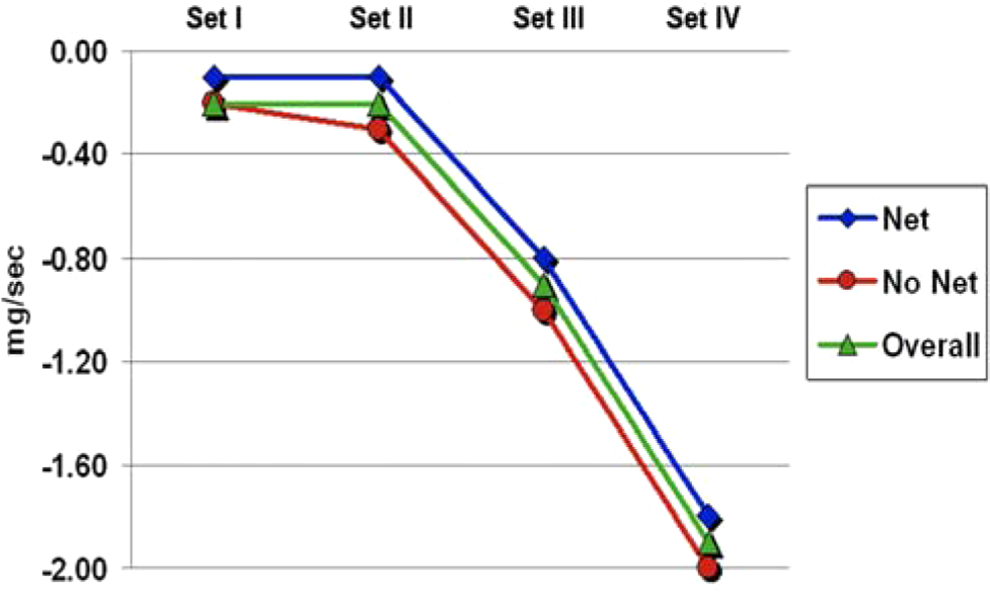
Influence of laser settings and entrapment device on fragmentation efficiency characterized by absolute weight reduction per second of lithotripsy.
VALIDATION OF A SIMULATION TOOL TO SUPPORT ROBOTIC-ASSISTED SURGICAL TRAINING
University of Washington, Seattle, WA
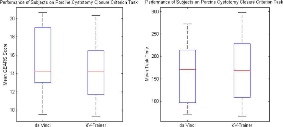
Performance of the two groups on a cystotomy closure task did not differ as measured by GEARS score nor task time.
Results Optics
Results Optics
MICROWAVE ABLATIONS AT 915 MHz IN EX VIVO AND IN VIVO PORCINE KIDNEYS
Joint Bioengineering and Endourology Developmental Surgical Laboratory, Division of Endourology, Laparoscopy, and Minimally-Invasive Surgery, Department of Urology, University of Miami Miller School of Medicine, Miami, FL, USA
In-vivo Ablation Data
Ex-vivo Ablation Data

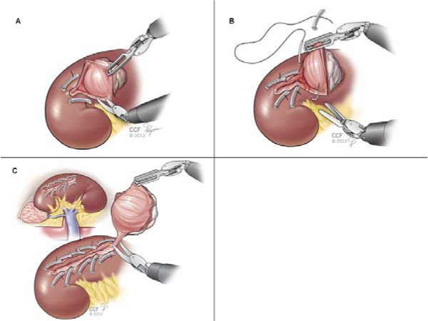
(A) 2-0 Vicryl suture with a knot and Hem-o-lok clip fixed to the free end is placed through the parenchyma, deep to the planned edge of resection and brought through the other side of the parenchyma, (B) Sequential 2-0 Vicryl stitches are placed adjacent to the tumor margin when parenchymal bleeding is encountered. This step is repeated as the tumor is gradually resected, (C) Completed excised tumor with tightened sequentially placed sutures fastened by Hem-o-lok clips to secure hemostasis.
Department of Urology, University of Washington School of Medicine
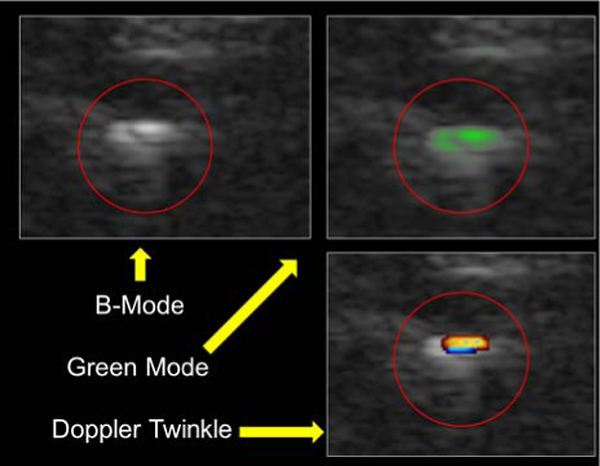
B-Mode and two experimental algorithms of the same stone.
Department of Urology, Singapore General Hospital

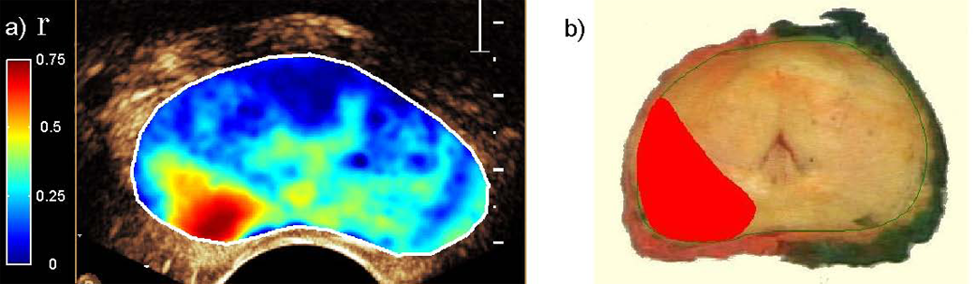
High flow areas and restricted diffusion at left PZ correlate with tumour area (Gleason 4+3 with microscopic extracapsular extension). MRI images (Clockwise from top left): DCE (F sequence) showing high flow (light blue) at left PZ, DWI sequence showing restricted diffusion at left PZ, DCE (V1 sequence) showing high flow (light blue) at left PZ), T2w sequence showing tumour nodule at left PZ. Prostatectomy whole mount slide: Areas of tumour at left PZ outlined in blue.
UMDNJ/New Jersey Medical School, Department of Urology, Newark, New Jersey USA
Biopsy-proven epithelial cancer detection was only slightly better than 50%. Patients presenting with microscopic hematuria post-treatment for bladder tumor could not consistently separate tumor recurrence from the healing process, as accuracy was below acceptable levels.
Harlachinger Krebshilfe e.V., Muenchen, Germany
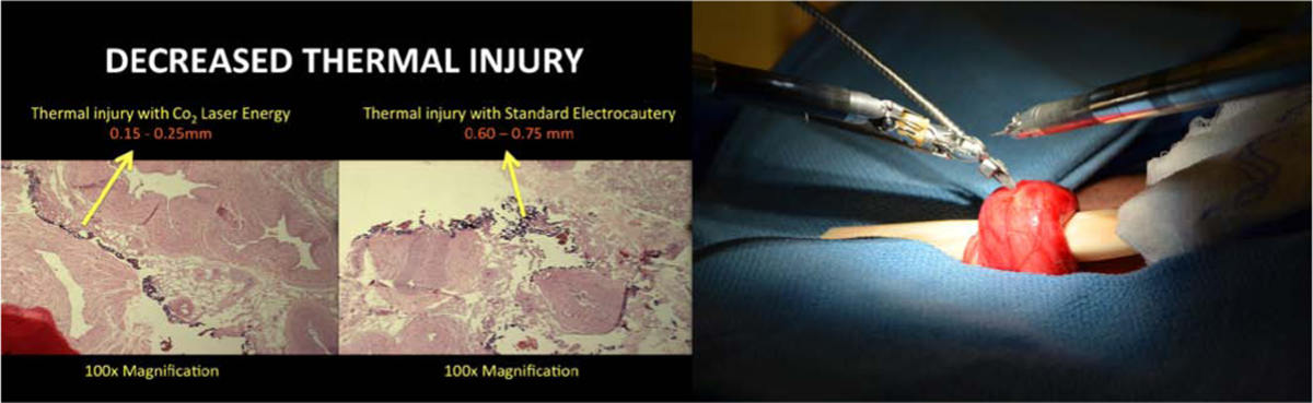
Robotic handling of the flexible CO2 laser fiber and pathologic identification of decreased thermal injury compared to standard monopolar electrocautery.
TOP 10 ABSTRACT
The BioRobotics Institute of Scuola Superiore Sant'Anna

The two fabricated prototypes: the LMA camera
University of Wisconsin School of Medicine and Public Health, Madison, WI
Brown University/RIH
TOP 10 ABSTRACT
LASER LITHOTRIPSY RETROPULSION VARIES WITH STONE MASS
ADVERSE EVENTS RESULTING FROM LASERS USED IN UROLOGY
AEs Reported in FDA MAUDE (1992–2012) and RLI (1970–2005) Databases
AEs Reported in FDA MAUDE (1992–2012) and RLI (1970–2005) Databases

TOP 10 ABSTRACT
ACTIVE REMOVAL OF BUBBLE SHIELDING IN SWL: AN IN VITRO STUDY
Department of Biomedical Engineering, University of Michigan, Ann Arbor, MI, USA

TOP 10 ABSTRACT
Steering mechanism with three segments connected in series.
With 3D path planning with optimized steering motion sequence, spiral scan trajectories are chosen to reduce the time for scan while maintaining the required percentage of overlap between neighboring image frames to help post-processing of the video. The total scanning process takes about three minutes to finish. In order to expediently review the bladder surface, a 3D stitching software which reconstructs the surface of the whole bladder from endoscopic video using structure from motion have been separately developed. The software was tested on endoscopic videos acquired from a phantom model and an excised pig bladder using a primitive robotic steering mechanism (Figure 2)[2][3].
Images captured from cystoscopic video within a bladder phantom, and the result of stitching and blending images together.
[1] Xianming Ye and W. Jong Yoon, Preliminary Design of a Bending Mechanism for Automated Cystoscope, The 8th IEEE International Conference on Automation Science and Engineering (CASE 2012), 257–262, August 20–24, 2012, Seoul, Korea.
[2] M. Burkhardt, T. Soper, W. Jong Yoon, and E. J. Seibel, Controlling the Trajectory of a Flexible Ultrathin Endoscope for Fully Automated Bladder Surveillance, IEEE/ASME Transactions on Mechatronics, 2013 (doi:10.1109/TMECH.2013.2237783).
[3] Timothy D. Soper; Michael P. Porter; Eric J. Seibel, Surface mosaics of the bladder reconstructed from endoscopic video for automated surveillance, IEEE Transactions on Biomedical Engineering. 2012; 59(6):1670–1680.

LED light source attached to cystoscope.
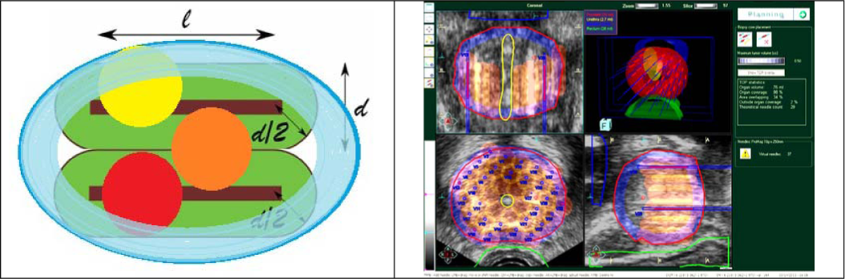
A BASIC NOVEL CONCEPT: PROSTATE-SPECIFIC COORDINATE SYSTEM
The assignment and location of the PCS are shown in Figure 1. This is based on selecting the center of the urethra at the base and apex points (B, A respectively in Figure 1a) on the central sagittal plane. The origin of the PCS is located in the middle of the AB segment and is aligned along the AB direction and the central sagittal plane. The directions of the PCS follow the LPS (Left-Posterior-Superior) standard anatomic system. The precision of defining the PCS was calculated as the radius of the minimal sphere enclosing the origins of the PCS.
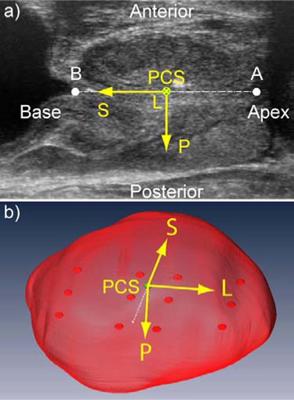
PCS Location: a) Central sagittal ultrasound through the prostate showing apex (A) and base (B) points, b) PCS with Left-Posterior-Superior coordinates.

CORRELATION BETWEEN THE PROSTATE SIZE AND LOCATION OF THE NEUROVASCULAR BUNDLES
Urology Robotics Laboratory, Johns Hopkins University, Baltimore, MD
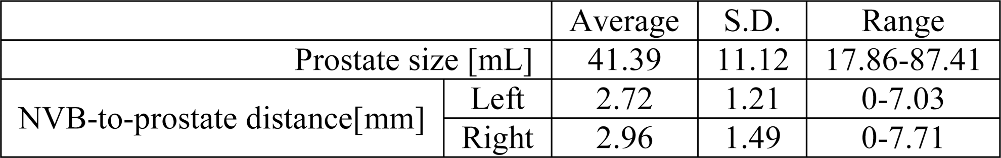
Prostate size was calculated using the width, height, and length measured in the transverse and sagittal slices. The NVB-to-prostate distance was measured in the mid-gland transverse B-mode and Doppler slice. The shortest distance from the periphery of the Doppler signal to the contour of the prostate was measured (Figure 1). Distances on both sides of the prostate were measured.

NVB-to-prostate distance.
Prostate Size and NVB-to-Prostate Distance

N-P Distance vs. Prostate volume.
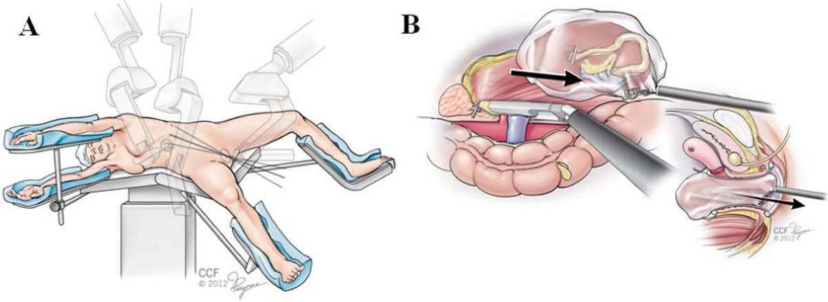
TRANSVAGINAL HYBRID NOTES ROBOTIC DONOR NEPHRECTOMY

Steering mechanism with three segments connected in series.
Department of Urology, University of Washington School of Medicine
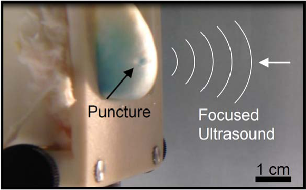
Puncture created in a bladder bleb after 180 seconds exposure of high-intensity ultrasound (indicated by white lines) focused onto the bleb wall.
Work supported by NIH 2T32DK00779-11A1, P01DK043881, and 2R01EB007643-05, and the National Space Biomedical Research Institute through NASA NCC 9–58.
Department of Urology, University of Washington School of Medicine

Evidence of crystal growth in porcine model of calcium oxalate lithiasis.
Work supported by NIH DK43881, DK092197 and NSBRI through NASA NCC 9–58.
Eindhoven University of Technology, Electrical Engineering Department, the Netherlands
Dispersion map
Virginia Urology, Richmond, VA
Harlachinger Krebshilfe e.V.
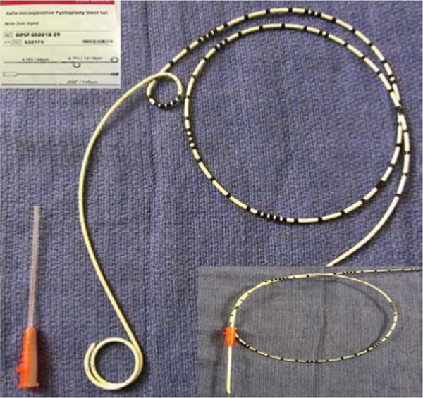
Modified Salle stent for robotic pyeloplasty.
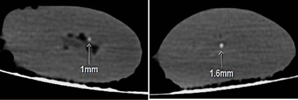
Stones demonstrated on HP-treated pigs at or near the renal papilla measuring up to 1.6 mm on CT, standard bone window.
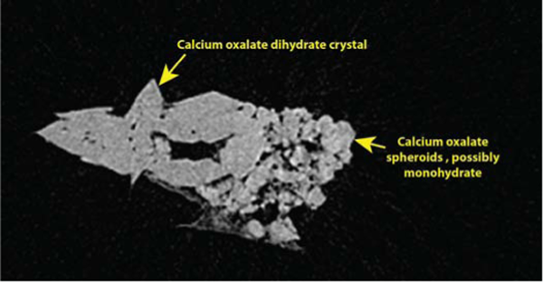
Micro CT demonstration of an extracted stone, confirming a mix of calcium oxalate dihydrate and monohydrate crystals.
A SCIENTIFIC CORRELATION BETWEEN ROBOTIC PROSTATE BIOPSY RESULTS WITH POST-RADICAL PROSTATECTOMY WHOLE-MOUNT PATHOLOGY SLIDES
Yong Loo Lin School of Medicine, National University of Singapore
Footnotes
*
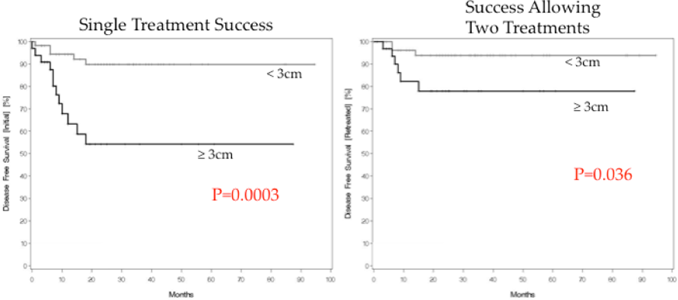
Thüroff, S., Chaussy, C., Evolution and outcomes of 3 MHz High intensity focused ultrasound therapy for localized prostate cancer over 15 years, The Journal of Urology (2013), DOI: 10.1016/j.juro.2013.02.010.