
Abstract
Introduction:
The features and usefulness of a novel wide-angle lens flexible cystoscope were assessed with a view to its practical application.
Materials and Methods:
A phantom bladder model, on which a total of 12 markers that resemble lesions were arranged, was created for this study. Twenty-six urologists at three institutions observed this phantom bladder model using a conventional flexible cystoscope and a novel wide-angle lens flexible cystoscope, and they compared the observation time, marker detection rate, number of misidentified marker locations, overlooked locations, and misidentified location sites of both devices. Specific observation procedures that make use of the features of a wide-angle lens flexible cystoscope were also investigated.
Results:
The observation time tended to be shorter with the wide-angle lens cystoscope than with a conventional cystoscope (104.9 seconds vs 113.6 seconds, p = 0.123). The marker detection rate was higher with the wide-angle lens cystoscope (90.2% vs 85.1%, p = 0.005). The number of marker location misidentifications did not differ between the two devices. Using a specifically designed observation procedure with the wide-angle lens cystoscope tended to further improve the marker detection rate (91.4% vs 88.1%, p = 0.157).
Conclusion:
Compared with a conventional cystoscope, a wide-angle lens cystoscope improved the lesion detection rate and has the potential to reduce observation time. The novel wide-angle lens flexible cystoscope is regarded as a useful device that offers advantages not available with a conventional cystoscope.
Introduction
R
Currently, a novel flexible cystoscope, with a wider viewing angle than conventional flexible cystoscopes, is being developed to achieve greater efficiency and reliability in the examinations mentioned above. This flexible cystoscope has a wider viewing angle of 140° in water, with reference to the ease of observation in the ureteral orifice, which is a feature of oblique rigid cystoscope. Therefore, on insertion into the bladder, there is the potential to more easily observe the bladder trigone and ureteral orifice than with a conventional flexible cystoscope. The viewing angle also makes it possible to bring the bladder sidewall into one visual field without having to bend the fiber from the position of the bladder neck, potentially yielding more information at a glance.
In this study, a phantom bladder model was used to assess the features and usefulness of this novel wide-angle lens flexible cystoscope.
Materials and Methods
Phantom bladder model
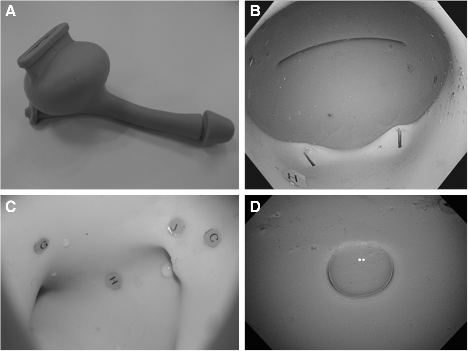
A specialized silicone phantom bladder model was created to evaluate a novel wide-angle lens flexible cystoscope. The phantom bladder model has a shape that includes not only the bladder but also the urethra, and it allows for actual insertion from the external urethral orifice. In the interior of the bladder, contiguous with the urethra, is a realistic reproduction of the shape of the bladder neck and bladder trigone, with air bubbles also reproduced in the dome of the bladder (Fig. 1). Inside the phantom bladder model, 12 red markers measuring 3 mm, listed with alphanumeric characters, were arranged at arbitrary locations as observation targets that resemble lesions. Four types of phantoms were created. The observation objects in each phantom were given different alphanumeric content; the objects were arranged with model A = C and B = D, where models A and B and models C and D were arranged with left and right mirror-inverted image alignment (Fig. 2); this was done to ensure that there would be no differences in difficulty between models during verification.
Phantom bladder model

The observation objects in phantom model
Novel wide-angle lens flexible cystoscope: olympus CYF-Y0013
CYF-Y0013 is a prototype scope at this stage that is being manufactured for in vitro studies. A commercially available flexible cystoscope (CYF-VHA; Olympus Corporation, Tokyo, Japan) from the same company has an underwater viewing angle of 80°. Compared with the conventional cystoscope, the newly developed CYF-Y0013 has an unaltered shaft diameter, and it has no major added modifications to the operation of the grip section and the like. The CYF-Y0013 is a videoscope equipped with a charge-coupled device image sensor, and it is also narrow-band imaging (NBI) compatible, as is the CYF-VHA. However, with the aim of easy observation of the ureteral orifice, such as with an oblique rigid cystoscope, and the aim of making it possible to observe half the region inside the bladder in one visual field on insertion as far as the bladder neck, the underwater viewing angle was expanded to 140° (Fig. 3). Fig. 4 shows differences in actual underwater viewing angles between phantom bladder models.

New wide-angle lens flexible cystoscope: Olympus CYF-Y0013.

The comparison of the underwater viewing angles between the CYF-VHA and the CYF-Y0013
Procedures
Twenty-six urologists at three institutions were chosen as subjects. Each subject was asked to perform the following two verifications within the format of using the conventional cystoscope (CYF-VHA) and the novel wide-angle cystoscope (CYF-Y0013) to actually observe inside the phantom bladder model.
Verification 1
Each subject was asked to observe the four different phantom bladder models with the CYF-VHA and with the CYF-Y0013, two times each, and to assess each cystoscope in terms of its (1) observation time, (2) total number of detected markers, (3) number of markers detected before retroflexed*, (4) number of misidentified marker locations, (5) overlooked locations, and (6) misidentified location sites. (*retroflexed: The act of inverting the flexible cystoscope shaft 180° and observing the bladder neck).
Subjects were randomly and evenly divided into an A group and a B group, so as to prevent bias in the observation results, whether from the order in which the CYF-VHA and the CYF-Y0013 were used or from a bias in the phantom bladder model being observed. A group observed models A and B with the CYF-VHA and then observed models C and D with the CYF-Y0013. B group observed models A and B with the CYF-Y0013 and then observed models C and D with the CYF-VHA. Because outpatient clinical practice is what was envisioned, observation time in the phantom was taken to be from the start of observation until when one observation pass of the inside of the bladder was complete, with 60 seconds as a benchmark. While observing inside the phantoms, subjects verbally indicated the alphanumeric characters of the markers that they detected, as well as their sites; the video and audio of these were recorded.
Verification 2
An observation procedure expected to harness the features of the wider-angle CYF-Y0013 was designed, and subjects were asked to use it to observe the inside of the phantoms, to assess whether there was an improvement in the marker detection rate. The subjects were asked to use the specifically designed observation procedure to observe the same two phantom bladder models that they observed with the CYF-Y0013 in verification 1. The assessment methods and items in this observation, being similar to verification 1, were compared with the results from when the CYF-Y0013 had previously been used in verification 1 (in verification 1, there was no specifically designed observation procedure).
Specifically designed observation procedure
The wider viewing angle makes it possible to observe from the air bubble to the ureteral orifice with less rotation. The following method was used in an effort to harness this property.
1. The cystoscope was inserted from the urethral orifice to the bladder neck, to observe around the trigone.
2. This was followed directly by observation of the posterior wall.
3. The cystoscope was flexed upward to check for an air bubble. The right ureteral orifice was observed so as to trace the curve of the right wall from the air bubble, and then it was returned to again observe the air bubble.
4. The left wall was observed in the same manner.
5. The shaft of the flexible cystoscope was flexed 180° to check for the internal urethral orifice.
6. The cystoscope was then twisted to observe the left and right ureteral orifices.
7. The cystoscope was pulled to simultaneously observe from the anterior wall to the air bubble.
8. The flexion of the cystoscope was restored, and finally the bladder as a whole was checked, and then the cystoscope was pulled out.
Statistical analysis
Significant differences in assessment items were tested using a nonparametric test, the Friedman test (level of significance p = 0.05), equivalent to repeated measures one-way analysis of variance. The analysis was performed with a statistical processing software IBM SPSS.
Results
Verification 1
Data were collected from 104 instances of observation of the phantom bladder models by the 26 subjects. The observation time tended to be shorter with the CYF-Y0013 than with the CYF-VHA (CYF-VHA: 113.6 ± 42.8 seconds vs CYF-Y0013: 104.9 ± 38.8 seconds, p = 0.123). The total number of markers detected (detection rate) was significantly higher with the CYF-Y0013 (CYF-VHA: 10.2 ± 1.1 [85.1%] vs CYF-Y0013: 10.8 ± 1.2 [90.2%], p = 0.005). The total number of markers detected (detection rate) before retroflexed was also significantly higher with the CYF-Y0013 (CYF-VHA: 8.1 ± 1.2 [67.1%] vs CYF-Y0013: 8.9 ± 1.3 [74.5%], p < 0.001). The number of misidentified marker locations did not differ between the two devices (Table 1).
Values are presented as mean ± standard deviation.
Level of significance p ≦ 0.05.
Easily overlooked marker sites did not show a characteristic difference between the two devices; both tended to have more at the bladder trigone site. Regarding sites where the marker location was easily misidentified, there were cases where the side wall was misidentified as the posterior wall with the CYF-Y0013.
Verification 2
Data were collected from 56 instances of observation of the phantom bladder models by the 14 subjects who observed it by the procedure that was clearly different from the specifically designed observation procedure in verification 1. There was no significant difference with the specifically designed observation procedure with the CYF-Y0013. However, there was a further increase in the total number of markers detected (detection rate) (no specifically designed procedure: 10.6 ± 1.4 [88.1%] vs specifically designed procedure: 11.0 ± 1.2 [91.4%], p = 0.157) and a decrease in the number of misidentified locations (no specifically designed procedure: 2.0 ± 1.7 vs specifically designed procedure: 1.4 ± 1.2, p = 0.835) (Table 2).
Values are presented as mean ± standard deviation.
Discussion
A review of the history of the development of the cystoscope shows that the conventionally used rigid cystoscope was followed by the first introduction of a flexible cystoscope in 1973. 4 Following continuous reductions of shaft diameter and improvements to image quality and ease of use, Olympus introduced the CYF-VHA, used as a comparison against the new device in this study, in 2011. Due to cost and the life cycle of equipment, it has not spread to all regions at the level of being in general urology outpatient care, 5 but more flexible examination has the important advantage of reducing pain, and patients prefer it to rigid cystoscopy. 6,7
As such, there is considerable significance in the further development and improvement of the flexible cystoscope. The focus here was on achieving a wider angle as one approach to developing the flexible cystoscope. The greatest difference in the characteristic viewing angles between a rigid cystoscope and a flexible cystoscope is that a rigid cystoscope uses an oblique lens, whereas a flexible cystoscope uses a fixed direct lens. With an oblique lens, it is easy to observe the bladder trigone or ureteral orifice, both of which are below a frontal view of the bladder, immediately after insertion into the bladder. Therefore, to achieve this same ease of use with a direct flexible cystoscope as well, the aim was to expand the underwater viewing angle to 140°. Currently, the viewing angle of flexible cystoscopes from manufacturers commonly used throughout the world is constant at 120° (∼80° for the underwater viewing angle). A novel wide-angle lens flexible cystoscope (Olympus CYF-Y0013) was assessed with two verification methods in the present study on the basis of the expectation that expanding the field of view to an underwater viewing angle of 140° may not only make it easier to observe the trigone and the ureteral orifice, but will also shorten examination time and reduce overlooked lesions due to the wider area that can be captured with one field of view.
Verification 1 was a comparison of actual use of the conventional cystoscope (CYF-VHA), with an underwater viewing angle of 80°, and the CYF-Y0013, with an underwater viewing angle expanded to 140°. As expected, this verification yielded the result that widening the viewing angle significantly improved the observation time, as well as the observation target detection rate. An especially noteworthy point is that the ability to detect many observation targets with the CYF-Y0013 before retroflexed appears to be a feature of the widened viewing angle. There did, however, tend to be a slightly higher number of misidentified locations with the novel wide-angle cystoscope. When the patterns of specific location misidentifications were aggregated, a common one was misidentification of the side wall as the posterior wall. In our view, this result occurs because the novel wide-angle cystoscope enables subjects who are used to the viewing angle of conventional cystoscopes to view a range beyond what they believe they are viewing. Thus, this problem will be naturally eliminated as users become accustomed to the wider angle, and having the wider angle will instead result in greater benefit. Regarding why the bladder trigone was an often-overlooked site in this verification, the subjects pointed out that the phantom bladder models used for the verification have a problematic shape in that the bladder trigone protrudes too far, farther than it does in the body, and this is thought to end up creating a blind spot that would not actually exist.
Next, because verification 1 had generally shown the advantages of the wider viewing angle, verification 2 served to search for an observation procedure for making the greatest use of these advantages. In verification 1, the subjects' patterns of observation within the phantom bladder models were broadly classified into two types. One was random observation, which involved randomly going on finding observation targets without fixed rules for the order of areas to be observed or the operation of the flexible fiber; the other was tracing observation, which involved finding observation targets in a process of observation that traces from the top to the side wall in a regular manner. Because we thought that the tracing observation procedure could make use of the characteristics of the wide-angle cystoscope, we devised the specifically designed observation procedure based on the tracing observation procedure. However, these verifications did not show a significant difference in the marker detection rate, but there was a tendency for it to improve. The specifically designed observation procedure, believed to take advantage of the wider-angle CYF-Y0013, may provide guidance for urologists who use it once it is actually available in clinical practice.
Some reports have focused on a high magnification as an approach for improving the visual field of the cystoscope, which is the reverse of the wide-angle lens approach that we have focused on. Detailed observation of the changes in vasculature on the mucosal surface in a high-magnification visual field, may help in the task of distinguishing between cancerous and noncancerous lesions. 8 On the other hand, when the observed object is viewed from the same distance, the wide-angle lens has a disadvantage in that the object appears smaller in the image than in the case of a nonwide angle lens. However, if a scope is brought close to a target site that is to be observed in detail with the CYF-Y0013, the object can be observed with the same high-definition high-image quality and resolution as with the CYF-VHA, which is a scope with the conventional angle of view. In addition, when necessary, it is also possible to observe the object by using NBI in combination with the CYF-Y0013. Based on the results of the present study, the CYF-Y0013 can be thought to provide a great advantage over high-magnification cystoscopes in situations such as outpatient screening examinations that need to scan the entire bladder in a short time and detect numerous lesions that a cancer is suspicious. Although it was outside the scope of verifications in this study, it can be thought that the CYF-Y0013 is not lacking in regard to its capacity for detailed observation and evaluation of lesions.
Conclusions
The CYF-Y0013 from Olympus, a novel flexible cystoscope with an underwater viewing angle that has been widened to 140°, has the potential to shorten observation time and improve the lesion detection rate compared with conventional flexible cystoscopes. In consideration of the fact that cystoscopy is frequently performed in urology outpatient care, these improvements are noteworthy, and the CYF-Y0013 is regarded as a useful device with advantages not available with conventional cystoscopes. Observation procedure that take advantage of the wider visual field also has the potential to allow all operators to reap these benefits.
Footnotes
Author Disclosure Statement
Olympus lent the equipment used in this study and provided advice, but no competing financial interests exist.