




Abstract
Introduction:
Morcellation of the adenoma after laser enucleation of the prostate (LEP) is both time-consuming and prone to complications. We have designed a novel polyethylene sack (ProSac) to improve the morcellation process following LEP. Both silicone and cadaver models were utilized to evaluate the safety and efficacy of ProSac.
Methods:
The inanimate model used tissue-mimicking silicone to accurately approximate bladder volume and compliance. The second model was developed using a fresh cadaver. Heat-fixed chicken breast was used to mimic enucleated prostatic adenoma. Morcellation of the simulated adenoma tissue was tested in both models with and without the ProSac. Morcellated tissue was removed from the filter, desiccated, and weighed after each run to establish morcellation efficiency. Visual inspection was used to evaluate for mucosal injury or bladder perforation. A hydromanometer was placed in the cadaveric bladder to measure bladder pressure.
Results:
The device was able to capture up to 30 g of tissue with good closure while maintaining good distention and visualization during morcellation. In the silicone model, morcellation efficiency with the device was 4.6 g/minute, while efficiency without the device was 2.6 g/minute (p = 0.03). In the cadaveric model, mean entrapment time was 22 ± 11 seconds. Morcellation efficiency with the device was 2.1 g/minute when excluding entrapment time and 1.9 g/minute including entrapment time. Without the ProSac, morcellation efficiency was 1.2 g/minute (p = 0.05). In both models, multiple mucosal injuries occurred without the device, while none occurred with the device. Bladder pressure was similar between study arms.
Conclusions:
The ProSac is a novel device that can provide additional safety during adenoma morcellation. It may also achieve clinically and statistically significant improvement in morcellation efficiency without increasing bladder pressure.
Introduction
B
LEP entails enucleating the transition zone adenomas (en bloc or in smaller pieces) and displacing these into the bladder. To remove the adenoma, tissue morcellation is necessary. Morcellators utilize a combination of suction with oscillating or reciprocating blades to mince prostate tissue into small pieces and suction these from the bladder. Morcellation can be a time-consuming and potentially morbid portion of LEP, often representing 15%–35% of total OR time, and increases with increasing gland size. 10 –12 The most significant concern during morcellation is the risk of a bladder injury. Engaging the bladder mucosa into the morcellator can lead to a bladder mucosal tear or in extreme cases, a bladder perforation. Bladder injuries are reported to occur at a rate of 2%–5%. 6,12 With the goal of making tissue morcellation following prostate enucleation safer and more efficient, we developed a novel device that can be deployed transurethrally and entrap the prostate adenoma.
Methods
ProSac design and use
Three identical prototypes of the ProSac were assembled for proof-of-concept testing. The ProSac prototype is made of an acrylic-coated nylon sleeve and entrapment area lined by a nitinol wire loop to allow for concentric closure of the distal end of the entrapment portion. An external view and internal view of the device are seen in Figures 1 and 2, respectively. The ProSac was designed to accommodate a nephroscope through its inner lumen. The nephroscope allows a three-prong grasper to be passed, which is used to draw the adenoma into the entrapment portion. From the distal end of the ProSac, the nitinol wire can be pulled, closing the entrapment portion around the adenoma. A morcellator is placed through the working channel of the nephroscope allowing morcellation to be completed entirely within the device. Once the adenoma pieces are smaller than the sheath, the sheath can be withdrawn and the remaining pieces discarded. Dual inflow of irrigation during morcellation is often recommended and was used during evaluation of the ProSac. 6
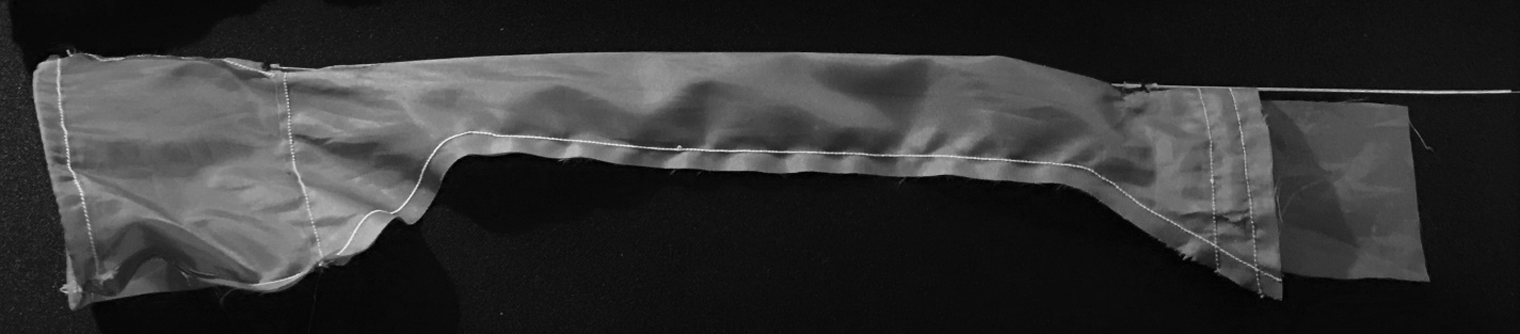
Longitudinal view of ProSac.
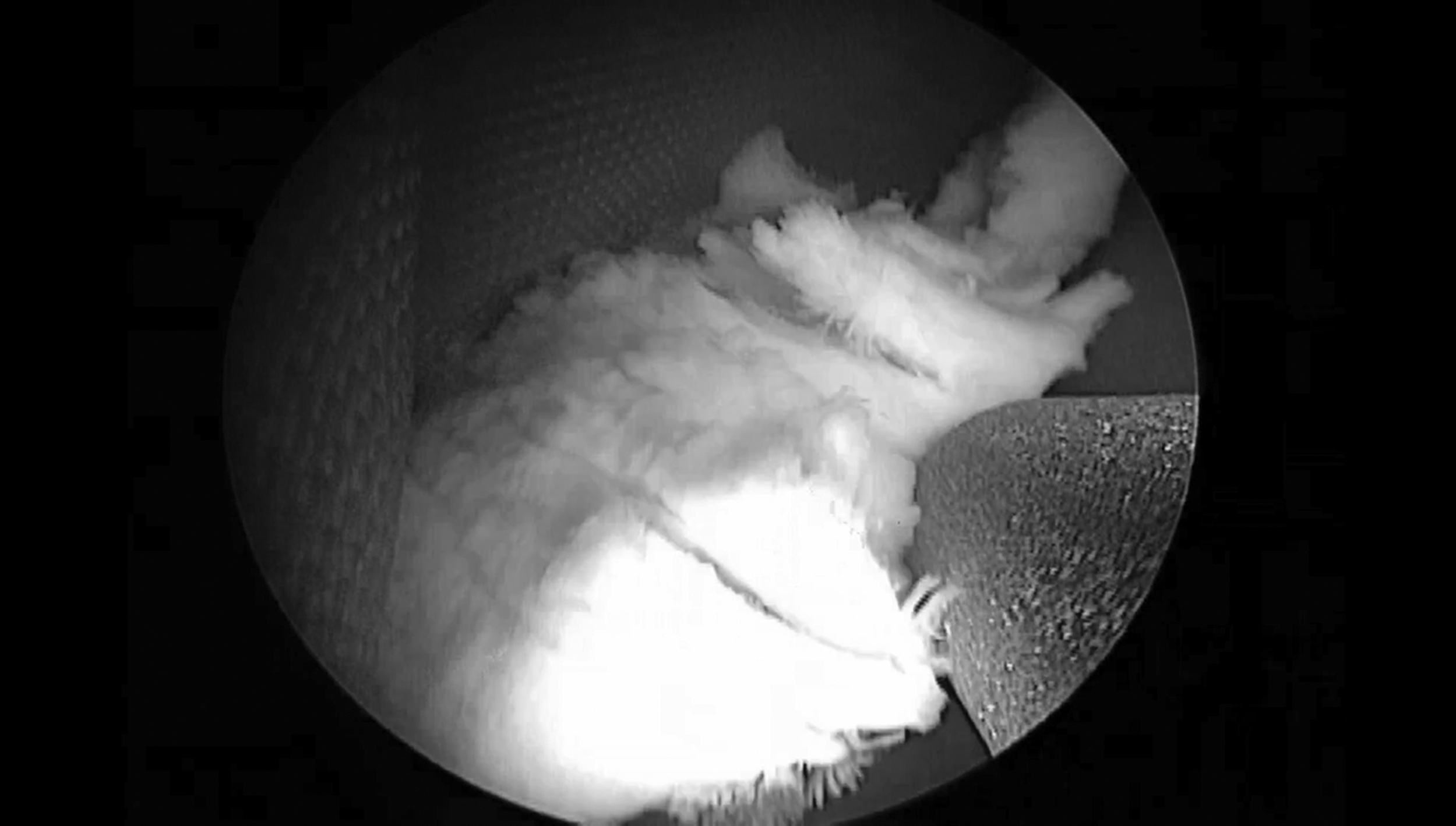
View of morcellation within the ProSac.
Phase I testing
The initial proof-of concept evaluation of the ProSac was performed with an in vitro nonbiologic model. A silicone bladder model was built that accurately approximated bladder volume and compliance. The model was made from Dragon Skin FX-Pro (Smooth-On, Macungie, PA), which is a platinum-cured tissue-mimicking silicone that approximates biologic tissue. Red paint was applied to the silicone and it was poured over a 350 cc clay mold of a bladder. A second flesh-colored layer of silicone was painted on the inside of the model to mimic bladder urothelium over the underlying detrusor. Heat-fixed chicken breast was used to mimic enucleated prostatic adenoma. Approximately 15–20 g of tissue was placed in the bladder model and five trials of morcellation with and without the ProSac were performed. The VersaCut™ tissue morcellator (Lumenis, Yokneam, Israel) was utilized for all runs. Morcellated tissue was removed from the filter, desiccated, and weighed after each run to establish morcellation efficiency. Separate bladder models were used for each series of runs. A single operator with HoLEP/morcellation experience performed all runs. The control arm and device arm were alternated to mitigate any learning curve associated with using the model. Following all runs, the models were disassembled, and total mucosal injuries were tabulated by visual inspection. Injury per minute of morcellation was calculated for each study arm.
Phase II testing
The second phase of testing utilized a cadaveric bladder model. A fresh male cadaver with no previous lower urinary tract surgery was obtained. Cystoscopy was performed to verify no pre-existing bladder injuries and to place a guidewire in the bladder. The urethra was dilated, and a 30F urologic sheath was placed across the urethra. An infraumbilical incision was made, and the anterior bladder was dissected. A small cystotomy was made using a retropubic approach to allow placement of heat-fixed chicken breast, which was used to simulate prostate adenoma. A hydromanometer was also placed in the bladder to measure bladder pressure. Five to 10-g pieces of simulated prostate adenoma were placed into the bladder. Five trials of morcellation were performed both with and without the ProSac. Two larger pieces (15–20 g) of simulated adenoma were placed in the bladder model and morcellated with the ProSac to demonstrate efficacy for larger amounts of tissue. Following each series of runs, morcellated tissue was removed from the filter and desiccated, to establish morcellation efficiency. Morcellation efficiency was calculated both with and without entrapment time added to morcellation time. Bladder pressure was recorded every 30 seconds of morcellation time. Total mucosal injuries and perforations were observed and recorded as they occurred.
Statistical analysis
Difference in mean morcellation time was analyzed using a Student's t-test for phase I and phase II testing. SPSS version 25 (International Business Machines Corporation, Armonk, NY) was used to perform data analysis. All p-values were two sided and statistical significance was defined as p < 0.05.
Results
Phase I results
The silicone bladder model easily accommodated the rigid nephroscope and the ProSac.
Mean adenoma morcellation time and morcellated tissue weight were 292 ± 16 seconds and 217 ± 65 seconds, 12.2 ± 3.6 g and 14.8 ± 1.2 g for the control arm and the device arm, respectively. Morcellation efficiency with the ProSac was 4.6 g/minute, while efficiency without the device was 2.6 g/minute (p = 0.031). This represents a 77% increase in morcellation efficiency with the ProSac (25% reduction in morcellation time). There were a total of three mucosal injuries in the bladder model for the control arm and none for the bladder model used for the device arm (see Table 1).
p = 0.031.
SD = standard deviation.
Phase II results
The device was effectively deployed into the cadaveric bladder via the 30F sheath. A 26F nephroscope was easily deployed through the ProSac and into the bladder. Results from each run of this study can be seen in Table 2. A three-pronged grasper entrapped the simulated prostate adenoma with mean entrapment time of 22 seconds ±11. The ProSac maintained adequate distention and visualization with average pressure in the bladder, 9.7 ± 4.4 cm-water, during morcellation. For small pieces (5–10 g), mean morcellation time (excluding entrapment time) was 191 ± 80 seconds with the ProSac and 265 ± 66 seconds without the ProSac. Mean morcellation efficiency for small pieces (5–10 g) with the ProSac was 2.0 g/minute if entrapment time is excluded. Morcellation efficiency without the ProSac was 1.1 g/minute.
p = 0.033.
For the larger pieces, only two runs could be performed with the ProSac and none without the ProSac as the cadaver model deteriorated to the point the bladder no longer was watertight. Total morcellated tissue for these two runs was 31.1 g. Mean morcellation efficiency for the larger pieces was 3.6 g/minute excluding entrapment time. For all morcellation runs with the ProSac, morcellation efficiency was 2.5 g/minute compared with 1.1 g/minute without the ProSac (p = 0.033). Without the device, five mucosal injuries and one bladder perforation occurred. No bladder mucosa injuries occurred with the device. Bladder pressure was continuously monitored via the hydromanometer and was recorded every 30 seconds during morcellation. Bladder pressure was similar between study arms (p = 0.006) (Fig. 3).
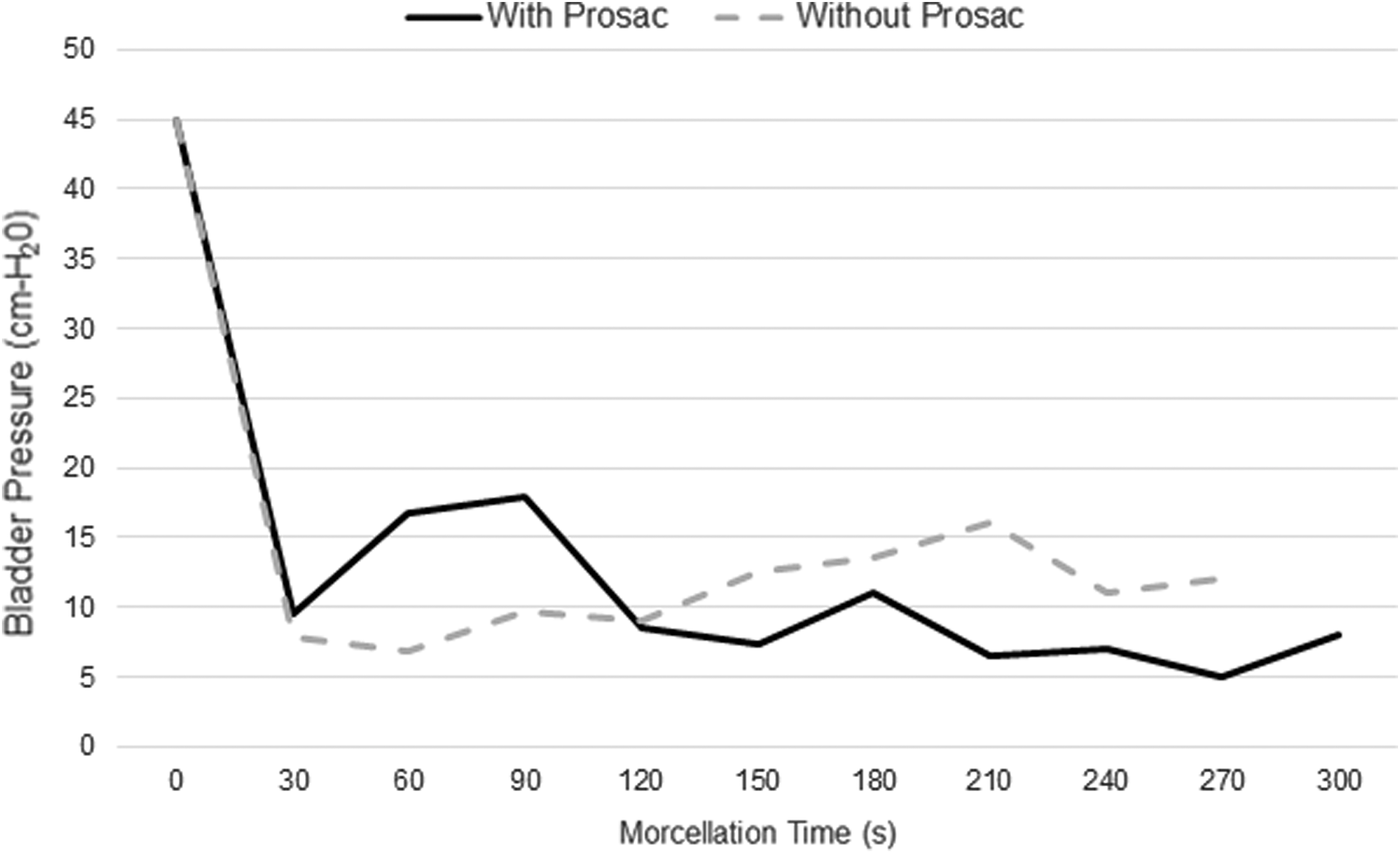
Intravesical pressure during morcellation.
Discussion
During prostate adenoma morcellation, bladder injury rates have been reported to be as high as 9% and bladder perforation rates in the range of 0.1%–1.5%. 13 Other studies have reported bladder injury rates ranging from 2% to 5% and the incidence of significant postoperative hematuria to be 3%–4%. 6,12 Despite the limited literature examining prostate adenoma morcellation in the context of LEP, there is a clear role for technologies that improve morcellation safety and efficiency. For this reason, we developed and tested a novel device to aid in tissue morcellation dubbed the ProSac.
The ProSac is based on a previously described device used for stone entrapment. 14,15 Similar to the ProSac, this early design utilized a thin, cylindrical, trumpet-shaped polyethylene sleeve and a nitinol wire to provide a mechanism to cinch the bag closed. However, several important modifications were made to address prostate adenoma morcellation. The device was modified to allow transurethral passage and accommodation of the morcellator. Acrylic-coated nylon was used as the material rather than polyethylene to make it more resistant to damage if caught within the jaws of the morcellation. The nylon mesh also has less memory compared with polyethylene and allows the distal end of the entrapment portion to cinch down when closed, creating a much tighter seal. The acrylic coating makes the nylon watertight, which is ideal, given the morcellator requires a significant amount of irrigation.
Using an entrapment sac in the bladder has been described for cystolithiasis. Miller and Park 16 performed transurethral bladder stone removal, utilizing a percutaneously placed entrapment sac into the bladder. Each stone was then guided into the entrapment sack and removed whole. While this was a small case series, the technique was effective and without complication in three of four cases. In one case, the sac tore and conversion to cystolithotomy was performed. Operative time was <1 hour for each case. 16 Hwang and colleagues 17 describe a similar procedure, however, CO2 gas is used as the optical transmission medium. Stones were ensnared within a transvesically placed entrapment sac and fractured with a pneumatic lithotripter. 17 While these techniques required percutaneous transversal access, the ProSac is designed to slide over the cystoscope obviating the need for percutaneous access.
Utilizing a silicone and cadaveric model, we demonstrated that the novel ProSac can entrap tissue transurethrally quickly and make morcellation safer and more efficient. We demonstrate a 77% and a 75% morcellation efficiency improvement in the silicone model and cadaver model, respectively. In addition, we found the ProSac could speed morcellation by allowing the last bit of tissue to be simply withdrawn once down to a relatively small size. This obviates the need to chase the last small pieces of adenoma throughout the bladder. Furthermore, the device decreased the number of injuries to the bladder in both models. There was also no difference in cadaveric bladder pressure during morcellation with or without the device. Notably, our proof-of-concept study has several strengths. We utilized a single operator experienced in HoLEP and morcellation, and the runs within control arm and device arm were alternated to mitigate any learning curve. Using a fresh cadaver model, we were also able to approximate the in vivo environment (bladder compliance, urethral diameter) as closely as possible without using the device in living patients.
The evaluation of this novel medical device also has elucidated several limitations.
In our experimentation, the simulated adenoma was limited to ∼15 g of tissue. In practice, enucleated prostate adenoma can be much larger. The device can accommodate upward of 50 g of tissue, however, the grasping device proved to be the limiting factor. The three-prong grasper used was designed to manipulate calculi during percutaneous nephrolithotomy and not for grasping prostate adenoma. Therefore, the size of the simulated adenoma was limited by the width of the grasper. If a purpose-built grasper was to be developed, the device would be able to manage much larger pieces of adenoma. We also found it was possible to catch the nylon mesh in the morcellator, although with little consequence. The nylon could be removed from the jaws of the morcellator by simply pulling the morcellator back with a tap of the actuator. This caused only a momentary pause in morcellation (1–2 seconds). We found the sac could get a small rent in the fabric while maintaining its integrity, and the bladder mucosa remained unharmed.
In addition, several factors influence morcellation efficiency, one being the type of morcellator used. Morcellators utilize either oscillating blades (Piranha; Wolf, Inc., Knittlingen, Germany) or reciprocating blades (VersaCut; Lumenis, Yokneam, Israel). A thorough evaluation of the morcellators currently in the market by Elshal and colleagues 13 determined that the rate of tissue retrieval was 6.2 g/minute for the Piranha and 2.1 for the VersaCut. 11 Our study only implemented the VersaCut morcellator, which may have artificially lowered the morcellation efficiency reported. Comparing both morcellators would have allowed for a more thorough evaluation of the device. This study also utilized heat-fixed chicken breast to simulate prostate adenoma, and results with actual prostate tissue may vary However, morcellation efficiency in our study was consistent with morcellation rate reported in the literature for similar devices, 10,12 indicating that our adenoma model simulated prostate tissue well.
Conclusions
The ProSac is a novel device that can provide improved safety during adenoma morcellation after LEP in preclinical models. Early proof-of-concept testing has further demonstrated improvements in morcellation efficiency. Further refinements to the device and more testing are needed.
Footnotes
Acknowledgment
Luminous provided the morcellator for evaluation of the ProSac.
Author Disclosure Statement
No competing financial interests exist.