




Abstract
Introduction and Objective:
Catheter-associated urinary tract infections are a major cause of patient morbidity and mortality. Despite many attempts to design biomaterials that might reduce the risk, none has had a profound impact on reducing the incidence of this most common nosocomial infection. Recent in vitro work, however, has shown promise for a silver-based biomaterial coating composed of methoxylated polyethylene glycol 3,4-dihydroxyphenylalanine (mPEG-DOPA3) in reducing uropathogen attachment and biofilm formation. The aim of this work was to investigate whether these results translate into a meaningful impact on infection development and bacterial adherence in an in vivo rabbit model.
Materials and Methods:
New Zealand white rabbits were randomized into groups of 12 and had the following catheters inserted: Group 1—uncoated polyurethane, Group 2—Coating A (mPEG-DOPA3 + 2 mg/mL AgNO3), and Group 3—Coating B (mPEG-DOPA3 + 10 mg/mL AgNO3). Each rabbit was challenged with 108 colony-forming units of Escherichia coli GR-12 instilled directly into the bladder at the time of catheter insertion and urine was monitored over 7 days for bacterial counts. Catheters were retrieved and evaluated for encrustation and attachment analysis, and tissues collected for histopathologic characterization and bacterial invasion.
Results:
Urinary bacterial colony counts were lower among rabbits in the Coating A group vs controls (4/11 vs 10/12, respectively) (p = 0.029), and there were fewer rabbits with invasive infections (3/12 vs 9/12, p = 0.02). More encrustation was observed among animals in the Coating B group vs controls (7.22 vs 2.69 mg/cm2, p = 0.033). There were no significant differences in tissue effects between groups.
Conclusions:
The use of a mPEG-DOPA3 urinary catheter coating effectively reduced urinary pathogen counts, while not causing adverse tissue effects in this model. Further clinical evaluation is warranted.
Introduction
Urinary catheters are among the most frequently used medical devices and greatly increase the risk for nosocomial urinary tract infections (UTIs). These devices interfere with host immune defenses and provide bacteria with an ideal surface on which to attach and form biofilms, rich communities of microorganisms that are resilient and often resistant to antibiotics. 1 –3 According to the Center for Disease Control, a nosocomial catheter-associated urinary tract infection (CAUTI) is defined as the new appearance of bacteriuria or funguria at >105 colony-forming units (CFUs)/mL (Ref. 4 ). Infections may be asymptomatic initially, but can progress to local symptoms or even systemic manifestations, including sepsis. Although CAUTI rates have decreased due to the development of best-practice guidelines outlining indications for catheter insertion and removal, maintaining aseptic technique and antibiotic stewardship, CAUTI still accounts for ∼80% of all hospital-acquired infections worldwide and cost the US health care system in excess of $300 million USD/year. 5 –8
From a biomaterial perspective, several approaches toward infection prevention have been researched, including antibiotic-releasing coatings, antifouling surface polymers, furanones, nanoparticles, and bacteriophages. 4,9 Our group has previously utilized a methoxylated polyethylene glycol (mPEG) 3,4-dihydroxyphenylalanine (DOPA) copolymer as an antifouling coating to reduce the formation of urinary conditioning films and uropathogen attachment. 10 A follow-up study demonstrated reduced bacterial attachment on this coating both in vitro and when placed in a rabbit bladder model and determined that cross-linking the polymers was most optimal. 11 Based on these initial experiences, we were encouraged to continue further refinements of the coatings to engineer a biomaterial that could also impart antimicrobial activity. Silver nitrate particles and quaternary amine coatings in combination have been shown to have strong antimicrobial effects. 12 –14 Using quaternary ammonium groups and silver particles in a cross-linked copolymer, we were able to develop a coating that demonstrated bactericidal activity against gram-positive and gram-negative organisms using an in vitro model. 15 Not only were planktonic bacteria killed, but we also observed a significant reduction in the number of attached organisms.
The aim of this study was to further assess a copolymer coating when applied to urinary catheters in an in vivo system. The well-established rabbit model was selected with the intention to measure urinary bacterial CFUs, bacterial attachment and encrustation on the catheters, and potential bacterial invasion into tissue. The primary outcomes in this analysis were urinary CFUs/mL and the degree of catheter encrustation comparing the new catheter-coating to an uncoated catheter. Local toxicity of the coating was also evaluated as a secondary outcome through examination of tissue pathology.
Methods
Thirty-six New Zealand white male rabbits were utilized in a study reviewed and approved by the animal use subcommittee at Western University. Two different coatings [Coating A (mPEG-DOPA3 + 2 mg/mL AgNO3) and Coating B (mPEG-DOPA3 + 10 mg/mL AgNO3)] were tested, both containing a different concentration of AgNO3 (2 or 10 mg/mL). The test devices were commercially available Universa* 5F ureteral stents (Cook Urological, Spencer, IN) that were externally coated with one of the two experimental coatings by DSM Biomedical (Exton, PA), sterilized and packaged separately. Preparation of the copolymers and silver cross-linking was performed as previously described. 15 A third group of animals received the same uncoated stent as a control. The number of animals was determined by a power analysis based on the number of recovered bacteria from urine samples in previous studies. 11,15 Given a desired power level of 0.8 and a p-value of 0.05, a sample size of randomized 12 animals/group would allow detection of a 30% to 40% difference between treatments. The veterinary surgical team and laboratory technician who undertook the postexperimental sample analysis were blinded to the treatments. Urethral catheterization was performed with a 5F, 15 cm length of catheter material under general anesthesia and was sutured to the skin to prevent migration, without occluding the catheter lumen. A portion of the stent was left protruding from the external meatus of varying length depending on the size of the animal, but usually 1 to 3 cm long. A syringe containing 108 CFU of Escherichia coli GR-12 (pyelonephritis clinical isolate) in a 1.5 mL saline solution was immediately instilled into the bladder, followed by a 0.5 mL saline flush.
Urine was collected by cystocentesis on days 1, 3, 5, and 7, after clamping the catheter for ∼30 minutes. Venous blood samples were also taken. Following anesthesia for sample collection on day 7, euthanasia and subsequent autopsy were performed to collect tissue. Representative segments of renal pelvis, distal ureter, and bladder were dissected for tissue bacterial count analysis. Histopathology samples were stored in formalin until analysis, while all others were held at −80°C. The presence of E. coli in the collected urine samples was quantified by dilution plating on Lysogeny Broth agar (Difco, MD). The agar plates were incubated at 37°C for 24 hours, after which colonies were counted to determine the CFU/mL. For analysis of bacterial attachment, catheters were cut into 1 cm segments and rinsed three times with sterile phosphate-buffered saline (PBS) to remove unattached bacteria. The segments were then resuspended in 1 mL PBS, sonicated for 20 minutes at 50/60 Hz (Branson 1200, Danbury, CT), and vortexed for 30 seconds to detach any bacteria, which were then plated.
The degree of encrustation was determined by taking 2 cm segments, air-drying them in open tubes for 24 hours, and then weighing on an analytical balance. Encrustations from the outer surfaces were scraped off manually using a scalpel, and the devices were then washed vigorously with water via pipette-mixing and vortexing to remove encrustations within the lumen and on the outer surface. The segments were air-dried for 24 hours and weighed again.
Representative tissue from the bladder, kidneys, and ureters were analyzed for tissue-associated bacterial attachment and internalization. Tissue samples were first rinsed with sterile PBS to remove unattached and loosely attached bacteria in 50 mL tubes. Samples were resuspended in varying volumes of PBS, depending on tissue size: 5, 1, and 10 mL PBS for bladder, ureter, and kidney, respectively. The tissue segments were subsequently homogenized at 30,000 rpm with a PT 2100 Homogenizer (Kinematica, Littau, Switzerland) until a suspension was established for every sample. The homogenate was then diluted and plated as above.
Multiple random biopsies were taken from within the bladder and renal pelvis from various sites. Tissue samples were suspended in a 10% neutrally buffered formalin solution. The collected tissues were processed in a paraffin wax tissue processor (Leica ASP3000; Leica Microsystems, Inc., Concord, Canada) and subsequently embedded into paraffin wax blocks before sectioning. The samples were sectioned into 5 μm sections (Microm HM335E Microtome; Thermo Fischer Scientific, Waltham, MA). The slides were stained with hematoxylin and eosin (Leica Autostainer XL). The sections were analyzed by a veterinary pathologist blinded to the study groups.
Kidney tissue was examined and scored for urothelial inflammation, urothelial fibrosis, ulceration, mucinous metaplasia, squamous metaplasia, suburothelial hemorrhaging, and suburothelial inflammation. For each parameter, the following scores were assigned: normal = 0, mild = 1, moderate = 2, or severe = 3, for a maximum score of 21. Bladder tissue was scored for urothelial inflammation, urothelial fibrosis, ulceration, mucinous metaplasia, squamous metaplasia, suburothelial hemorrhaging, lamina propria fibrosis, lamina propria inflammation, muscle fibrosis, and muscle integrity. A similar scoring scheme was used with a maximum score of 30.
Statistical analysis was performed using SPSS 22 software (IBM Corp., Armonk, NY). Significance was assessed at p < 0.05. For categorical variables such as the qualitative outcomes of the urine culture (positive or negative), Chi-squared and Fisher's exact test were used with a Bonferroni correction to correct for multiple groups. A positive urine culture was defined as >105 CFU/mL. As preliminary work showed that the coated surfaces should resist infection more than uncoated surfaces, 11 we likewise expected results showing lower CFUs in the animals with the experimental coatings and therefore used a one-sided Fisher's exact test for post hoc between group analyses where applicable. Continuous variables such as the quantitative outcomes of the urine cultures, encrustation, and bacterial tissue counts were analyzed using analysis of variance with the post hoc Dunnett's t-test to control for multiple group comparison. For data not normally distributed, nonparametric tests were applied such as Mann–Whitney U and Kruskal–Wallis where appropriate. Means and ranges are reported when nonparametric tests are performed.
Results
Rabbit urine samples were analyzed for bacterial growth and results are shown in Table 1. Urine samples were unavailable at some time points due to an inadequate volume at the time of collection. There was no statistical difference in the number of animals having a positive (>105 CFU/mL) E. coli urine sample on days 1, 3, and 5. By day 7, however, there were fewer rabbits with a positive urine culture in the coating A group (4/11) compared to the control group (10/12) (p = 0.029) (Fig. 1). When analyzing the number of E. coli CFU/mL present in the culture-positive samples, there were no significant differences across the three groups.

Percentage of rabbits with a positive Escherichia coli urine culture during study (*p < 0.05).
Rabbits with Positive Urine Samples with Escherichia coli GR-12 (Means and Ranges at Different Time Points)
Fisher's exact test.
Kruskal–Wallis test.
In the first group of 12 animals tested, migration of the catheters out of the bladder occurred in 2 animals. This was resolved by using a wingtip to improve anchoring of the distal catheters. Bacterial attachment data are therefore not available for these catheters.
When comparing the amount of E. coli attached to the different catheters, there were no significant differences between groups (Fig. 2). Of note, there was a reduction of bacterial numbers on the distal section of coating A vs control (27.5 vs 2.52 × 10⁴ CFU/cm2, respectively), and a higher number present on coating B vs control, most prominently on the middle segment (2.43 × 10⁶ vs 1.48 × 10⁴ CFU/cm2, respectively), although not statistically significant.
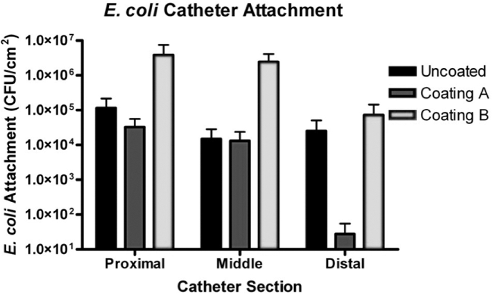
Attachment of Escherichia coli to catheter surfaces. CFU = colony-forming unit.
Encrustation was present on all devices regardless of whether coated or not. The uncoated group had a mean encrustation of 2.69 mg/cm2, ranging from 0.14 to 6.73 (Fig. 3A). The catheters from coating A and coating B had mean encrustations of 4.74 mg/cm2 (2.39–11.08 mg/cm2) and 7.22 mg/cm2 (1.48–16.9 mg/cm2), respectively. Overall, we found a significant difference in the amount of encrustation among the three groups (p = 0.016). While the Group A catheter encrustation did not differ from the control group (p = 0.301), catheters from Group B had significantly more encrustation compared to the controls (p = 0.033).
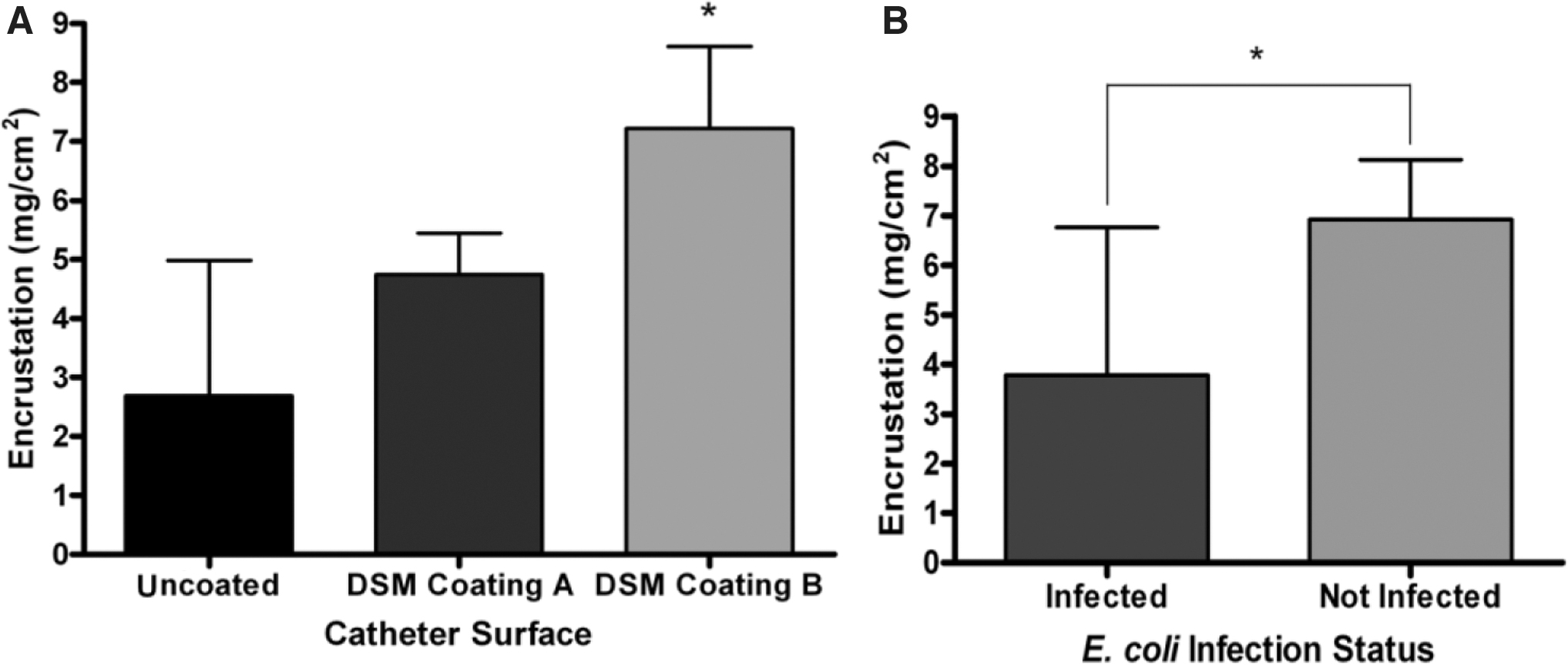
Encrustation of rabbit catheters across all three study groups
The number of rabbits having E. coli within bladder, ureter, or kidney tissue in each of the groups is demonstrated in Table 2. There were fewer rabbits in the Coating A group that had E. coli in bladder tissue specimens compared to the control group (p = 0.020) and is consistent with the urine culture results. Only three of the ureters analyzed demonstrated presence of E. coli, while no bacteria were seen in any of the kidney samples.
Number of Rabbits with Escherichia coli Found Within Bladder, Ureter, and Kidney Tissues
Evaluation of the tissues revealed the presence of consistent pathologic changes across all three study groups, including mucinous metaplasia (100% of rabbits), squamous metaplasia (77.8%), urothelial fibrosis (72.3%), lamina propria inflammation (66.7%), suburothelial hemorrhage (44.5%), ulceration (38.9%), and urothelial inflammation (36.1%). There were no significant differences in pathologic features or the severity of change between any of the groups. Although not statistically significant, the greatest difference was seen in the prevalence of urothelial fibrosis in the bladder, with the control group having a higher proportion compared to rabbits in Coating B group (91.7% vs 50%, respectively) (p = 0.069).
Discussion
Attempting to reduce CAUTIs is an important health care issue, but has been an elusive goal. While considerable attention has been directed toward the development of materials that are more biocompatible and have antimicrobial and antifouling properties, the ideal urinary catheter possessing all three of these important characteristics does not yet exist. Our previous work exploring the antifouling properties of mPEG-DOPA3 revealed a potential role for this material and led us to consider combining its properties with an anti-infective agent. 15 Silver has been extensively studied and has been shown to possess antimicrobial properties via several mechanisms, including cell membrane dysfunction, protein denaturation, and oxidative stress. 16 –20
Our results revealed that animals implanted with Coating A had lower CFU/mL bacterial counts after E. coli inoculation at day 7. Contrary to our expectations, the devices with Coating B, having five times the amount of antimicrobial material, did not have a significant effect on reducing infection, although there was a trend toward a beneficial effect. Whether a longer period of exposure would have produced different effects is unclear.
Not unexpectedly given the model we used with a portion of the catheter externalized, contamination with other bacterial species was noted. The initial rabbit model description for investigating CAUTI described housing the animals in a restricted-mobility Plexiglass cage and fed mainly with intravenous liquids. 21,22 To fulfill local animal use committee guidelines, however, the rabbits required a free range of mobility. Although the externalization of the catheter did lead to other bacterial species entering the bladder, our model is likely more representative of the human urethral catheter scenario where it is well known that external bacteria can enter the bladder even in a closed system.
Bacterial attachment numbers in the control and study coatings were not statistically different between coatings, although there was a slight decrease associated with Coating A in line with the reduced urinary CFU/mL results. Interestingly, some uninfected rabbits on day 7 were found to have E. coli attached on their catheter surfaces. We hypothesize that this can be accounted for by the occurrence of inoculated bacteria attaching on the catheter surface quite soon after inoculation, before planktonic bacteria are eradicated by the antibacterial effects of the catheter coating. The inoculated coliforms may then get covered by encrustation material, which provides protection against the bactericidal effect. The higher bacterial CFU numbers seen on Coating B, where encrustation was the highest, support this interpretation.
Analysis of the degree of encrustation demonstrated that both coating A and B had more encrustations than the uncoated devices, and coating B devices had significantly more. A subanalysis revealed that there was significantly more encrustation on devices from uninfected vs infected urines. Rabbits that had E. coli-positive infection status on the day of extraction had an average catheter encrustation of 3.79 mg/cm2, compared to 6.93 mg/cm2 in the uninfected rabbits (p = 0.022) (Fig. 3B). Further analysis demonstrated that the presence of non-E. coli infection was linked to significantly more encrustation than in the absence of infection (6.40 vs 3.13 mg/cm2, respectively). The mean and standard deviations of the encrustations in each of the groups are shown in Table 3. These findings are compatible with earlier work from our group. 11
Mean Catheter Encrustation (mg/cm2) in Relationship to Surface Type and Infection Status
Kruskal–Wallis test.
Analysis of variance.
Mann–Whitney U-test.
SD = standard deviation; UTI = urinary tract infection.
The development of encrustation on urinary device surfaces is dependent on multiple factors, including the indwelling time, urinary pH, and the presence of predisposing and inhibitory proteins, found in the urine and in the conditioning film. 23 –27 It is believed that the high degree of encrustation that occurs in healthy rabbits is due to their alkaline urine and linked to their diet. 28 Baseline urine pH values were typically measured at 8.5 to 9.0. By day 7, we found lower urine pH values in rabbits infected with E. coli. The paradoxical finding of increased encrustation in the coated groups in our study is likely due to the noninfected urine in these animals. Interestingly, this phenomenon may have contributed to the finding of decreased bacterial attachment on the distal segments of coating A. During poststudy sample processing, there was visibly more encrustation noted distally on coating B devices relative to coating A and control, and this may have been due to relatively alkaline urine earlier in the study. This in turn would have hindered the coating's bactericidal and antifouling properties. In contrast, coating A had less encrustation distally, which would have allowed the polymer to retain its anti-attachment properties as seen in prior in vitro work. 15
Similar to the number of infected rabbits, there were fewer rabbits in the coating A group that had bacteria attached to or invading bladder tissue compared to the other groups. Considering that fewer rabbits had E. coli infections by day 7 in the coating A group, this was expected. Tissue infection when observed was localized to the lower urinary tract in all the animals. Animals in each group were found to have inflammatory tissue changes, suggesting that the effects were related to the presence of the catheter itself, and not unique to the mPEG-DOPA3 silver-containing coating. In fact, a higher rate of mild urothelial fibrosis of the bladder was seen in the uncoated group compared to rabbits with coated devices, suggesting that this change is more related to the effects of E. coli infections.
Conclusion
In this rabbit model, the application of a novel silver-containing, polymer-based coating on a urinary catheter has been shown to be associated with reduced E. coli bacterial growth rates in urine and urinary tract tissues, without evidence of local toxicity. Although the current research is an important step in assessing the antimicrobial and antifouling effects of this coating, we acknowledge that the urinary milieu in rabbits is different from that of human's. As such, we cannot extrapolate these findings to clinical practice, but do believe our results warrant further research using other models that may more closely resemble the human urinary environment.
Footnotes
Author Disclosure Statement
No competing financial interests exist.
Funding Information
This project was funded by the U.S. National Institute of Health, Small Business Innovation Grant R44DK080547.