
Abstract
During the past decade, extended-spectrum cephalosporin resistance has increased among human isolates of Salmonella enterica serovar Heidelberg, the fourth most common serotype in the United States. We therefore characterized 54 Heidelberg isolates with decreased susceptibility (minimum inhibitory concentrations ≥2 mg/L) to ceftriaxone or ceftiofur; 49 (90.7%) contained the CMY-type β-lactamase (bla CMY) gene. The 49 bla CMY-positive human Heidelberg isolates demonstrated a high degree of relatedness; 4 clusters (25 isolates total) had indistinguishable XbaI and BlnI patterns by pulsed-field gel electrophoresis and were indistinguishable from 42 retail meat Heidelberg isolates. Further characterization of 15 of these isolates demonstrated that all of the bla genes were bla CMY-2 and plasmid-encoded, and most (11/15) of the plasmids were approximately 100 kb in size and belong to the incompatibility group I1 (IncI1). All five IncI1 plasmids tested by plasmid multilocus sequence typing analysis were ST12. This report suggests that extended-spectrum cephalosporin resistance among human Heidelberg isolates is mediated by the spread of a common IncI1 bla CMY-2 plasmid, which may have a preference for a particular genetic background.
Introduction
In the United States, antimicrobial resistance among Salmonella isolated from humans is monitored by the National Antimicrobial Resistance Monitoring System (NARMS) at Centers for Disease Control and Prevention (CDC). S. enterica serovar Heidelberg is the fourth most prevalent Salmonella serotype isolated from humans in the United States (CDC, DFBMD, 2006; CDC, 2009). In the past decade, the proportion of human Heidelberg isolates in NARMS resistant to ceftiofur, an ESC used in food animals (Hornish and Kotarski, 2002) in the United States, increased from 0.4% (1/251) in 1996–1998 to 9.2% (21/227) in 2005–2006 (CDC, unpublished data). In the United States, ESC resistance usually indicates the presence of a CMY-type β-lactamase (bla CMY) gene (Dunne et al., 2000). In this study, we report the identification and characterization of ESC-resistant Heidelberg isolated from humans in the United States.
Materials and Methods
Isolate collection and testing
Isolates of Salmonella from specimens collected from ill persons were collected by clinical laboratories in the United States and forwarded to state public health laboratories. Participating state public health laboratories, after serotyping, submitted every 10th (1996–2002) or every 20th (2003–2006) non-Typhi Salmonella isolated along with the patient's age, sex, state of residence, and specimen collection date to the CDC NARMS laboratory for susceptibility testing. Broth microdilution (Sensititre®; Trek Diagnostics, Westlake, OH) was used to determine the minimum inhibitory concentrations (MIC) for 14–17 antimicrobial agents, including amikacin, ampicillin, amoxicillin-clavulanic acid, cefoxitin, ceftiofur, ceftriaxone, cephalothin (last tested in year 2003), chloramphenicol, ciprofloxacin, gentamicin, kanamycin, nalidixic acid, streptomycin, sulfamethoxazole (before 2004), sulfisoxazole (in years 2004–2005), tetracycline, and trimethoprim–sulfamethoxazole. Resistance was defined by Clinical and Laboratory Standards Institute interpretive standards, when available. For ceftiofur, where no Clinical and Laboratory Standards Institute standards exist, the resistance breakpoint used in NARMS is 8 mg/L. Testing was performed according to the manufacturer's instructions and with the following quality control strains: Escherichia coli American Type Culture Collection (ATCC) 25922, Salmonella aureus ATCC 29213, E. coli ATCC 35218, and Pseudomonas aeruginosa ATCC 27853. Salmonella serovar Heidelberg isolates submitted from 1996 to 2006 that exhibited decreased susceptibility (≥2 mg/L) to ceftiofur and/or ceftriaxone were included in the study.
NARMS retail meat monitoring was conducted by the U.S. Food and Drug Administration Center for Veterinary Medicine (FDA-CVM) in collaboration with FoodNet (
Polymerase chain reaction amplification and sequencing of bla CMY
For each isolate, DNA was isolated by lysing the bacteria at 95°C and collecting the supernatant after centrifugation for 10 minutes at 20,000 g (Sorvall RC5B Plus, SS-34 rotor; Thermo Fischer Scientific Inc., Waltham, MA). Polymerase chain reaction (PCR) reactions contained 1× Hot Start PCR Master Mix (Qiagen Inc., Valencia, CA), 0.4 μM of each primer, 2 μL template DNA, and sterile PCR water to a final volume of 50 μL. Thermal cycling was performed using the following conditions: 15 minutes at 95°C, followed by 30 cycles of 95°C for 30 seconds, 56°C for 30 seconds, and 72°C for 90 seconds. To determine the presence or absence of the bla CMY genes, primers ampC1 and ampC2, previously shown to amplify the bla CMY gene, were used (Winokur et al., 2001). These primers and internal primers CMYSEQ1 (5′-GGTTGCAGGACGCGTCTG-3′) and CMYSEQ2 (5′-CCGCAATGGACTCCGGGC-3′) were used to sequence the entire bla CMY gene. Sequencing was performed using Big Dye version 3.1 (Applied Biosystems, Foster City, CA) and sequence reactions are cleaned with Centri-sep plates (Princeton Separations, Adelphia, NJ). The reactions are electrophoresed through POP-7 polymer (Applied Biosystems) on a 3730 DNA Analyzer (Applied Biosystems) equipped with a 48-capillary 50-cm array. Sequence analysis was performed using Lasergene 8 software (Dnastar Inc., Madison, WI). Isolates that were PCR negative for bla CMY were screened for additional β-lactamases (bla CTX-M, bla OXA, bla PSE, bla SHV, and bla TEM) using PCR with the previously described primer sets (Rasheed et al., 1997; Brinas et al., 2002; Bonnet et al., 2003; Chen et al., 2004).
Plasmid purification and characterization
Purified plasmid DNA was used to transform E. coli, to separate the bla CMY plasmids from other plasmid types before plasmid inc/rep testing, PFGE, and plasmid multilocus sequence typing (pMLST). Plasmids carrying bla CMY were purified using the QiaFilter Midi kit (Qiagen Inc.), following a modified manufacturer's protocol. Modifications included using 2 mL (half the recommended value) of buffers P1, P2, and P3, eluting DNA from the columns using the elution buffer (QF) that had been warmed to 55°C, and resuspending the final DNA pellet in 200 μL nuclease-free water. This protocol usually yields 4–8 μg of purified plasmid DNA. Contaminating genomic DNA was removed from the plasmid DNA preparation by treatment with Plasmid-safe DNase (Epicentre Biotechnologies, Madison, WI), according to manufacturer's protocol. For electroporation, 2 μL of each plasmid preparation was added to 40 μL of E. coli DH10B electro-competent cells (Invitrogen, Carlsbad, CA) in a cuvette (0.1 cm gap; Invitrogen) and electroporated at 1250 V, 100 ohms, and 25 μF using a Genetronics BTX ECM 630 Electro Cell Manipulator (Inovio Biochemical Corp., Blue Bell, PA). Cells were recovered with the addition of 1 mL super optimal culture (SOC) medium, transferred to 15 mL conical Falcon tubes (Becton Dickinson Biosciences, Franklin Lake, NJ), and incubated at 37°C for 1 hour. Cells were plated on Luria-Bertani (LB) agar plates containing 100 mg/L of ampicillin (Sigma-Aldrich, St. Louis, MO). Plasmids were re-purified from a single bla CMY PCR-positive transformant to isolate a single plasmid from each isolate. Purification was performed as described above with the additional modification of growing the cells overnight in 25 mL of LB broth with 100 mg/L of ampicillin. Plasmid size was determined using a plasmid PFGE protocol. In brief, plasmids were purified using the QiaFilter Midi kit plasmid purification kit (Qiagen Inc.). Ten to 20 μL of each plasmid was loaded onto 1.0% SeaKem Gold agarose gels (Lonza, Walkersville, MD) prepared in 0.5× Tris-borate ethylenediaminetetraacetic acid (EDTA) buffer, and the plasmids were separated by PFGE using a CHEF Mapper (Bio-Rad Laboratories, Hercules, CA) under the following conditions: 14°C, 6.0 V/cm, run time of 16 hours with a pulsing time linearly ramped from 6.27 to 21.7 seconds. Plasmids from E. coli strains PDK9 and V517, which contain several plasmids that range in size from 2.2 to 220 kb, were also extracted and used as size standards on the gels (Macrina et al., 1978).
Plasmid inc/rep typing was performed as previously described (Carattoli et al., 2005). pMLST was performed as previously described (García-Fernández et al., 2008). In brief, targets were sequences within pilL, sogS, ardA, trbA, pndC, and repI1. PCR reactions contained 1× Hot Start PCR Master Mix (Qiagen Inc.), 0.4 μM of each primer, 1 μL template DNA, and sterile PCR water to a final volume of 50 μL. Thermal cycling was performed using the following conditions: 15 minutes at 95°C, followed by 30 cycles of 95°C for 30 seconds, 56°C for 30 seconds, and 72°C for 90 seconds. Sequencing was performed by a 3730 DNA Analyzer, and sequence analysis was performed using Lasergene 8 software (Dnastar Inc.). Sequences were submitted to the pMLST Web page (
PFGE
PFGE was performed according to the CDC PulseNet protocol (Ribot et al., 2006). Heidelberg isolates were grown overnight on Trypticase Soy Agar with 5% defibrinated sheep blood (Becton Dickinson Biosciences). Bacterial cell concentration was adjusted by diluting with sterile cell suspension buffer (100 mM Tris, 100 mM EDTA, pH 8.0) to a turbidity measurement of 0.48–0.52 (Dade Microscan Tubidity Meter; Siemens Healthcare Diagnostics Inc., Deerfield, IL). Agarose-embedded cells were lysed by proteinase K treatment and extensively washed. Agarose plugs containing genomic DNA were digested with 50 U of XbaI and BlnI restriction enzymes (New England Biolabs, Ipswich, MA) and incubated at 37°C for 2 hours. The fragments were then separated by PFGE using a CHEF Mapper (Bio-Rad Laboratories) with the following conditions and reagents: 1% SeaKem Gold agarose in 0.5% Tris-borate EDTA buffer, voltage at 6 V/cm, run time at 18 hours with switch times ranging from 2.16 to 63.8 seconds, temperature at 14°C. S. enterica serovar Braenderup H9812 was used as a molecular reference marker. Gel images were captured using the GelDoc XR system (Bio-Rad Laboratories) and Quantity one 1D analysis software (Bio-Rad Laboratories). Pattern analysis and unweighted pair group method with arithmetic mean (UPGMA) dendrogram generation were performed using BioNumerics software (Applied Maths, Saint-Martens-Latem, Belgium) with the Dice coefficient and tolerance of 1.5%.
Results
Identification of Heidelberg isolates with decreased cephalosporin susceptibility
From 1996 to 2006, 1040 Heidelberg isolates were screened for antimicrobial susceptibility by NARMS. Of these, 54 isolates displayed decreased susceptibility (MIC ≥ 2 mg/L) to ceftiofur and/or ceftriaxone. Ceftiofur MICs for the 54 isolates ranged from 2 to >16 mg/L; ceftriaxone MICs ranged from 0.25 to 32 mg/L. In the United States, ESC resistance usually indicates the presence of a bla CMY (Dunne et al., 2000). Forty-nine (91%) of 54 isolates were positive by PCR analysis for the bla CMY gene. Of the five isolates that were PCR negative for bla CMY, all five were PCR negative for additional β-lactamases (bla CTX-M, bla OXA, bla PSE, bla SHV, and bla TEM).
PFGE genotyping of bla CMY-positive Heidelberg isolates
PFGE was performed on all 49 bla CMY-positive human isolates. Similarity by PFGE varied among the isolates from approximately 74% to 100% (Fig. 1, bar at top left). We defined isolates with indistinguishable (100% similarity) XbaI and BlnI PFGE patterns as being part of a cluster (Fig. 1). One large cluster (cluster C) contained 18 isolates from six different testing years. There were three additional clusters (A, B, and D, containing three, two, and two isolates, respectively).
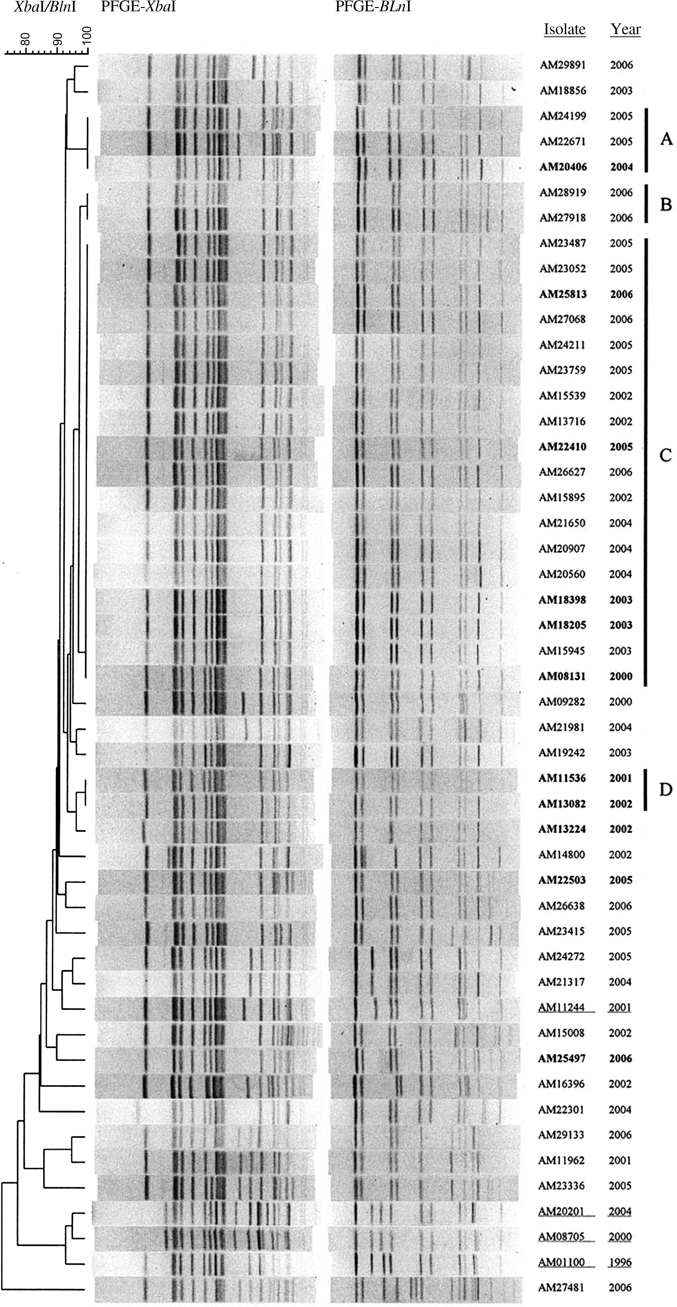
PFGE patterns of bla CMY-positive Salmonella enterica serovar Heidelberg isolated from humans. Dendrogram of percent genetic similarity by PFGE among isolates was generated using BioNumerics based on XbaI and BlnI restriction digestion. Percent similarity is located above dendrogram. Isolates (National Antimicrobial Resistance Monitoring System #) and the year of isolation are listed to the right of the PFGE pattern images. Letters A, B, C, and D designate four clusters of isolates with indistinguishable two-enzyme PFGE patterns. Cluster A contained three isolates with XbaI pattern JF6X01.0047 and BlnI pattern of JF6A26.0059. Cluster B contained two isolates (XbaI pattern JF6X01.0022 and BlnI pattern JF6A26.0085) and cluster D contained two isolates (XbaI pattern JF6X01.0080 and BlnI pattern JF6A26.0009). Cluster A, C, and D also have indistinguishable patterns from three clusters of Heidelberg isolates submitted to PulseNet by the Food and Drug Administration Center for Veterinary Medicine. Isolates in bold carry bla CMY-2 on IncI1 plasmids, while isolates underlined carry bla CMY-2 on IncA/C plasmids. PFGE, pulsed-field gel electrophoresis; Inc, incompatibility group.
Previous studies performed by the U.S. FDA-CVM examined Heidelberg isolated from retail meats in the United States, and the PFGE profiles were available for comparison in the PulseNet National database (Zhao et al., 2008). A comparison of the PFGE profile for the 49 bla CMY-positive Heidelberg human isolates with Heidelberg retail meat isolates showed two-enzyme PFGE pattern matches for 42 retail meat isolates; cluster A (3 human isolates) matched 11 retail isolates, cluster C (18 human isolates) matched 22 retail isolates, and cluster D (2 human isolates) matched 9 retail isolates. Sample sources for the Heidelberg retail meat isolates included chicken breast (n = 31) and ground turkey (n = 11).
Characterization of bla CMY-positive plasmids
To determine if the bla CMY genes were encoded on plasmids or chromosomally located, plasmids were purified from the 49 bla CMY-positive isolates. PCR analysis of plasmid-safe DNase-treated plasmid preparations suggested that all the 49 bla CMY genes were carried on plasmids. Plasmids from 15 of the 49 bla CMY-positive isolates were analyzed. From transformation experiments, all 15 transformants were PCR positive for bla CMY, and antimicrobial susceptibility testing of the transformants confirmed the transfer of the plasmid and bla CMY-associated resistances (Table 1). Sequence analysis of all 15 amplicons confirmed that the gene was bla CMY-2. Eleven of the 15 donors and corresponding transformants displayed only the bla CMY-mediated resistance profile (nontransformed DH10B cells are naturally resistant to streptomycin) (Table 1). All 15 plasmids were >100 kb in size and 12 out of 15 plasmids appeared identical in size (∼100 kb) (Table 1).
SMX and FIS are equivalent drugs.
MIC, minimum inhibitory concentrations; AMP, ampicillin; AUG, amoxicillin-clavulanic acid; AXO, ceftriaxone; TIO, ceftiofur; CHL, chloramphenicol; FIS, sulfisoxazole; GEN, gentamicin; KAN, kanamycin; SMX, sulphamethoxazole; STR, streptomycin; TET, tetracycline; Inc, incompatibility group.
PCR-based inc/rep typing demonstrated that 11 of the 15 plasmids were incompatibility group I1 (IncI1) and the remaining 4 were IncA/C (Table 1). pMLST of five IncI1 plasmids from isolates that were distinguishable by two-enzyme PFGE analysis (AM13082, AM20406, AM22410, AM22503, and AM25497) demonstrated that all five were ST12.
Based on the PFGE data, isolates carrying IncI1 plasmids tended to be within clusters; 8 out of 11 isolates are in cluster A, C, or D (Fig. 1, isolates in bold). In contrast, isolates containing IncA/C plasmids (AM11244, AM20201, AM08705, and AM01100) were less similar to other isolates within the entire group (Fig. 1, isolates underlined), and none were found in clusters.
Eight human isolates found in clusters, one from cluster A (AM20406), five from cluster C (AM25813, AM22410, AM18398, AM18205, and AM08131), and two from cluster D (AM13082 and AM11536) were further characterized in this study and found to have plasmid-encoded bla CMY-2 genes. All eight plasmids were IncI1 and approximately 100 kb in size (Table 1). Six of the 42 FDA Heidelberg isolates that were indistinguishable from cluster A, C, or D, were ceftiofur-resistant and were PCR positive for a bla CMY gene (Zhao et al., 2008). With the exception of a single ampicillin-resistant isolate containing a bla TEM gene, the remaining FDA-CVM isolates were ampicillin and ceftiofur susceptible and were not PCR tested for the presence of the bla genes.
Discussion
In the United States, Salmonella serovar Heidelberg is the most common serotype isolated from retail meat, the third most common serotype among chickens, and the fourth most common serotype isolated from humans (FDA, 2007; USDA, 2008; CDC, 2009). Heidelberg is commonly isolated from chicken purchased in grocery stores (Zhao et al., 2008). Not surprisingly, poultry is believed to be the major reservoir of human infections with Heidelberg and has caused large outbreaks of foodborne illness (Currie et al., 2005; Chittick et al., 2006; Vincent et al., 2007; Smith et al., 2008). A previous study determined that ESC resistance among a subset of earlier (1996–1998) NARMS isolates was due exclusively to a plasmid-encoded bla CMY gene (Carattoli et al., 2002). Our study suggests that the increase in cephalosporin resistance we observed among Heidelberg isolates is likely being mediated by the transmission of plasmids encoding the bla CMY-2 gene, the most common type of bla CMY among Salmonella in the United States (Philippon et al., 2002). The majority of this resistance is likely being mediated by the transmission of a similar, if not identical, 100 kb, IncI1, ST12, single resistance determinant (bla CMY-2) plasmid.
While 91% of our ESC-resistant Heidelberg isolates contain a bla CMY gene, five isolates were negative for the gene. Three of these isolates were ampicillin susceptible, suggesting that these isolates are unlikely to express a β-lactamase enzyme and the decreased susceptibility may be due to other mechanisms of resistance, such as active efflux of antimicrobial agents (Kallman et al., 2009). Based on their resistance profiles, the remaining two isolates likely have unidentified extended spectrum β-lactamases. Also, in our plasmid-transferring experiments, three transformants (DH-08131, DH-13244, and DH-25497) showed a fourfold difference from the original isolates in ceftriaxone susceptibility (Table 1). These observed differences suggest that either bla CMY expression or plasmid copy number differences may contribute to ceftriaxone susceptibility levels. Another possibility is that bla CMY may not be the only determinant for ceftriaxone susceptibility (i.e., changes in efflux systems or outer membrane porins).
Besides the carriage of a common bla CMY plasmid, our PFGE data suggest that many of our bla CMY Heidelberg isolates are genetically similar (>90%), which may be due to clonal expansion of particular isolates, along with the advantage of certain genetic backgrounds to acquire and maintain these bla CMY-encoding plasmids. The XbaI patterns of the largest cluster (C) of NARMS human isolates matches 41.8% of all Heidelberg isolates, and the BlnI pattern matches 45.7% of all Heidelberg isolates in the PulseNet database. The high frequency of these patterns among Heidelberg isolates suggests that we have a large number of human isolates with identical PFGE patterns and that human isolates may be genetically similar in general. Visual comparison of our bla CMY Heidelberg human isolate patterns to FDA-CVM Heidelberg retail meat isolate patterns in the PulseNet National database identified three clusters of isolates with PFGE patterns indistinguishable by two enzymes. The largest cluster, C, contained 18 of the isolates studied here and 22 retail meat isolates. This data demonstrate that a similar genetic background is present among human and retail meat isolates.
It is interesting to note that all of the IncI1 plasmids characterized in this study carried no additional resistance determinants, while all of the IncA/C plasmids carried at least one additional determinant, a phenomenon previously reported among both clinical and agri-food Heidelberg isolates from Canada (Andrysiak et al., 2008). More importantly, since all of the IncI1 plasmids identified in this study were found to encode a single resistance determinant, bla CMY-2, this increases the possibility that Heidelberg containing this plasmid are being selected for. Further research is needed to explore the link between cephalosporin uses among food animals and the selection for bla CMY plasmids among Heidelberg isolates.
In Europe, IncI1 plasmids were associated with the spread of several other bla genes, mostly ESBL genes identified in strains isolated from human and poultry sources. For instance, the bla TEM-52 gene on IncI1 plasmids disseminates in France and Belgium among S. enterica serotypes Agona, Derby, Infantis, and Paratyphi B (Cloeckaert et al., 2007; Marcadé et al., 2008) and IncI1 plasmids carrying the bla CTX-M-1 gene were identified in E. coli from poultry fecal samples collected in 10 slaughterhouses located in seven districts in France and in human patients (Girlich et al., 2007). The recurrence of IncI1 plasmids in isolates from poultry strongly suggests a potential link among poultry and humans for the dissemination of this gene variant.
IncI1 plasmids are so common in Enterobacteriaceae that a subtyping scheme has been proposed based on pMLST (
Besides the resistance determinants IncI1 plasmids may encode, additional factors could explain the diffusion of these plasmids. IncI1 plasmids have been shown to carry genes encoding type-IV pili, a known virulence factor of Shiga toxin–producing E. coli (Kim and Komano, 1997), and an additional study has demonstrated that IncI1 plasmids are more common in pathogenic than commensal E. coli (Johnson et al., 2007). Type-IV pili are considered a virulence factor, and the association of epidemic ability and resistance determinants may have favored the dissemination of plasmids belonging to this plasmid family.
Conclusions
Cephalosporins are used in both animal and human medicine, and the increase in cephalosporin resistance among Salmonella is concerning. This report suggests that ESC resistance among human Heidelberg isolates is mediated by the spread of a common IncI1 bla CMY-2 plasmid, which may have a preference for a particular genetic background. Clearly, additional studies are needed to ascertain the origin of these bla CMY-2 plasmids, the antimicrobial and nonantimicrobial selective pressures that have mediated their proliferation within Salmonella serovar Heidelberg populations, and the degree to which different strains are successful in production environments and in causing human illness.
Footnotes
Acknowledgments
We thank the NARMS-participating public health laboratories for submitting the isolates, Anne Whitney for DNA sequencing, and Maria Karlsson for her critical review. This work was supported by an interagency agreement between CDC and the FDA-CVM.
Disclosure Statement
No competing financial interests exist.