
Abstract
The use of luminescent plasmids in bacteria may serve as a viable model for the real-time validation of various pre-harvest interventions on the colonization or shedding patterns of Escherichia coli O157:H7 within cattle. The objective of this study was to determine if the growth characteristics of E. coli O157:H7 in mixed ruminal and fecal microbial fluid cultures would be altered when transformed with one of the two luminescent plasmids: pAK1-lux (PAK) or pXEN-13 (XEN). Transformants harboring the luminescent plasmids were compared to the non-transformed parental strain (wild type [WT]) after incubating in mixed ruminal or fecal microbial fluid media for 6 h in triplicate (n=3). The transformants and WT exhibited similar growth rates. Within mixed ruminal microbial fluid fermentations and mixed fecal microbial fluid, all transformants grew similarly (p=0.28) through the 6-h study. The reflective light unit (RLU; photons/pixel per second) photonic emissions of each plasmid within ruminal fluid differed at 0 h (p=0.002) and 2 h (p=0.02) and within fecal fluid at 0 h (p=0.009) and 2 h (p=0.04). The RLU remained the same within rumen fluid at 4 h (p=0.22) and 6 h (p=0.80) and within fecal fluid at 4 h (p=0.06) and 6 h (p=0.29). Growth of E. coli O157:H7 transformed with the bioluminescent plasmids was not altered in comparison to the WT, suggesting that both plasmids may serve as useful models for in vivo studies.
Introduction
A limiting factor to the development of pre-harvest intervention strategies has been the inability to physically track STEC or E. coli O157:H7 in real-time. Since contamination on beef carcasses could potentially originate from fecal material, contents from punctured gastrointestinal tracts and from feces on hides, a means of tracking this pathogen through the use of biophotonics would provide a valuable model. Detection of bioluminescent cells is extremely sensitive when using an intensified charged-coupled device camera, with as few as 50 colony forming units (CFU) being distinguishable (Siragusa et al., 1999). Salmonella transformed with luminescent plasmids that constitutively express the luxCDABE operon have been reported to have no alterations in growth kinetics compared to parental strains (Beyer and Bohm, 1996) and no significant alterations biochemically, serologically, or structurally (Chen and Griffiths, 1996). Additionally, the stability of these plasmids within E. coli MM294 and Salmonella Typhimurium has also been evaluated. A study conducted by Moulton et al. (2009a) with E. coli MM294 reported a continual decline in the percentage of bacteria emissions from 100% to 84% over a 24-h period without antibiotic pressure and a decline from 100% to 66% over an 8-day (d) period (i.e., a 30% loss in emitting bacteria in the absence of antibiotic selection). Moulton et al. (2009b) also reported that with or without antibiotic pressure, there was a continual decline in emissions. For a period of 4 d, there was no difference in the decline with or without antibiotic pressure; however, after 6 d, there was a greater decline in emissions for bacteria without antibiotic pressure. In a similar study, Moulton et al. (2009b), evaluating various plasmids (pCGLS-1, pAK1-lux, and pXEN-1) within Salmonella Typhimurium, reported a continual decline in percent of emissions for all three plasmids (very similar to Moulton et al., 2009a).
The ability to track bioluminescent pathogens in vivo through the gastrointestinal tract has previously been successful in pigs and mice (Burns-Guydish et al., 2005; Moulton et al., 2009b). Employing bioluminescence in live animal models will facilitate tracking of pathogens (Contag et al., 1997) and can indicate the physical location of these pathogens (Siragusa et al., 1999), thus enhancing the knowledge base of researchers with regard to pathogenic processes and transmission (Contag et al., 1995). However, limited information is available with regard to the capability and utilization of this technology for monitoring E. coli O157:H7 colonization and shedding in vivo. In particular, it is not known whether bioluminescent plasmids can be retained within the stressful, competitive environment of the gastrointestinal tract. Therefore, the objectives of this study were to analyze the non-transformed parental strain E. coli O157:H7 wild type (WT), transformed separately with two different bioluminescent plasmids—pAK1-lux (PAK) and pXEN-13 (XEN)—in order to determine whether the growth of E. coli O157:H7 was altered in bovine mixed ruminal microbial fluid or fecal microbial fluid media in relation to the WT. The present study is a preliminary step in the validation of the use of bioluminescent E. coli O157:H7 as a real-time model for foodborne pathogen contamination.
Materials and Methods
Bacterial cultivation conditions
The WT (ATCC 43888) is an O157:H7 serotype that does not possess the Shiga toxin I or Shiga toxin II genes. This bacterium was routinely cultured on Tryptic Soy Agar (TSA; Difco Co., Corpus Christi, TX) at 37°C. For transformation of WT with the plasmid XEN (Caliper Life Sciences, Hopkinton, MA), cells were washed three times with 10% glycerol and electroporated with 50 ng of the XEN plasmid (Sambrook and Russell, 2001). The WT was transformed with PAK as previously described (Moulton et al., 2009b). Both transformed E. coli O157:H7 strains were selected for growth on TSA with 50 μg/mL of ampicillin (AMP) at 37°C; bioluminescence was confirmed by analyzing transformed strains with a Berthold/NightOwl camera equipped with the WinLight 32 software, version 2.51.11901 (Berthold Technologies, Oak Ridge, TN). In order to select for the WT in a mixed microbial population, this strain was passed through a series of increasing concentrations of novobiocin (NOVO) until the cells were made resistant to 10 μg/mL NOVO; it was not possible to confer AMP resistance to the WT without introducing genetic manipulations.
Pure culture growth
Overnight cultures of the WT, the WT containing PAK, and WT containing XEN were cultured in triplicate at 37°C with constant agitation (140 rpm) in TSB supplemented with 50 μg/mL AMP where appropriate. Overnight cultures (2 mL) were pelleted for 2 min at 10,000 x g, washed once with 2 mL of 1×phosphate buffer solution (PBS) in order to remove the antibiotics, and resuspended in 2 mL of TSB. All three strains were then diluted 1:100 in TSB, and 200-μL aliquots were transferred, in duplicate, to a 96-well microtiter plate and sealed to prevent evaporation. The two transformed strains were also grown in the presence of 50 μg/mL AMP in a 96-well microtiter plate in duplicate. The absorbance at 600 nm for cultures grown in either the presence or absence of antibiotics at 37°C was measured hourly for 24 h using a BioTek (Winooski, VT) Synergy HT multi-mode microplate reader and analyzed with Gen5 data analysis software. Maximum specific growth rate was measured during exponential phase, while absorbance at 600 nm was less than 0.6 units and calculated as previously described (Callaway and Russell, 1999).
Mixed ruminal and fecal microbial contents collection
Ruminal contents (1000 mL) were collected from the rumen ventral sac of a 362-kg cannulated steer at the Henry Leveck Animal Research Center at Mississippi State University. Fecal contents (6000 mL) were collected rectally from six Holstein cows at the Joe Bearden Dairy Research Center at Mississippi State University. Particles were separated from the ruminal and fecal fluid by passing contents through nylon paint strainers as previously described (Leyendecker et al., 2004). After separation, rumen and fecal fluids were incubated for 30 min at 37°C, resulting in the formation of three visible layers. The middle layer of both ruminal and fecal fluids were removed and utilized as microbial inocula (Cotta and Russell, 1982). The base medium utilized for both rumen and fecal fluid contained (per liter): 6.0 g KH2PO4, 12.0 g (NH4)2SO4, 12.0 g NaCl, 2.5 g MgSO4•7H2O, 1.6 g CaCl2•2H2O, and 0.04 g cysteine HCl. After sterilizing the base medium by autoclaving, 33.0 mL of an 8% solution of Na2CO3 and 333 mL of ruminal fluid or fecal fluid were added and then homogenized by mixing. The pH was adjusted to 6.5 with 1 M of NaOH solution and bubbled with CO2. Both fully prepared media were incubated in an orbital shaker at 140 rpm at 37°C for 12 h prior to use.
Mixed ruminal and fecal microbial fermentations
Overnight cultures of the WT, WT containing PAK, and WT containing XEN grown in TSB supplemented with the appropriate antibiotic (AMP or NOVO) were washed once with PBS to remove antibiotics, diluted 1:100 in 2 mL of TSB, and allowed to incubate with agitation (140 rpm) at 37°C until mid-log phase (absorbance ∼0.50) as described above. Cultures (in triplicate) were pelleted by centrifugation at 10,000×g for 2 min, resuspended in an equal volume of either ruminal or fecal fluid medium, and allowed to incubate with constant agitation (140 rpm) at 37°C for 6 h. At each 120-min interval, four 100-μL aliquots of each culture were transferred to a 96-well plate (Costar® Black Sterile Polystyrene; Corning Inc., Corning, NY) and subsequently imaged. Photonic emissions were recorded following a 2-min acquisition phase and were measured based on reflective light unit (RLU; photons/pixel per second [ph/pix s]). Additionally, aliquots of each culture were collected at 0 h and continuously at 120-min intervals for 6 h, serially diluted in 1×PBS, and plated onto the appropriate selective media: WT on TSA supplemented with 10 μg/mL NOVO and WT containing PAK or XEN on TSA supplemented with 50 μg/mL AMP. Plates were then incubated at 37°C for 12 h prior to viable plate count analysis. Plates were also imaged using a Berthold/NightOwl camera to validate luminescence.
Statistical analysis
Data were analyzed as a completely randomized design with repeated measures using PROC MIXED in SAS (version 9.2; SAS Institute Inc., Cary, NC). Experimental unit was defined as tube, and significance was declared at p<0.05. Pair-wise differences among least squares means at various sample times were evaluated with the PDIFF statement.
Results and Discussion
Growth of WT, and WT containing PAK or XEN
The growth of WT transformed with either the PAK or XEN plasmid was monitored over a 24-h period (Fig. 1). The maximum specific growth rates of the PAK (1.05 h−1) and the XEN (1.02 h−1) containing-transformants were similar to the WT (0.99 h−1; Fig. 1A). The growth patterns of XEN and WT visually appeared similar. Growth of both the PAK and XEN containing-transformants was also examined in the presence of the selective pressure of AMP (Fig. 1B). The maximum specific growth rates were similar for both the PAK and XEN transformants (1.41 h−1 and 1.44 h−1, respectively).
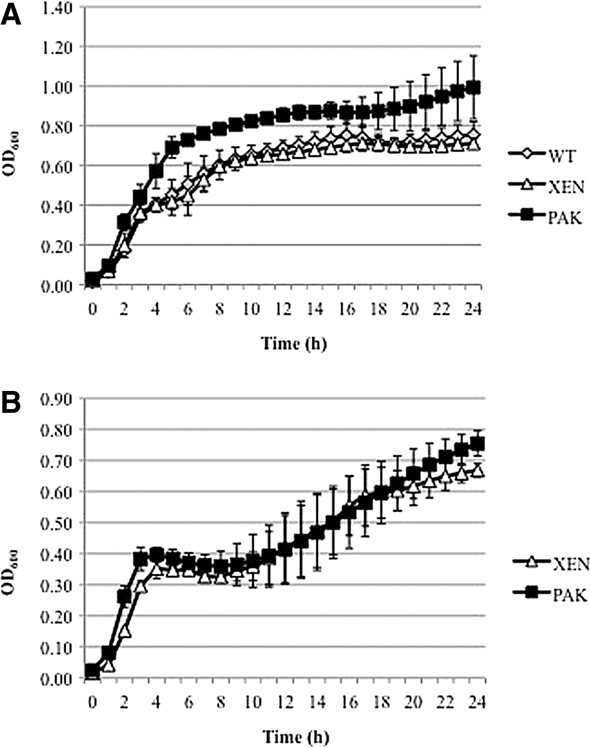
Growth of Escherichia coli O157:H7 transformed with either the bioluminescent plasmid pAK1-lux or pXEN-13. The absorbance at 600 nm of cultures of the non-transformed parental strain (wild type) (WT; ♦) and WT transformed with either the plasmid pXEN-13 (▲) or pAK1-lux (■) was monitored over 24 h in either the absence (
Bioluminescence studies have suggested correlations with in vitro enzyme activity; the amount of luciferase protein and lux mRNA generated directly correlates with cell viability (Close et al., 2010). The two plasmids utilized in this study were selected on the basis of harboring the luxCDABE operon. The PAK plasmid was developed as a wide range host plasmid for gram-negative bacteria (Karsi et al., 2006). Research with this plasmid has suggested stability and luminescence without antibiotic pressure for at least 8 d, though the percentage of photonic emissions decreases past 0 d (Moulton et al., 2009a). The XEN plasmid carries the original Photorhabdus luminescens operon for production of luminescence in gram-negative bacteria (Harms et al., 2009). The XEN plasmid has been introduced into E. coli strains previously and reported to emit photonic emissions within the mouse model (Harms et al., 2009). In terms of emission of photons over time, Moulton et al. (2009b) reported a gradual 30% decline in bacterial emissions over an 8-d period (100% on d 0 to 66% on d 8) in the absence of antibiotic pressure. Moulton et al. (2009b) suggested that the results of their study indicate that bioluminescent plasmids may permit real-time monitoring for 1–6 d in situations where antibiotic pressure may not be feasible. The data reported within this study, coupled with the results of Moulton et al. (2009b), suggest that transformants will function similarly to the parental strain and greater than 65% retention of the plasmid up to 6 d post-inoculation.
Growth in ruminal fluid fermentation
Many studies have demonstrated that E. coli O157:H7 can be cultured from the rumen, but it does not grow rapidly or extensively in this environment (Grauke et al., 2002). The PAK and XEN transformants grew similarly (p=0.21) to the WT within mixed ruminal microbial fluid (Fig. 2). Growth was monitored by viable plate counts, and populations were recorded in log10 (CFU/mL) populations. There was no difference (p=0.18) observed within populations between the WT and XEN transformed strains throughout the time period analyzed. The only difference observed within the study was at 4 h, when PAK had greater populations than XEN (p=0.004), but was not different from the WT (p=0.10). There was no difference (p=0.11) observed in the two transformants compared with the controls when grown in mixed microbial ruminal fluid fermentations, thus suggesting that both transformants can survive the harsh rumen conditions and grow similarly to WT.
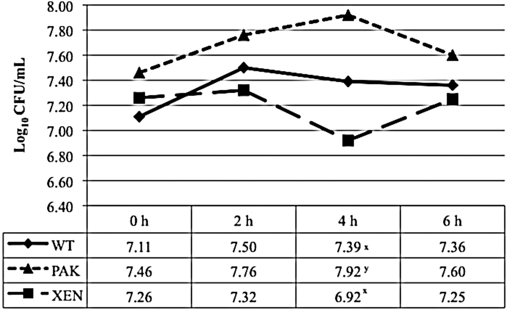
The least squares (LS)–means of Escherichia coli O157:H7 in bovine mixed ruminal microbial incubations. Growth of the non-transformed parental strain (wild type) (WT; ♦), or WT containing the plasmid pAK1-lux (PAK; ▲) or pXEN-13 (XEN; ■) was monitored by viable plate counts during a 6-h growth period in bovine mixed ruminal fluid medium. Values represent the LS-means (log10 CFU/mL) from three independent replicates. The x,y-values without a common superscript letter wihin a column differ (p<0.05). Standard deviations: WT=0.35; PAK=0.15; XEN=0.16.
Growth in fecal fluid fermentation
E. coli O157:H7 can contaminate beef carcasses via fecal spread from hide or spilled digesta, and it has been suggested that 30% of cattle harbor E. coli O157:H7 or other STEC in their feces, depending on the season (Callaway et al., 2003; Elder et al., 2000). The WT and the two transformants grew similarly (p=0.28) within mixed fecal microbial fluid fermentations (Fig. 3). There was no difference (p=0.14) observed within populations between the WT and XEN for the duration of the study. The only difference observed was at 6 h, when XEN exhibited greater populations than PAK (p=0.01), but was not different than the WT (p=0.18). Overall, the transformants and WT grew similarly in the presence of a mixed fecal microbial fluid environment throughout the duration of the study. These data suggest that the transformants function similarly within the fecal fluid media. However, further research is needed to determine if the transformants will be capable of surviving the competitive fecal environment similarly to the WT within the animal.
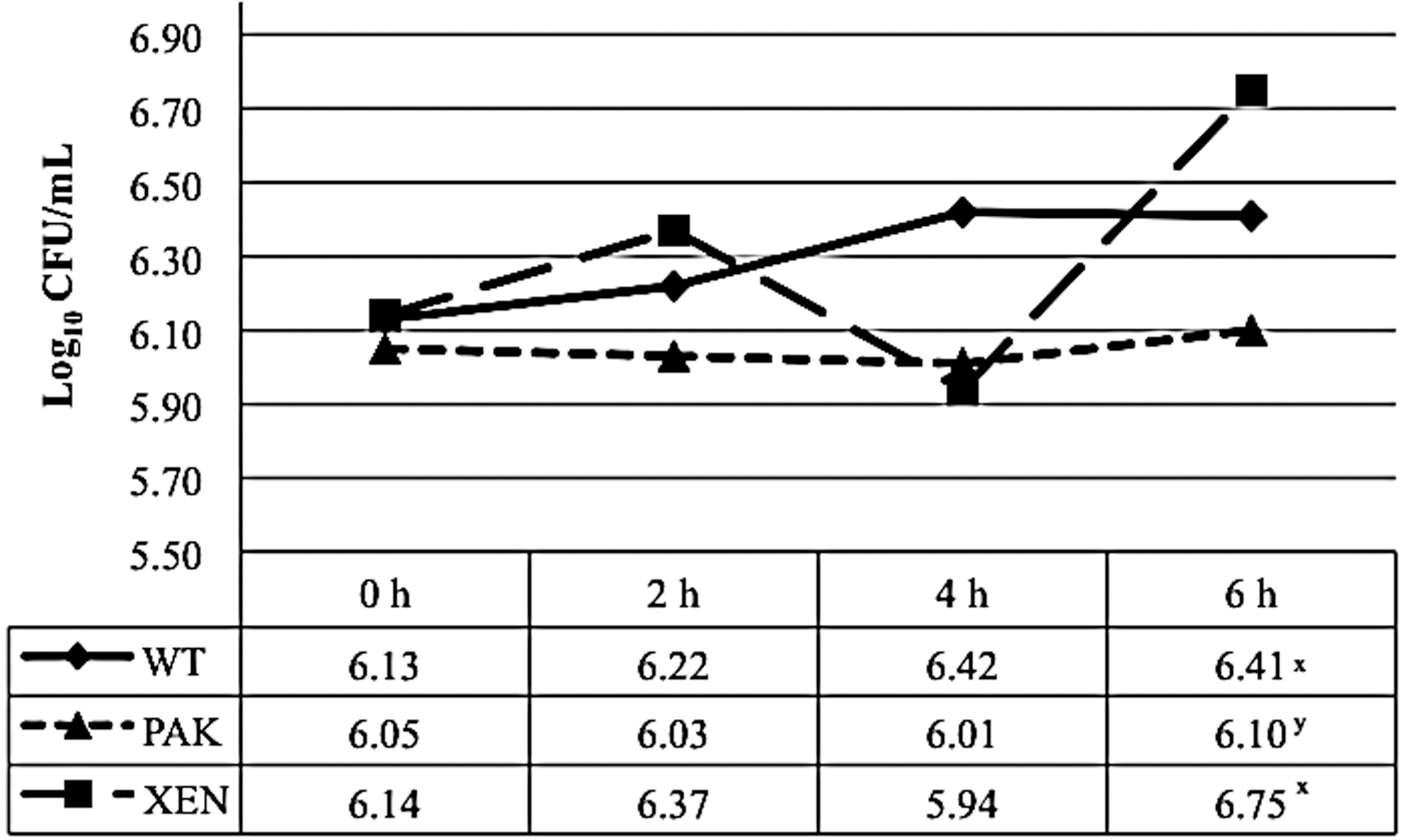
The least squares (LS)–means of Escherichia coli O157:H7 in bovine mixed fecal microbial incubations. Growth of the non-transformed parental strain (wild type) (WT; ♦), or WT containing the plasmid pAK1-lux (PAK; ▲) or pXEN-13 (XEN; ■) was monitored by viable plate counts during a 6-h growth period in bovine mixed fecal fluid medium. Values represent the LS-means (log10 CFU/mL) from three independent replicates. The x,y-values without a common superscript letter wihin a column differ (p<0.05). Standard deviations: WT=0.25; PAK=0.05; XEN=0.98.
Comparison of photonic emissions by PAK and XEN
Transforming bacteria with the bioluminescent plasmids has been suggested to be a successful means of tracking bacteria in real time (Moulton et al., 2009b). Photonic emissions gradually decreased from 0 h for both plasmids within ruminal fluid, indicating that there was a time effect (p=0.001; Table 1). The RLU values for PAK ranged from 16,226 to 880.2 ph/pix s. The XEN plasmid had the greatest RLU values at 0 h (292.3 ph/pix s) and the least at 6 h (86.3 ph/pix s). The PAK plasmid had greater RLU values, yet was only different from XEN at 0 h (p=0.002) and 2 h (p=0.02). RLUs decreased over time for both plasmids with similar emission values at 4 h (p=0.22) and 6 h (p=0.80), indicating that there was a tendency (p=0.07) for a transformant effect. This decrease in RLUs is most likely a result of increased turbidity of the rumen media and/or a decrease in availability of nutrients, not a decrease in retention of the plasmid. Moulton et al. (2009b) and Broadway (2011) have both reported retention of the plasmids for 8 and 10 d, respectively.
Within mixed fecal microbial fluid medium, both plasmid RLU values decreased from 0 h, indicating that time impacted photonic emissions (p=0.001; Table 1). The PAK plasmid ranged from 5513.7 to 1404.4 ph/pix s. The XEN plasmid RLU values ranged from 112.9 to 34.6 ph/pix s. The two plasmids were different from 0 h (p=0.009) and 2 h (p=0.04), with a tendency to be different at 4 h (p=0.06). At 6 h, the plasmids were not different (p=0.29), further indicating a tendency for transformant (p=0.06) to impact photonic emissions, while also indicating a transformant-by-time interaction (p=0.001). Again, this decrease in RLUs is most likely related to increased turbidity (decreasing the ability to capture the emission of photons) or decreased availability of nutrients, and not a loss of the plasmid.
Conclusion
These data indicate that the PAK and XEN transformants are stable and can persist in rumen and fecal-like environments. Furthermore, while grown in these harsh, competitive environments, these transformants still displayed luminescence and could be a useful method for pathogen tracking in real-world environments.
Footnotes
Acknowledgments
We would like to thank Dr. Peter Ryan and Dr. Scott Willard for their knowledge and support. We would also like to thank the Biophotonic Initiative USDA-ARS 58-6402-3-0120 for funding to acquire the imaging equipment. This work was supported by the MAFES Special Research Initiative at Mississippi State University.
Disclosure Statement
No competing financial interests exist.