




Abstract
Salmonella, one of the most important pathogens transmitted by food, especially poultry, has the ability to form biofilms on surfaces. Its adhesion can be influenced by different physicochemical properties of these surfaces, while Salmonella uses fimbriae and produces cellulose as the main matrix components of biofilms. Their synthesis is co-regulated by a LuxR-type regulator, the agfD (aggregative fimbriae, curli), and adrA genes, respectively. Thus, this study investigated the production of biofilm by Salmonella spp. isolated from raw poultry (breast fillet), purchased in Botucatu, Sao Paulo, Brazil, on glass, polyvinyl chloride, and stainless steel at different temperatures (16°, 20°, 28°, and 35°C). We analyzed the frequency of the agfD and adrA genes and the rdar morphotype at 28°C and 35°C in isolated strains. We found Salmonella in 112 of 240 poultry samples (46.7%), and 62 strains previously isolated from the same kind of food were included in the study on biofilm development, gene expression, and rdar morphotype. All of them were positive for both genes, and 98.3% were able to produce biofilm in at least one temperature. The rates of rdar morphotype at 28°C and at 35°C were 55.2% (96 strains) and 2.3% (4 strains), respectively. Glass was the best material to avoid biofilm production, while Salmonella grew even at 16°C on stainless steel. These results point out the need for more effective sanitizing processes in the slaughter plants in order to avoid the permanence of these bacteria in food and eventual human foodborne diseases.
Introduction
R
Persistence of Salmonella spp. on equipment and instruments of the food industry can be an important cause of foodborne diseases, since strains with higher ability to produce biofilm seem to be more resistant to trisodium phosphate and chlorination (Xu et al., 2010; Hasegawa et al., 2011). In fact, Vestby et al. ( 2009) argue that biofilm produced by Salmonella strains may be an important factor for its longevity in the food-processing environments.
Polyvinyl chloride (PVC) is a hydrophobic surface that favors bacterial adhesion, whereas microorganisms are less likely to adhere to hydrophilic surfaces, such as stainless steel and glass (Simões et al., 2008). In a food-processing plant, pipes and parts of the poultry water feeders are usually made of PVC (Trachoo et al., 2002), whereas stainless steel is often used because of its mechanical strength, and resistance to corrosion by chemicals and sanitizing agents. Its versatility for manufacturing allows a wide application in sinks and machinery (Boulanger-Peterson, 1996). Finally, glass is also widely used in butcher shops, coating reservoirs, and for pots and boards at home.
Salmonella has demonstrated the ability to form biofilms on abiotic surfaces such as plastic, rubber, cement, glass, and stainless steel (Joseph et al., 2001; Solano et al., 2002; Prouty and Gunn, 2003; Arnold and Yates, 2009; Hurrell et al., 2009; Moretro et al., 2009). Aggregative fimbriae (curli) play an important role in biofilm formation and the aggregative fimbriae genes (agf ), also known as csg, are organized in two operons, agfBAC and agfDEFG, being involved in their synthesis (Collinson et al., 1996). Cellulose is another important component to develop biofilms and is controlled by bcsA, bcsB, bcsZ, and bcsC (bacterial cellulose synthesis) genes.
The production of fimbriae is co-regulated by a LuxR-type regulator gene, agfD, which indirectly acts on the gene adrA, in order to regulate the cellulose production (Römling, 2002; Zakikhany et al., 2010). Therefore, when cellulose synthesis is associated with the presence of curli fimbriae, Salmonella colonies develop a distinctive phenotype on Luria Bertani (LB) agar plates, showing a characteristic morphology of red, dry, and rough (rdar morphology) at 28°C, but not at 37°C (Solano et al., 2002; Gerstel and Römling, 2003). This morphology provides higher resistance to desiccation and the disinfection processes, increasing the survival of microorganisms (Anriany et al., 2001; Scher et al., 2005; White et al., 2006). Under these conditions, Salmonella displays the rdar morphology, and the agfD gene seems to be necessary for the maturation of biofilms and is responsible for regulating the expression of all major constituents of this matrix (Grantcharova et al., 2010). Other studies have reported that this gene may be susceptible to environmental stimulation such as temperature, nutrients, oxygen tension, and pH (Gerstel and Römling, 2003).
Thus, in this study we examined the presence of two genes responsible for biofilm formation in Salmonella spp. isolated from raw poultry and their capacity to produce biofilm at different temperatures and on different materials (glass, PVC, and stainless steel). This study was developed to test the hypothesis that most Salmonella strains isolated from poultry samples are able to form biofilms and to find the best conditions to avoid its development. Our results show that 46.7% of samples were contaminated by Salmonella spp., 98.3% of which are able to produce biofilms on diverse surfaces and under varied temperatures. Glass was the least favorable material for its development independent of the tested temperature.
Materials and Methods
Samples
Two hundred forty samples of raw poultry were purchased in supermarkets and butcher shops in Botucatu, Sao Paolo, Brazil and immediately transported in a refrigerated isothermal box with ice (8°C). The samples were collected in the morning and kept at 4°C until processing in the afternoon. Sixty-two additional strains of Salmonella, previously isolated from poultry, were used for the biofilm-producing assay.
Isolation and identification
All culture media, except when specified, were from Oxoid (Oxoid, Basingstoke, UK). The detection of Salmonella was performed according to Andrews et al. (2001), and the identification was done using the API-20E System (bioMérieux, l'Etoile, France). Salmonella spp. strains were confirmed by agglutination tests using polyvalent somatic and flagellar antisera (Probac, São Paulo, Brazil).
Detection of biofilm-producing genes in Salmonella
Salmonella strains were inoculated into brain heart infusion (BHI) broth at 35°C/24 h. Following incubation, 1 mL was transferred to microtubes and centrifuged at 10,000×g for 10 min. The supernatant was disposed and the pellet was resuspended in 1 mL of phosphate-buffered salt solution (PBS; 0.01 M, pH 7.2). This step was repeated twice, and the pellet was resuspended in 200 μL of lysis buffer (50 mM Tris-HCl, 1 mM EDTA, 0.025% Tween, 0.2 mg proteinase K), and incubated in a water bath at 56°C for 1 h and then at 95°C for 10 min. A new centrifugation step was performed at 13,000×g for 5 min and the supernatant was used for the PCR reaction (Arnold et al., 2004).
PCR reactions were performed using a total volume of 25 μL per sample (2.5 μL of 10X PCR buffer, 2.5 mM magnesium chloride, 200 mM of each dNTP [Fermentas, St. Leon Rot, Germany], 1.25 U of Taq DNA polymerase [Fermentas], 10 pmol of each primer [Table 1], autoclaved ultrapure water [qs] [Milli-Q Plus, Millipore, Billerica, MA], and 3 μL of DNA sample). PCR was performed in a GeneAmp PCR System 9700 thermocycler (Applied Biosystem, Wellesley, MA) offset for an initial cycle at 94°C for 5 min for initial denaturation, followed by 35 cycles of 94°C/30 s, 60°C/30 s, and 72°C/30 s. The final temperature for extension was 72°C/4 min. Nucleic acid was replaced by ultrapure water as negative control, and a standard strain of Salmonella Typhimurium ATCC 14028 was used as positive control. The primer pair agfD (forward: TGCGGACTCGGTGCTGTTGT; reverse: CAGGAACACGTGGTCAGCGG; 123 bp) was designed using the program Primer Blast (
p<0.001.
SS, stainless steel; PVC, polyvinyl chloride; NP, no producer of biofilm.
Verification and quantification of biofilm production
The production of biofilm was tested on three different materials: stainless steel (AISI 304), glass, and PVC, taking in account that stainless steel comprises almost all surfaces in the process of poultry slaughtering; glass is used as cutting surfaces, and PVC is the main component of poultry drinking fountains.
Preparation of plates
We used circular chips of stainless steel (1-cm diameter), PVC squares (1 cm2), and glass slides (1.3-cm diameter). These materials were properly washed, dried, and placed into Petri dishes for sterilization in an autoclave. Then they were placed into the wells of a 24-well culture plate.
Preparation and inoculation of the culture plate
Salmonella strains were incubated in BHI broth and incubated at 35°C for 24 h. The cultures had been diluted using Densichek (bioMérieux, l'Etoile, France), and an aliquot of 600 μL in a final concentration of 108 colony-forming units of bacteria was distributed in triplicate into the wells of the 24-well plate and incubated at 16, 20, 28, or 35°C for 96 h. These temperatures were chosen because 16°C is required by Brazilian sanitary service at the prechiller plant (MAPA, 1998); 20°C is the real average temperature of the water in the prechiller; 28°C is the optimum temperature for the development of rdar morphology, and 35°C is the optimum temperature for Salmonella growth.
Quantification of biofilm production
After the incubation time, the chips were transferred to a new 24-well plate, in order to prevent the quantification of biofilm that eventually formed on the bottom and walls of the plastic plate. These chips were washed three times with PBS for the removal of unfixed cells and dyed with 1% crystal violet for 15 min. Dyeing solution excess was removed and the plates were washed again with PBS. Next, the biofilm was resuspended in 300 μL of glacial acetic acid for 15 min, which ensured the dissolution of crystal violet for colorimetric measurement. Two hundred microliters of each dyed supernatant was transferred to a 96-flat-bottomed-well microplate, and optical density (OD) was read in an enzyme-linked immunosorbent assay reader (Babsystems, Multiskan EX) at 560 nm. Noninoculated BHI broth was used as a negative control (ODc) (Stepanovic et al., 2004), and an average of three wells was used to correct the absorbance value. BHI broth inoculated with Salmonella Typhimurium ATCC 14028 was used as a positive control (Kim and Wei, 2009; modified).
Salmonella strains were classified as strong, moderate, or weak biofilm producer, according to Stepanovic et al. (2000), as follows: OD ≤ODc=no biofilm producer; ODc<OD≤(2X ODc)=weak biofilm producer; (2XODc)<OD≤(4XODc)=moderate biofilm producer; (4XODc)≤OD=strong biofilm producer. (ODc: 0.152).
Analysis of rdar colony morphology
Colony morphology was observed on LB agar without salt, supplemented with Congo red (40 μg/mL) and Coomassie Brilliant Blue (20 μg/mL). The Salmonella strains were grown in BHI broth at 35°C for 18 h, plated on this agar, and incubated at 28°C and 35°C for 96 h, with daily reading of colony morphology (Malcova et al., 2008).
Scanning electron microscopy
In order to visualize biofilms attached to the glass, stainless steel, and PVC slides, these supports were fixed in 3% glutaraldehyde–PBS solution (pH 7.4) and dehydrated with acetone. The samples were prepared as described by Austin and Bergeron (1995). The samples were examined using a scanning electron microscope (Quanta 200; Fei Company) under a 30-kV acceleration.
Results
We first observed that 112 of 240 samples of raw poultry (46.7%) presented Salmonella spp. An additional 62 strains previously also isolated from poultry increased our sampling to 174 Salmonella strains. We observed that 171 (98.3%) of these strains were able to produce biofilm on at least one kind of material and at one of the tested temperatures. As can be observed in Table 1, most of strains were classified as weak biofilm producers, and just a small number of them produced strong biofilms.
The agfD and adrA genes were identified in all of the isolated strains (Fig. 1). However, the rdar morphology was observed in only 96 (55.2%) strains at 28°C and just 4 (2.3%) produced this morphology at 35°C (p<0.0001), indicating that 28°C is the best temperature to visualize the biofilm production in Petri dishes. Therefore, rdar morphotyping cannot be used as a marker of the presence of these genes.
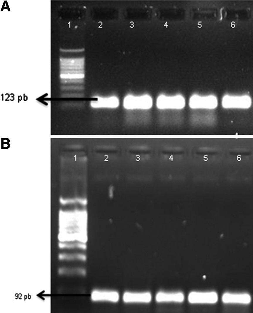
Polymerase chain reaction amplification for detection of
Biofilm production was dependent on the temperature and the material (Table 1), and glass was consistently the best material to resisted biofilm formation, demonstrating significantly lower production at three of the four temperatures analyzed. Images obtained by scanning electron microscopy confirmed biofilm formation by analyzed strains (Fig. 2).
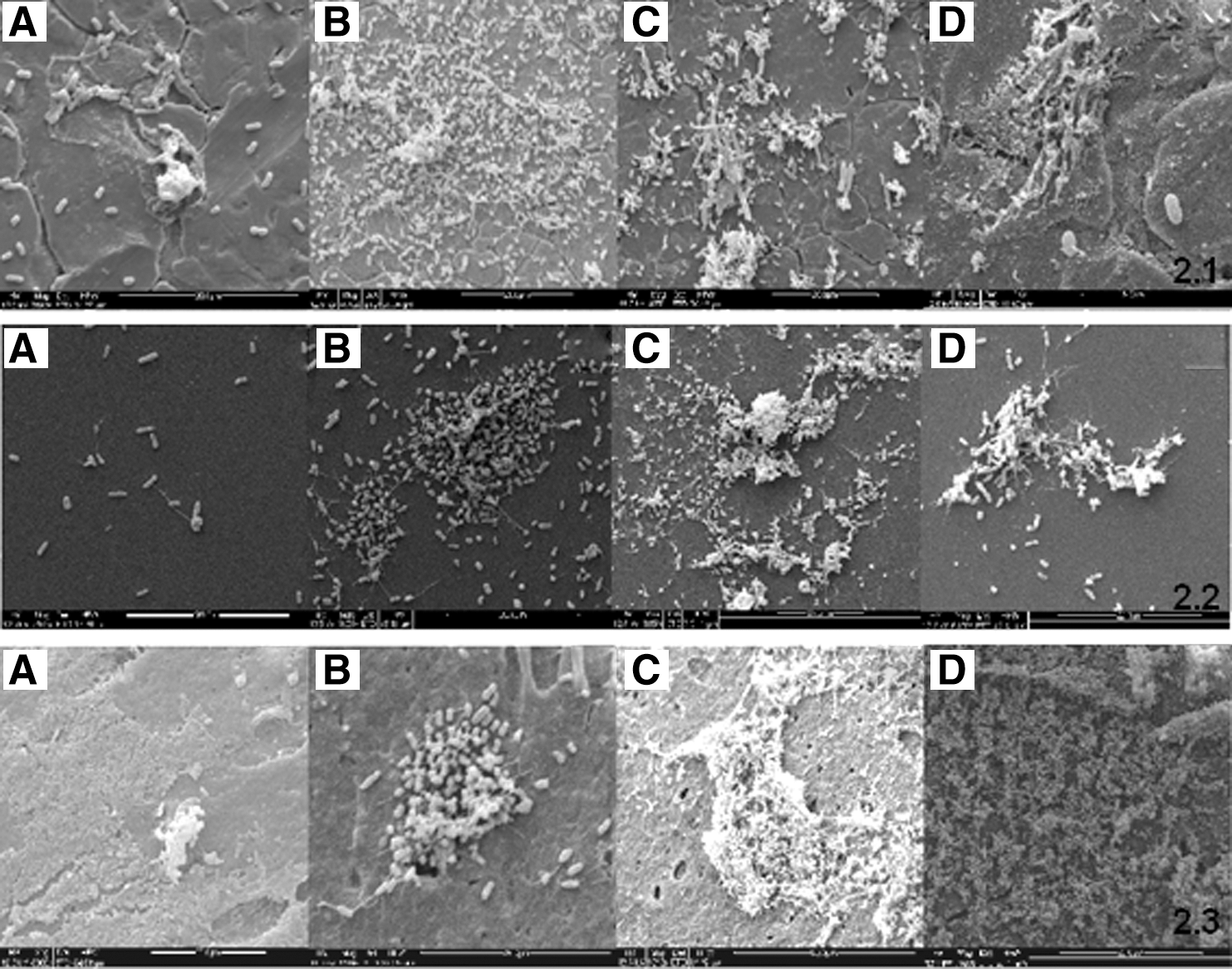
Biofilms of Salmonella spp. produced in stainless steel (2.1), glass (2.2), and polyvinyl chloride (2.3) at
Discussion
A large majority of strains, 171/174 (98.3%), produced biofilms on at least one kind of material and at one of the four tested temperatures. A high percentage of biofilm-producing strains was also observed by Solano et al. (2002), who found a similar rate (97%), while Lu et al. (2011) found a lower percentage (63%).
It is interesting to note that Solano et al. (2002) tested Salmonella Enteritidis and Lu et al. (2011) tested Salmonella Pullorum. Unfortunately, the serovars of the strains in this work were not identified, but since all the 174 strains were isolated from poultry, it can be considered that most of them are probably Salmonella Enteritidis, the most common serotype in Brazil (Oliveira et al., 2007). According to Gerstel and Römling (2001), most serovars with broad host range such as Salmonella Typhimurium and Salmonella Enteritidis highly express the rdar morphotype below 30°C, while others, such as Salmonella Typhi and Salmonella choleraesuis, restricted to certain hosts did not express this morphology (Römling et al., 2003).
Although we have observed a strong association between agfD and adrA genes and the production of biofilm, just a limited proportion of strains was able to produce the characteristic rdar morphology on LB culture medium. The lower percentage of rdar colonies at 28°C (2.3%) could be explained by the observation that thin aggregative fimbriae production in Salmonella spp. is regulated by environmental conditions that play a role on the agfD promoter for triggering the cascade of biofilm production (Römling et al., 1998). In addition, according to Gerstel and Römling (2003), oxygen and pH variation can also interfere on biofilm formation by Salmonella Typhimurium, and directly influence the expression of rdar morphology. Another possibility is that the rdar morphology test is not sensitive enough to detect weak biofilm-producer strains.
Römling et al. (1998) observed that the expression of thin aggregative fimbriae occurs at 37°C only in the absence of iron. Solano et al. (2002) showed that only Salmonella Typhimurium expressed cellulose production at 35°C, but they used a different method, the air–liquid interface test. We did not identify the serotype of our Salmonella spp. strains, but considering that Salmonella Enteritidis is considered the more frequent serovar in Brazil, it is possible that we have had a low frequency of Salmonella Typhimurium.
The influence of the temperature on biofilm production varied with each surface material. Glass was the surface that presented the lower susceptibility to colonization, independent of the temperature, whereas stainless steel better avoided the biofilm production at 28°C, and PVC at 20°C. The high frequency of strains producing biofilm on stainless steel at 16°C is worrying, because the permanency of Salmonella on this surface in the beginning of industrial processing can be an important source of poultry contamination and a potential cause of foodborne diseases.
Our results are in contrast with those reported by Sinde and Carballo (2000), who observed that Salmonella adhered more easily to hydrophobic materials than to stainless steel (more hydrophobic). Joseph et al. (2001) also observed higher production of biofilm by Salmonella spp. on plastic than on stainless steel.
Stepanovic et al. (2003) tested 30 strains of Salmonella spp. and observed a similar proportion of biofilm-forming strains (97%) on polystyrene plates at 30°C after 48-h incubation. Their isolates also grew at 37°C (93%) and 22°C (90%). The authors found higher percentages of strains presenting capacity to form strong biofilm than we did, since just 9 of our 174 Salmonella strains formed strong biofilm. Interestingly, those authors observed that, despite the fact that the temperature of 22°C showed a low percentage of strains capable of producing the matrix (90%), this temperature had the highest number of biofilm-forming-capacity strains (30%).
The differences between our results and those reported by Stepanovic et al. (2003) may be explained by the different methodology and materials used, but mainly because of the different sources of samples, since they tested strains isolated from humans and other nonspecified foods. In 2004, Stepanovic et al. performed a new study on the capacity of Salmonella to produce biofilm on polystyrene, using 122 strains of Salmonella isolated from different sources and cultured in different media. They found that only 1.6% of the strains produced a strong biofilm at 28°C in BHI broth. In the present work, under the same conditions, we observed 1.7%, of strains growing on stainless steel and glass, but PVC (the most similar to polystyrene material) was not colonized.
Most published works included in the broad literature reviewed (Römling et al., 1998; 2000; Stepanovic et al., 2003; Malcova et al., 2008) used polystyrene, a hydrophobic material like PVC, but which is not approved for use in Brazilian food industries. In addition, the published studies do not provide the degree of hydrophobicity of the PVC and polystyrene, making it impossible to perform a real comparison between the materials.
In addition, studies that tested stainless steel or glass used few strains, normally two or three, since they were focusing the genetic manipulation of biofilm producer strains (Latasa et al., 2005; Malcova et al., 2008; Kim and Wei, 2009).
In conclusion, the agfD and adrA genes were widely dispersed in this genus, and were strongly associated with the ability to produce biofilm. Although all the isolates had both analyzed genes, the biofilm formation was not associated with rdar morphology of bacterial colonies. Since there were some strains that did not produce biofilms, we suggest that other genes may also be involved in this process.
Footnotes
Acknowledgments
We are grateful to FAPESP (Fundação de Amparo à Pesquisa do Estado de São Paulo) for financial support (Process 2010/06436-7).
Disclosure Statement
No competing financial interests exist.